- Phare
- Validé par KD/KO
Anticorps Polyclonal de lapin anti-RIPK1-Specific
RIPK1-Specific Polyclonal Antibody for WB, IHC, ELISA
Hôte / Isotype
Lapin / IgG
Réactivité testée
rat, souris et plus (3)
Applications
WB, IHC, IP, CoIP, ELISA, IF
Conjugaison
Non conjugué
111
N° de cat : 17519-1-AP
Synonymes
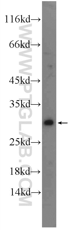
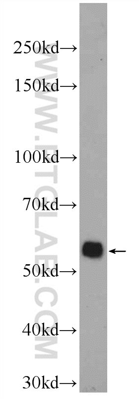
Galerie de données de validation
Applications testées
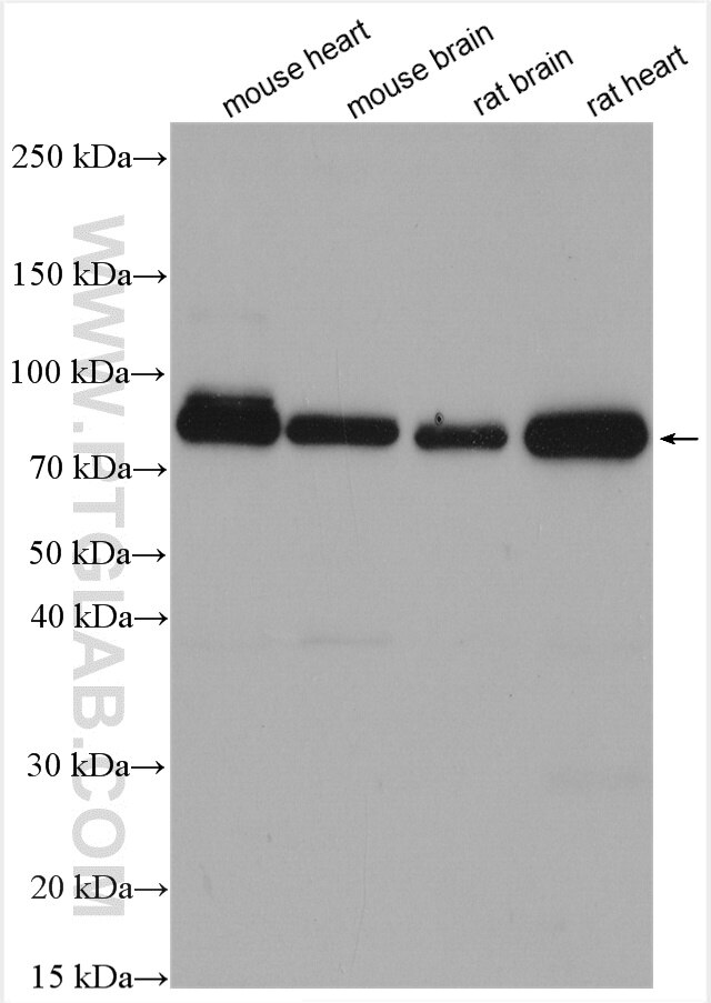
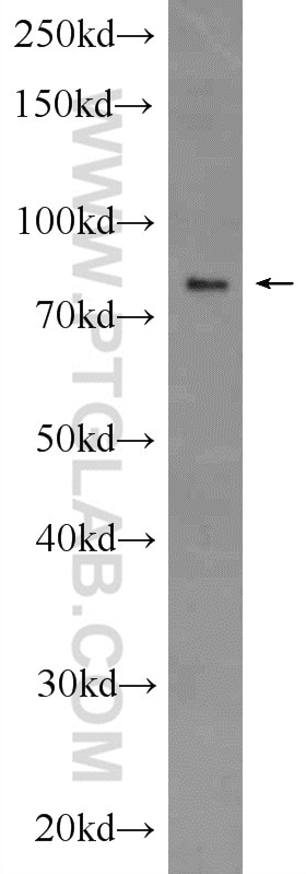
| Résultats positifs en WB | tissu cardiaque de souris, tissu cardiaque de rat, tissu cérébral de rat, tissu cérébral de souris, tissu hépatique de rat |
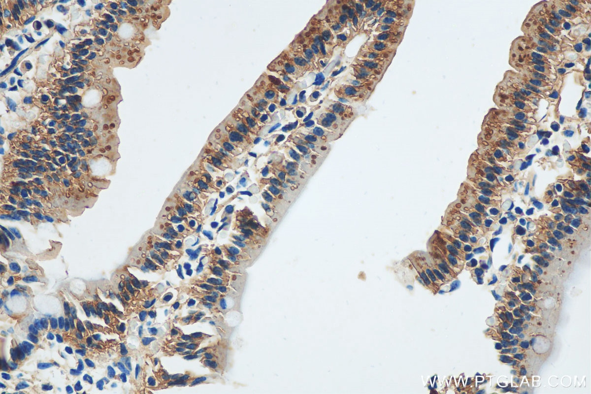
| Résultats positifs en IHC | tissu d'intestin grêle de souris, il est suggéré de démasquer l'antigène avec un tampon de TE buffer pH 9.0; (*) À défaut, 'le démasquage de l'antigène peut être 'effectué avec un tampon citrate pH 6,0. |
Dilution recommandée
| Application | Dilution |
|---|---|
| Western Blot (WB) | WB : 1:500-1:3000 |
| Immunohistochimie (IHC) | IHC : 1:50-1:500 |
| It is recommended that this reagent should be titrated in each testing system to obtain optimal results. | |
| Sample-dependent, check data in validation data gallery | |
Applications publiées
| KD/KO | See 1 publications below |
| WB | See 99 publications below |
| IHC | See 13 publications below |
| IF | See 14 publications below |
| IP | See 4 publications below |
| CoIP | See 2 publications below |
Informations sur le produit
17519-1-AP cible RIPK1-Specific dans les applications de WB, IHC, IP, CoIP, ELISA, IF et montre une réactivité avec des échantillons rat, souris
| Réactivité | rat, souris |
| Réactivité citée | rat, Humain, levure, porc, souris |
| Hôte / Isotype | Lapin / IgG |
| Clonalité | Polyclonal |
| Type | Anticorps |
| Immunogène | Peptide |
| Nom complet | receptor (TNFRSF)-interacting serine-threonine kinase 1 |
| Masse moléculaire calculée | 76 kDa |
| Poids moléculaire observé | 70-80 kDa |
| Numéro d’acquisition GenBank | NM_003804 |
| Symbole du gène | RIPK1 |
| Identification du gène (NCBI) | 8737 |
| Conjugaison | Non conjugué |
| Forme | Liquide |
| Méthode de purification | Purification par affinité contre l'antigène |
| Tampon de stockage | PBS avec azoture de sodium à 0,02 % et glycérol à 50 % pH 7,3 |
| Conditions de stockage | Stocker à -20°C. Stable pendant un an après l'expédition. L'aliquotage n'est pas nécessaire pour le stockage à -20oC Les 20ul contiennent 0,1% de BSA. |
Informations générales
RIPK1(Receptor-interacting serine/threonine-protein kinase 1) is primarily involved in mediating TNF-R1-induced cell activation, apoptosis, and necroptosis and belongs to a novel class of kinases that function in cell survival and cell death mechanisms(PMID:22685397 ). It has 2 isoforms produced by alternative splicing. It also can exist as a protein with a lower molecular weight of about 25 kDa and 45 kDa in HAEC and HUVEC(PMID:22685397).
Protocole
| Product Specific Protocols | |
|---|---|
| WB protocol for RIPK1-Specific antibody 17519-1-AP | Download protocol |
| IHC protocol for RIPK1-Specific antibody 17519-1-AP | Download protocol |
| Standard Protocols | |
|---|---|
| Click here to view our Standard Protocols |
Publications
| Species | Application | Title |
|---|---|---|
Acta Pharm Sin B A new perspective of triptolide-associated hepatotoxicity: the relevance of NF- κ B and NF- κ B-mediated cellular FLICE-inhibitory protein. | ||
Nat Commun Regulation of RIPK1 activation by TAK1-mediated phosphorylation dictates apoptosis and necroptosis. | ||
Sci Total Environ Evaluation of 6-PPD quinone toxicity on lung of male BALB/c mice by quantitative proteomics | ||
Cancer Res Proteomic mapping and targeting of mitotic pericentriolar material in tumors bearing centrosome amplification. | ||
Environ Pollut Polystyrene nanoplastics and cadmium co-exposure aggravated cardiomyocyte damage in mice by regulating PANoptosis pathway |
Avis
The reviews below have been submitted by verified Proteintech customers who received an incentive forproviding their feedback.
FH Yuan (Verified Customer) (01-07-2020) | This RIPK-1 antibody was tried on the MCF7 tamoxifen resistant (5uM) cell line, the 74 kDa band was very faint, but the 45 kDa band is clear. In addition, some unspecific bands near 45 kDa showed up too.
|
