- Phare
- Validé par KD/KO
Anticorps Polyclonal de lapin anti-AMPK Alpha
AMPK Alpha Polyclonal Antibody for WB, IP, IHC, ELISA
Hôte / Isotype
Lapin / IgG
Réactivité testée
Humain, rat, souris et plus (5)
Applications
WB, IHC, IP, ELISA, IF
Conjugaison
Non conjugué
213
N° de cat : 10929-2-AP
Synonymes
Galerie de données de validation
Applications testées
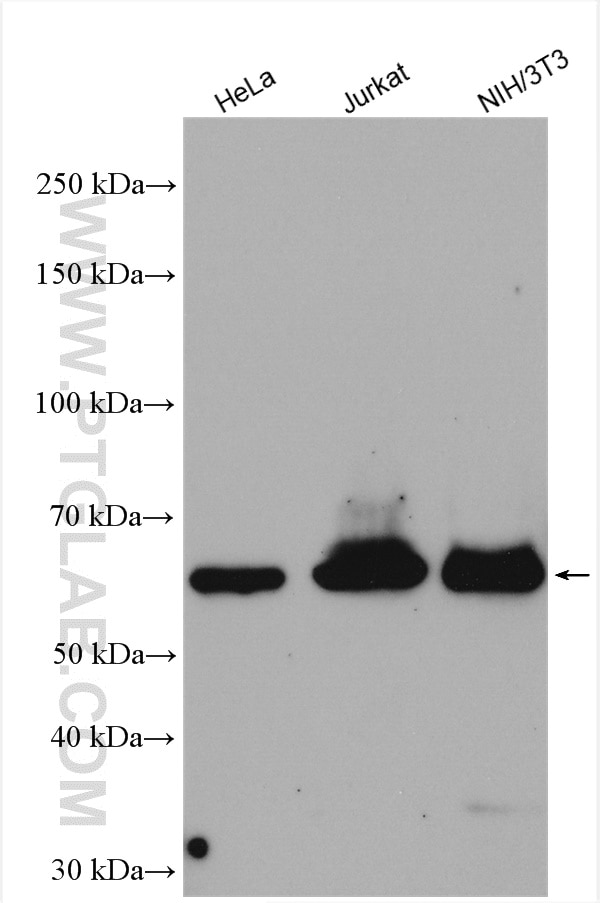
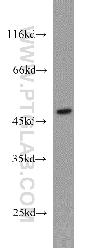
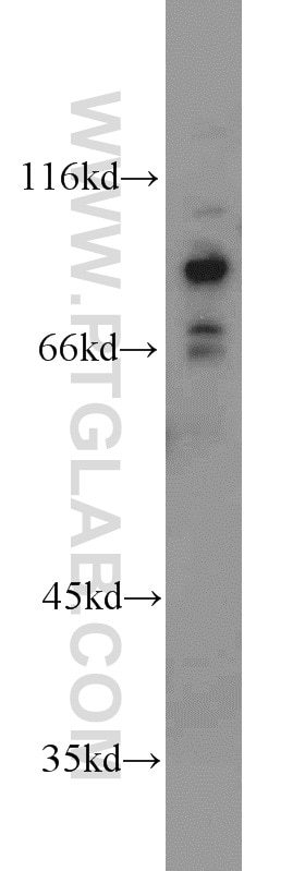
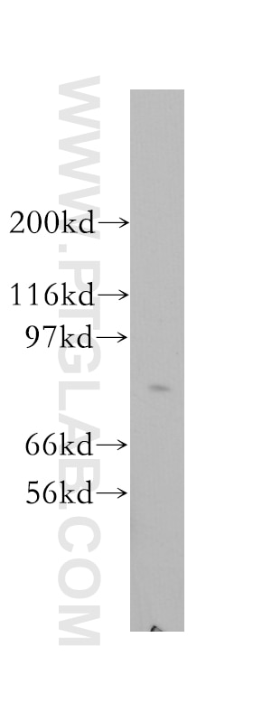
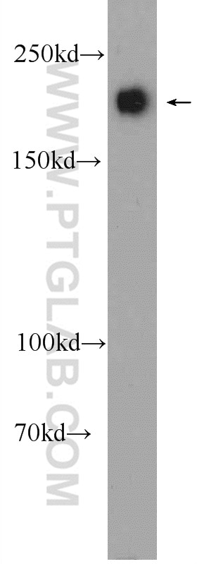
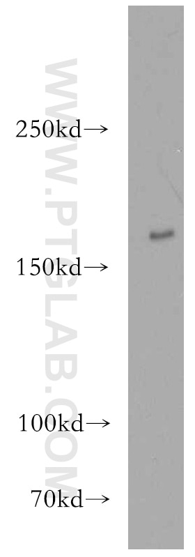
| Résultats positifs en WB | cellules HeLa, cellules Jurkat, cellules NIH/3T3 |
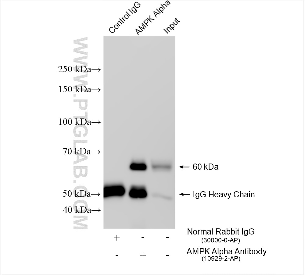
| Résultats positifs en IP | cellules HeLa, |
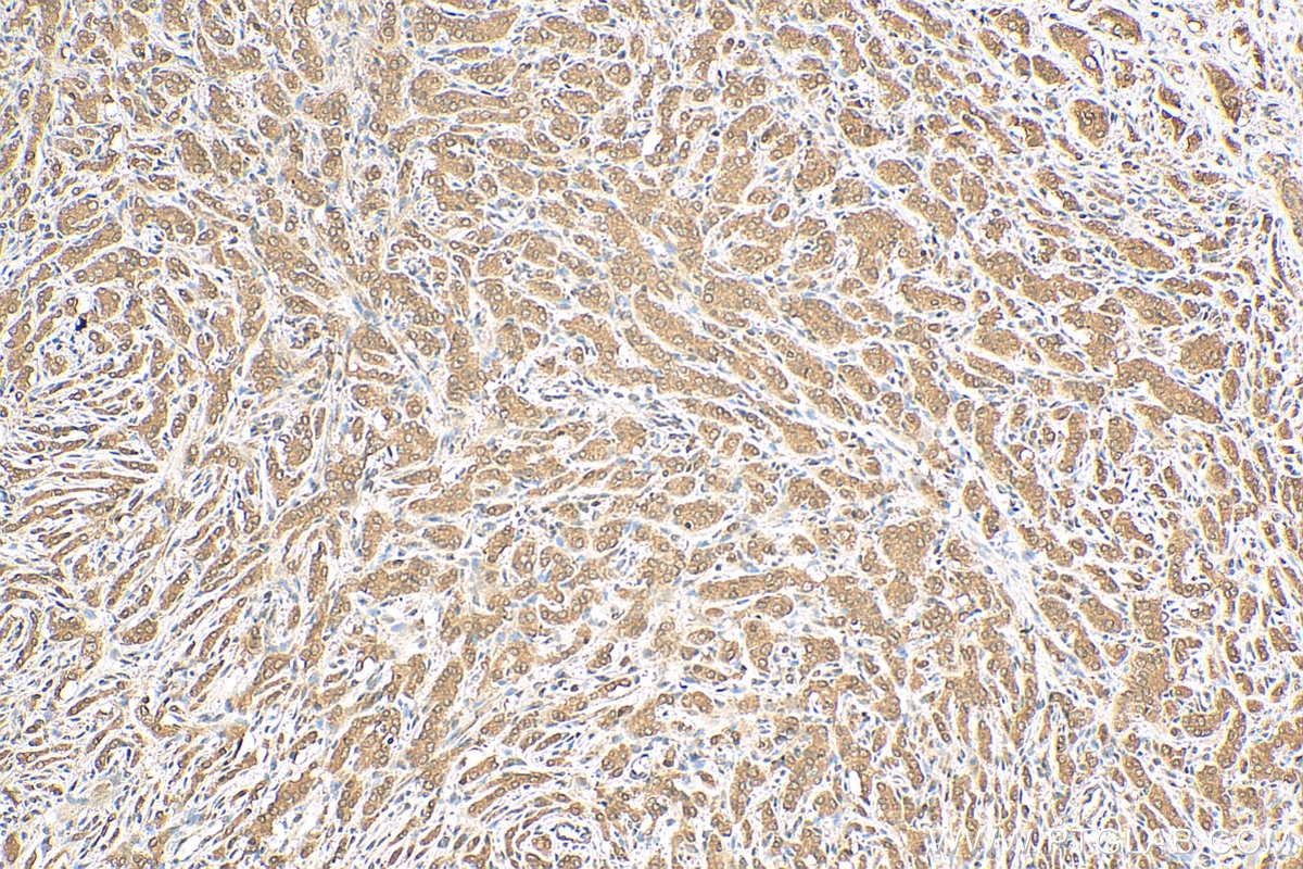
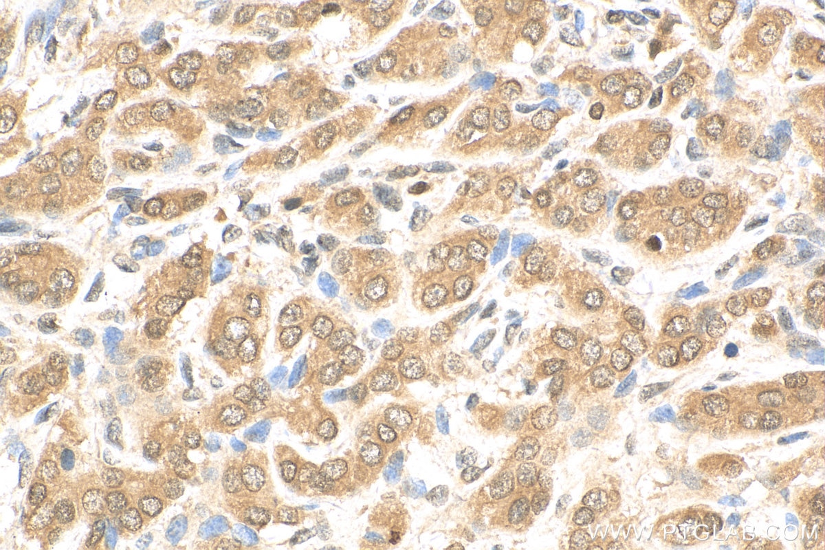
| Résultats positifs en IHC | tissu de cancer de la prostate humain, il est suggéré de démasquer l'antigène avec un tampon de TE buffer pH 9.0; (*) À défaut, 'le démasquage de l'antigène peut être 'effectué avec un tampon citrate pH 6,0. |
Dilution recommandée
| Application | Dilution |
|---|---|
| Western Blot (WB) | WB : 1:2000-1:10000 |
| Immunoprécipitation (IP) | IP : 0.5-4.0 ug for 1.0-3.0 mg of total protein lysate |
| Immunohistochimie (IHC) | IHC : 1:50-1:500 |
| It is recommended that this reagent should be titrated in each testing system to obtain optimal results. | |
| Sample-dependent, check data in validation data gallery | |
Applications publiées
| KD/KO | See 12 publications below |
| WB | See 205 publications below |
| IHC | See 5 publications below |
| IF | See 7 publications below |
| IP | See 1 publications below |
Informations sur le produit
10929-2-AP cible AMPK Alpha dans les applications de WB, IHC, IP, ELISA, IF et montre une réactivité avec des échantillons Humain, rat, souris
| Réactivité | Humain, rat, souris |
| Réactivité citée | rat, Chèvre, Humain, Lapin, poisson-zèbre, porc, souris, fish |
| Hôte / Isotype | Lapin / IgG |
| Clonalité | Polyclonal |
| Type | Anticorps |
| Immunogène | AMPK Alpha Protéine recombinante Ag1367 |
| Nom complet | protein kinase, AMP-activated, alpha 1 catalytic subunit |
| Masse moléculaire calculée | 63 kDa |
| Poids moléculaire observé | 64 kDa |
| Numéro d’acquisition GenBank | BC012622 |
| Symbole du gène | AMPK Alpha 1 |
| Identification du gène (NCBI) | 5562 |
| Conjugaison | Non conjugué |
| Forme | Liquide |
| Méthode de purification | Purification par affinité contre l'antigène |
| Tampon de stockage | PBS avec azoture de sodium à 0,02 % et glycérol à 50 % pH 7,3 |
| Conditions de stockage | Stocker à -20°C. Stable pendant un an après l'expédition. L'aliquotage n'est pas nécessaire pour le stockage à -20oC Les 20ul contiennent 0,1% de BSA. |
Informations générales
The mammalian 5-prime-AMP-activated protein kinase (AMPK) appears to play a role in protecting cells from stresses that cause ATP depletion by switching off ATP-consuming biosynthetic pathways. PRKAA1 is also named as AMPK1, ACACA kinase, HMGCR kinase. It is a mammalian homologue of sucrose non-fermenting protein kinase (SNF-1), which belongs to a serine/threonine protein kinase family. It has 2 isoforms with molecular mass of 63-66 kDa produced by alternative splicing.10929-2-AP can recognize both AMPK Alpha 1 and AMPK Alpha 2.
Protocole
| Product Specific Protocols | |
|---|---|
| WB protocol for AMPK Alpha antibody 10929-2-AP | Download protocol |
| IHC protocol for AMPK Alpha antibody 10929-2-AP | Download protocol |
| IP protocol for AMPK Alpha antibody 10929-2-AP | Download protocol |
| Standard Protocols | |
|---|---|
| Click here to view our Standard Protocols |
Publications
| Species | Application | Title |
|---|---|---|
Autophagy Periplocin suppresses the growth of colorectal cancer cells by triggering LGALS3 (galectin 3)-mediated lysophagy | ||
Autophagy The AMPK-MFN2 axis regulates MAM dynamics and autophagy induced by energy stresses.
| ||
Autophagy A plant nonenveloped double-stranded RNA virus activates and co-opts BNIP3-mediated mitophagy to promote persistent infection in its insect vector. | ||
J Hazard Mater Mitigation of honokiol on fluoride-induced mitochondrial oxidative stress, mitochondrial dysfunction, and cognitive deficits through activating AMPK/PGC-1α/Sirt3. | ||
J Hazard Mater Graphene oxide disrupted mitochondrial homeostasis through inducing intracellular redox deviation and autophagy-lysosomal network dysfunction in SH-SY5Y cells. |
Avis
The reviews below have been submitted by verified Proteintech customers who received an incentive forproviding their feedback.
FH Priya (Verified Customer) (06-21-2023) | Used this antibody for Caco2 cells andmice tissue
|
FH Priya (Verified Customer) (06-21-2023) | Used this antibody for Caco2 cells andmice tissue
|
FH LUNFENG (Verified Customer) (01-27-2020) | GOOD
|
FH Jing (Verified Customer) (01-27-2020) | tested in mouse heart lysate, with clear single band around 60 kd.
|
FH Jie (Verified Customer) (01-27-2020) | showed a single band at the correct size with mouse heart tissue section
|
FH Yan (Verified Customer) (01-03-2020) | We tested this antibody in mouse heart tissue extract. It worked very well and showed a single band around 60kDa.
|
