- Phare
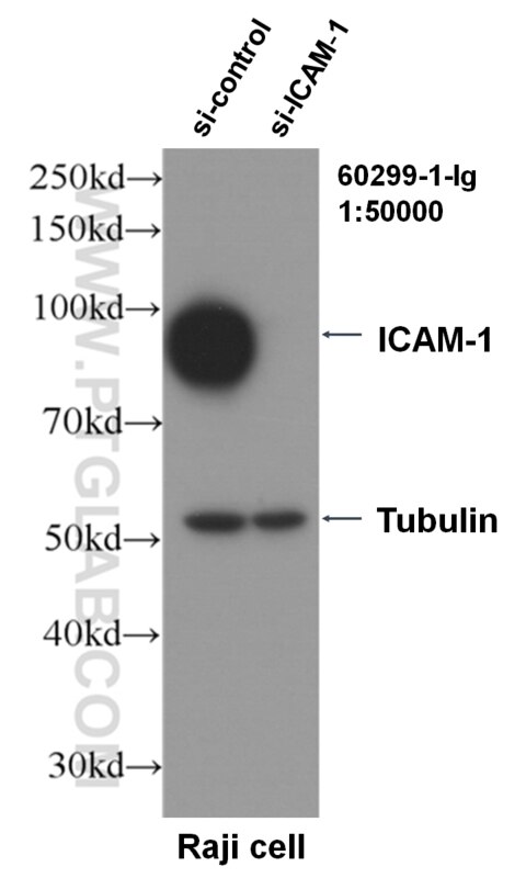
- Validé par KD/KO
Anticorps Monoclonal anti-ICAM-1
ICAM-1 Monoclonal Antibody for FC, IF, IHC, WB, ELISA
Hôte / Isotype
Mouse / IgG2b
Réactivité testée
Humain
Applications
WB, IHC, IF, FC, ELISA
Conjugaison
Non conjugué
CloneNo.
2F9A8
N° de cat : 60299-1-Ig
Synonymes
Galerie de données de validation
Applications testées
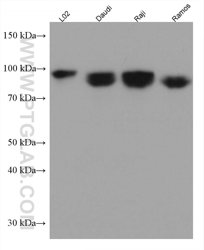
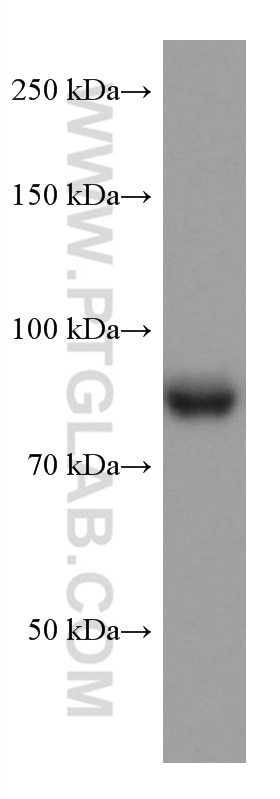
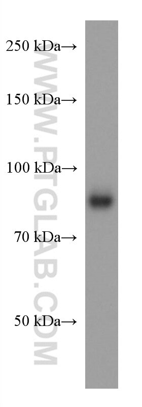
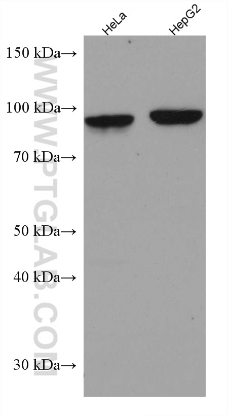
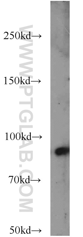
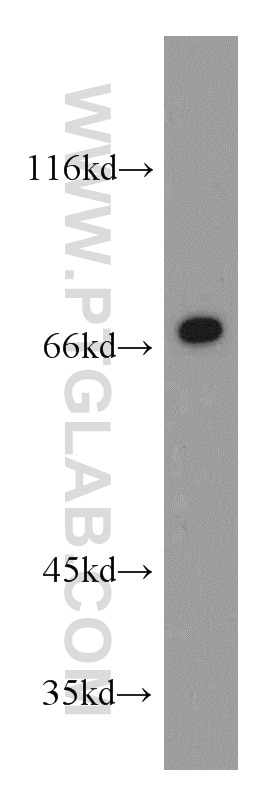
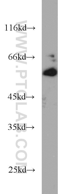
| Résultats positifs en WB | cellules L02, cellules Daudi, cellules HeLa, cellules HepG2, cellules Raji, cellules Ramos |
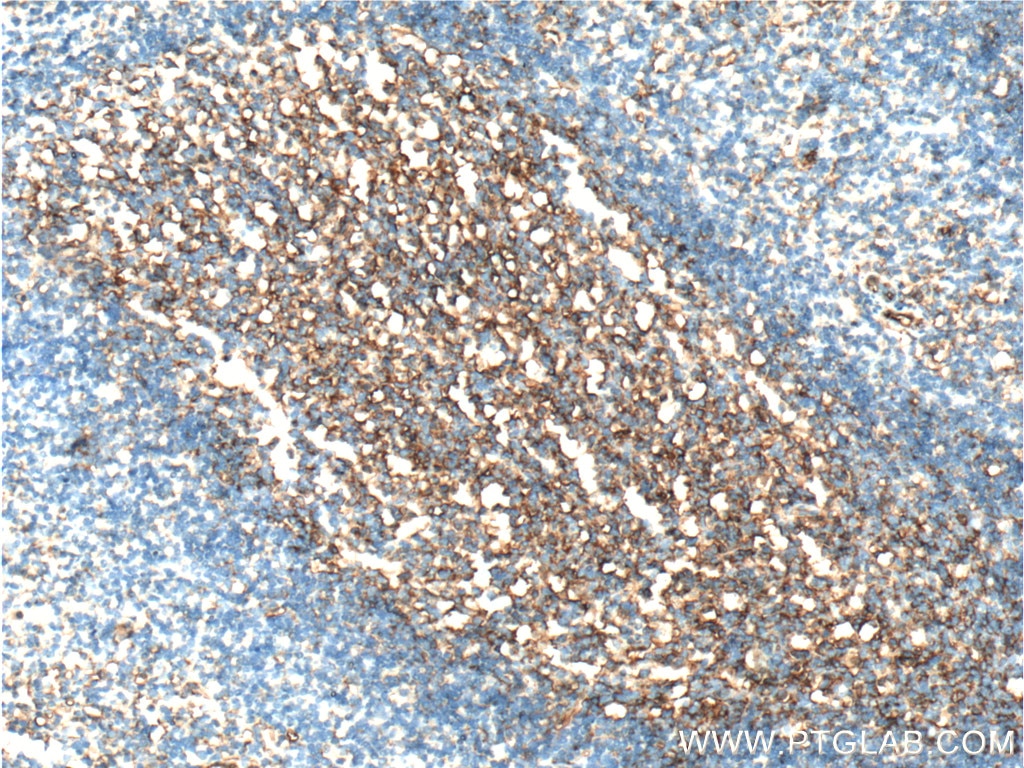
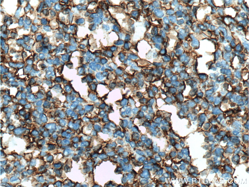
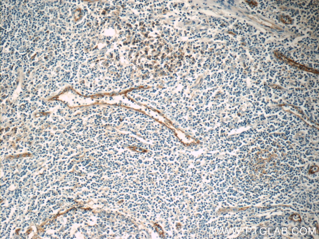
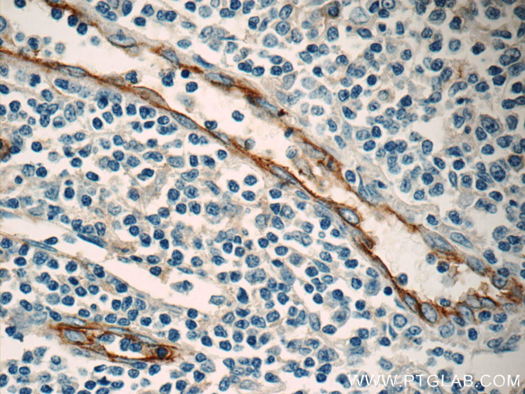
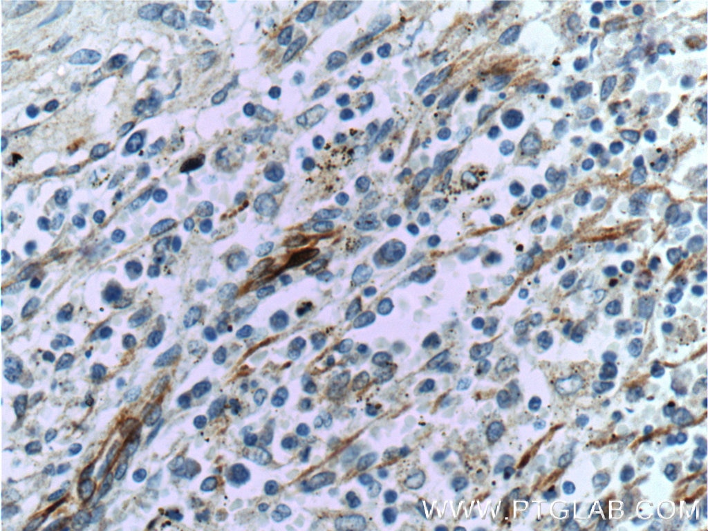
| Résultats positifs en IHC | tissu d'amygdalite humain il est suggéré de démasquer l'antigène avec un tampon de TE buffer pH 9.0; (*) À défaut, 'le démasquage de l'antigène peut être 'effectué avec un tampon citrate pH 6,0. |
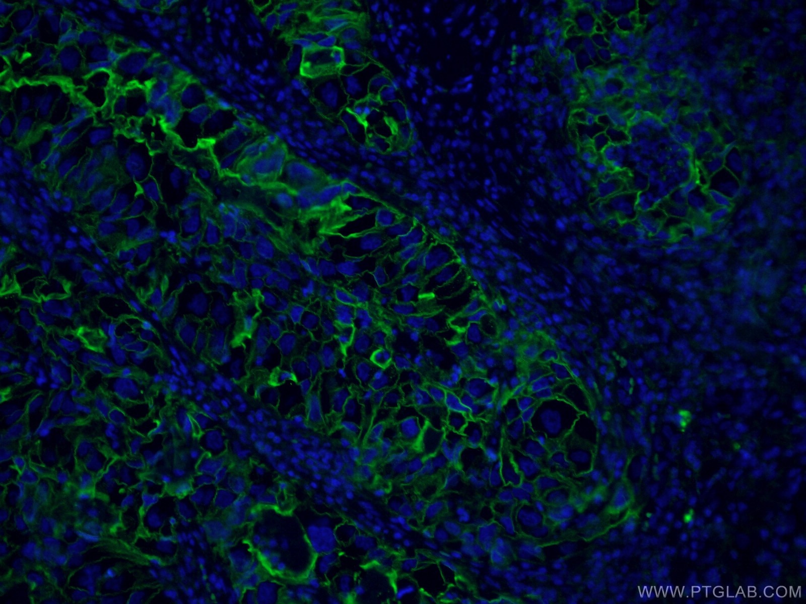
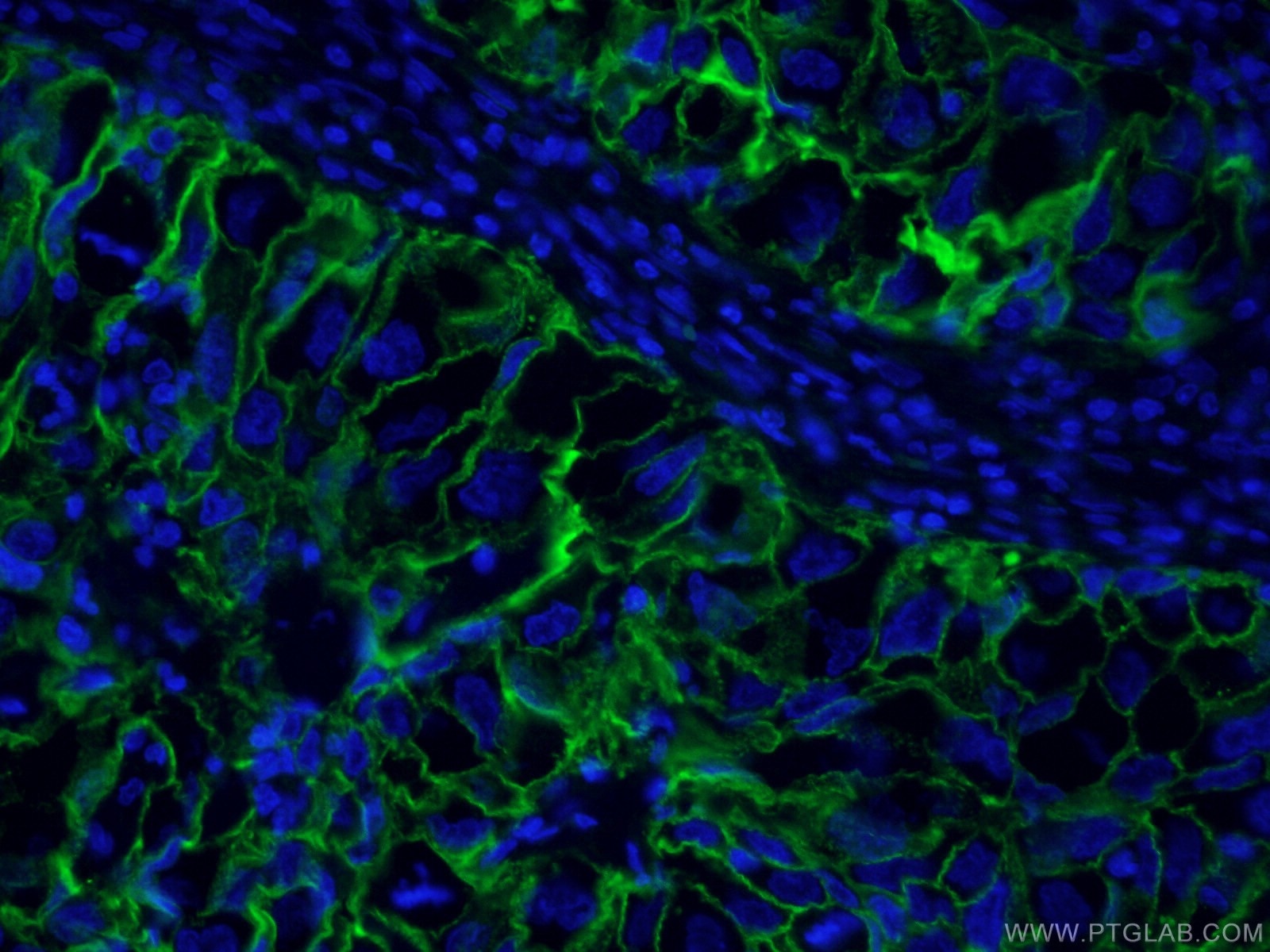
| Résultats positifs en IF | tissu de cancer du poumon humain, |
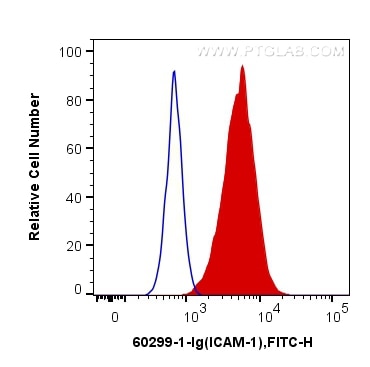
| Résultats positifs en cytométrie | cellules Daudi, |
Dilution recommandée
| Application | Dilution |
|---|---|
| Western Blot (WB) | WB : 1:5000-1:50000 |
| Immunohistochimie (IHC) | IHC : 1:50-1:500 |
| Immunofluorescence (IF) | IF : 1:50-1:500 |
| Flow Cytometry (FC) | FC : 0.40 ug per 10^6 cells in a 100 µl suspension |
| It is recommended that this reagent should be titrated in each testing system to obtain optimal results. | |
| Sample-dependent, check data in validation data gallery | |
Applications publiées
| WB | See 32 publications below |
| IHC | See 12 publications below |
| IF | See 10 publications below |
Informations sur le produit
60299-1-Ig cible ICAM-1 dans les applications de WB, IHC, IF, FC, ELISA et montre une réactivité avec des échantillons Humain
| Réactivité | Humain |
| Réactivité citée | Humain |
| Hôte / Isotype | Mouse / IgG2b |
| Clonalité | Monoclonal |
| Type | Anticorps |
| Immunogène | ICAM-1 Protéine recombinante Ag8309 |
| Nom complet | intercellular adhesion molecule 1 |
| Masse moléculaire calculée | 90 kDa |
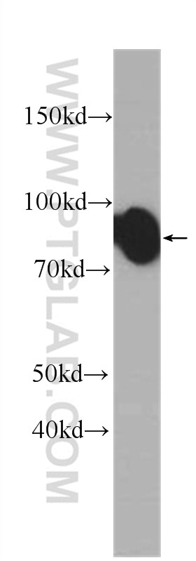
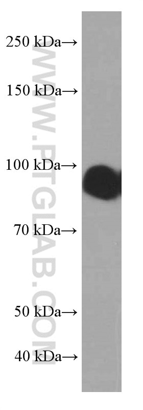
| Poids moléculaire observé | 85-95 kDa |
| Numéro d’acquisition GenBank | BC015969 |
| Symbole du gène | ICAM1 |
| Identification du gène (NCBI) | 3383 |
| Conjugaison | Non conjugué |
| Forme | Liquide |
| Méthode de purification | Purification par protéine A |
| Tampon de stockage | PBS avec azoture de sodium à 0,02 % et glycérol à 50 % pH 7,3 |
| Conditions de stockage | Stocker à -20°C. Stable pendant un an après l'expédition. L'aliquotage n'est pas nécessaire pour le stockage à -20oC Les 20ul contiennent 0,1% de BSA. |
Informations générales
Where is ICAM-1 expressed?
Intercellular Adhesion Molecule 1 (ICAM-1), also known as Cluster of Differentiation 54 (CD54) is a transmembrane glycoprotein constitutively expressed at low levels in endothelial cells, pericytes and on some lymphocytes and monocytes1. It is located at the cytoplasmic membrane, with a large extracellular region of mainly hydrophobic amino acids joined to a small transmembrane region and a cytoplasmic tail. It has a molecular weight of 75 to 115 kDa depending on the level of glycosylation.
What is the function of ICAM-1?
ICAM-1 is important in both innate and adaptive immune responses as an adhesion molecule. Although it is constitutively expressed, in the presence of pro-inflammatory cytokines such as TNFα the endothelial cells are activated and upregulate expression of ICAM-12. In blood vessels lined with endothelial cells, leukocytes that are rolling over the surface are able to bind to ICAM-1 and transmigrate through the endothelial barrier and into the tissue. The initial binding of the leukocytes to ICAM-1 causes a Ca2+ release that initiates endothelial cell contraction and weakening of the intercellular tight junctions3, 4. This protein can be used as an indicator of endothelial activation and of vascular inflammation.
What is the role of ICAM-1 in disease?
Beyond the role in the immune response, ICAM-1 has also been identified as the target of attachment for the human rhinovirus, the cause of the common cold. Binding of the virus to ICAM-1 causes the viral capsid to uncoat and leads to release of the genetic material5.
Hubbard, A. K. & Rothlein, R. Intercellular adhesion molecule-1 (ICAM-1) expression and cell signaling cascades. Free Radic. Biol. Med. 28, 1379-86 (2000).
Long, E. O. ICAM-1: getting a grip on leukocyte adhesion. J. Immunol. 186, 5021-3 (2011).
Lawson, C. & Wolf, S. ICAM-1 signaling in endothelial cells. (2009).
Lyck, R. & Enzmann, G. The physiological roles of ICAM-1 and ICAM-2 in neutrophil migration into tissues. Curr. Opin. Hematol. 22, 53-59 (2015).
Xing, L., Casasnovas, J. M. & Cheng, R. H. Structural analysis of human rhinovirus complexed with ICAM-1 reveals the dynamics of receptor-mediated virus uncoating. J. Virol. 77, 6101-7 (2003).
Protocole
| Product Specific Protocols | |
|---|---|
| WB protocol for ICAM-1 antibody 60299-1-Ig | Download protocol |
| IHC protocol for ICAM-1 antibody 60299-1-Ig | Download protocol |
| IF protocol for ICAM-1 antibody 60299-1-Ig | Download protocol |
| FC protocol for ICAM-1 antibody 60299-1-Ig | Download protocol |
| Standard Protocols | |
|---|---|
| Click here to view our Standard Protocols |
Publications
| Species | Application | Title |
|---|---|---|
Gastroenterology Proteomic characterization identifies clinically relevant subgroups of gastrointestinal stromal tumors | ||
Nat Commun CDK4/6 inhibition triggers ICAM1-driven immune response and sensitizes LKB1 mutant lung cancer to immunotherapy | ||
EBioMedicine Interactions between neutrophil extracellular traps and activated platelets enhance procoagulant activity in acute stroke patients with ICA occlusion. | ||
Redox Biol Targeting mitochondria-inflammation circle by renal denervation reduces atheroprone endothelial phenotypes and atherosclerosis. | ||
Int J Biol Sci Direct targeting of sEH with alisol B alleviated the apoptosis, inflammation, and oxidative stress in cisplatin-induced acute kidney injury | ||
J Cell Biol Hematopoietic progenitors polarize in contact with bone marrow stromal cells in response to SDF1. |
