

Tested Applications
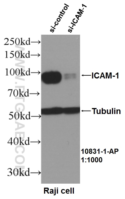
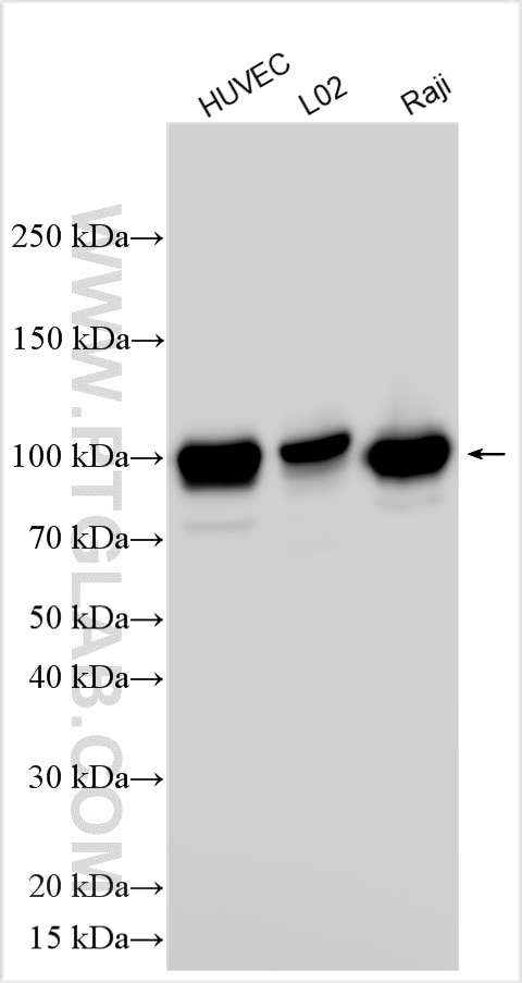
| Positive WB detected in | HUVEC cells, Raji cells, L02 cells |
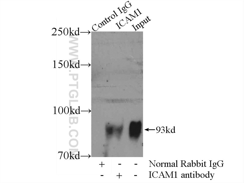
| Positive IP detected in | Raji cells |
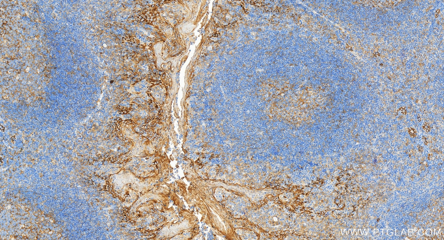
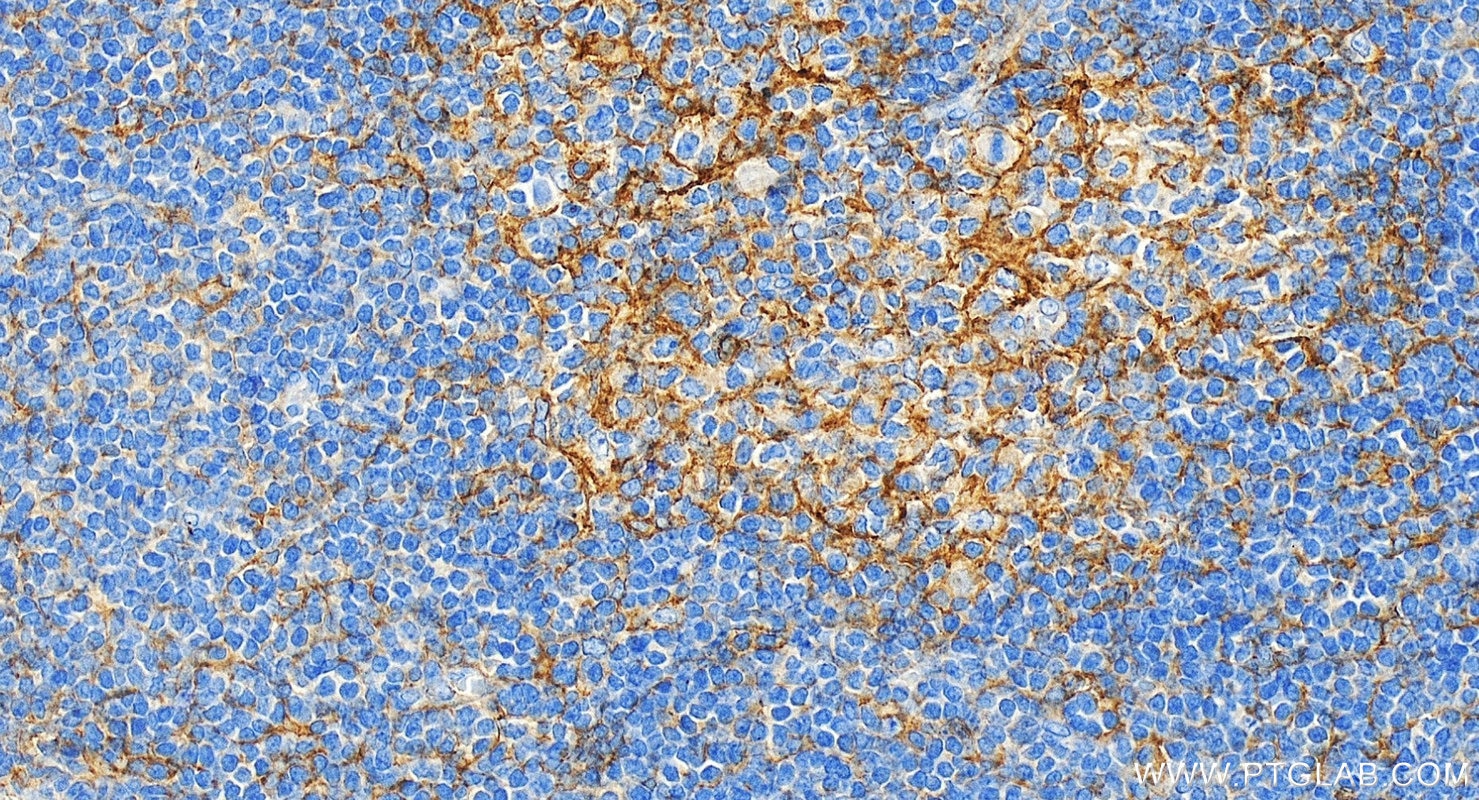
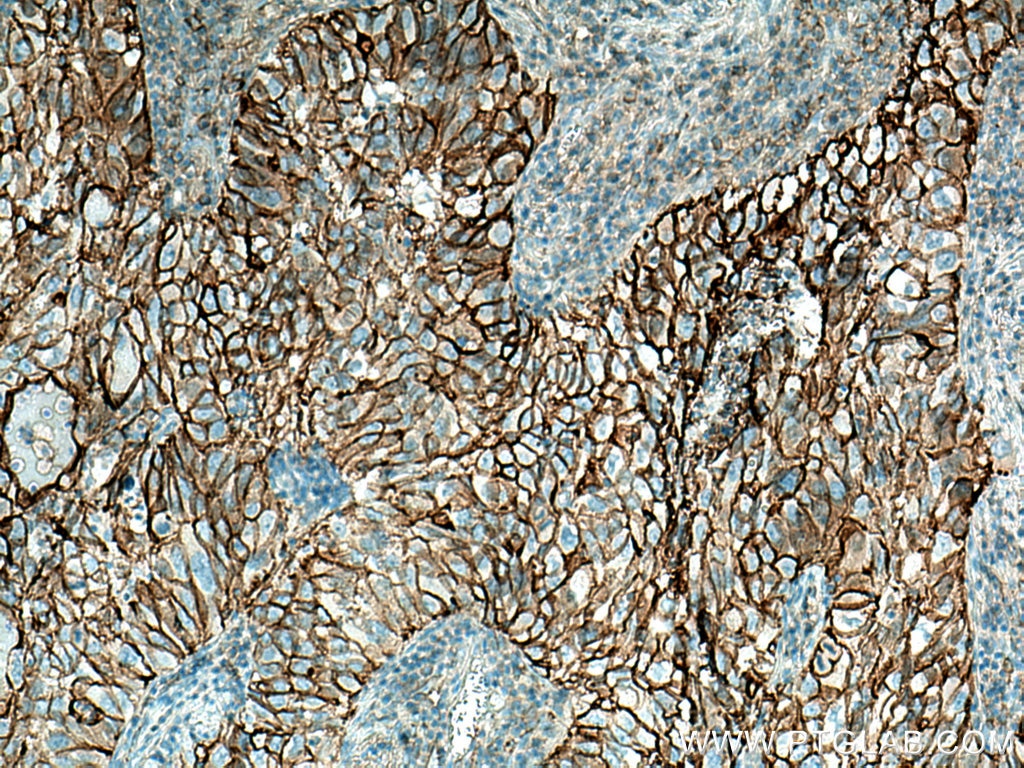
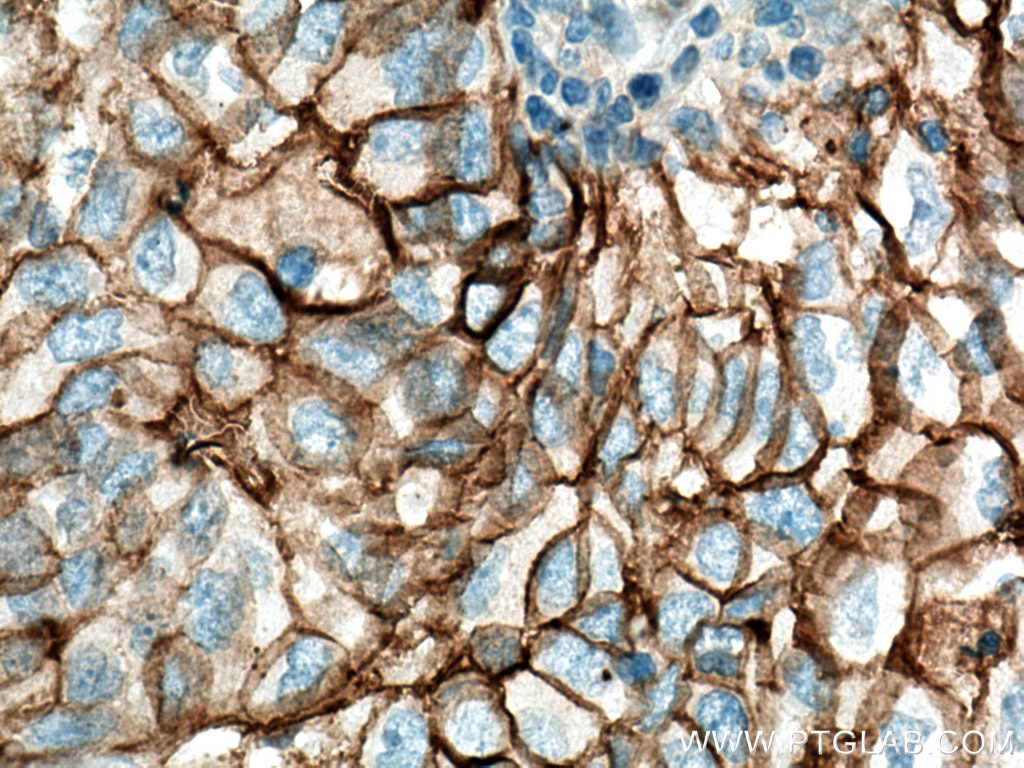
| Positive IHC detected in | human tonsillitis tissue, human lung cancer tissue Note: suggested antigen retrieval with TE buffer pH 9.0; (*) Alternatively, antigen retrieval may be performed with citrate buffer pH 6.0 |
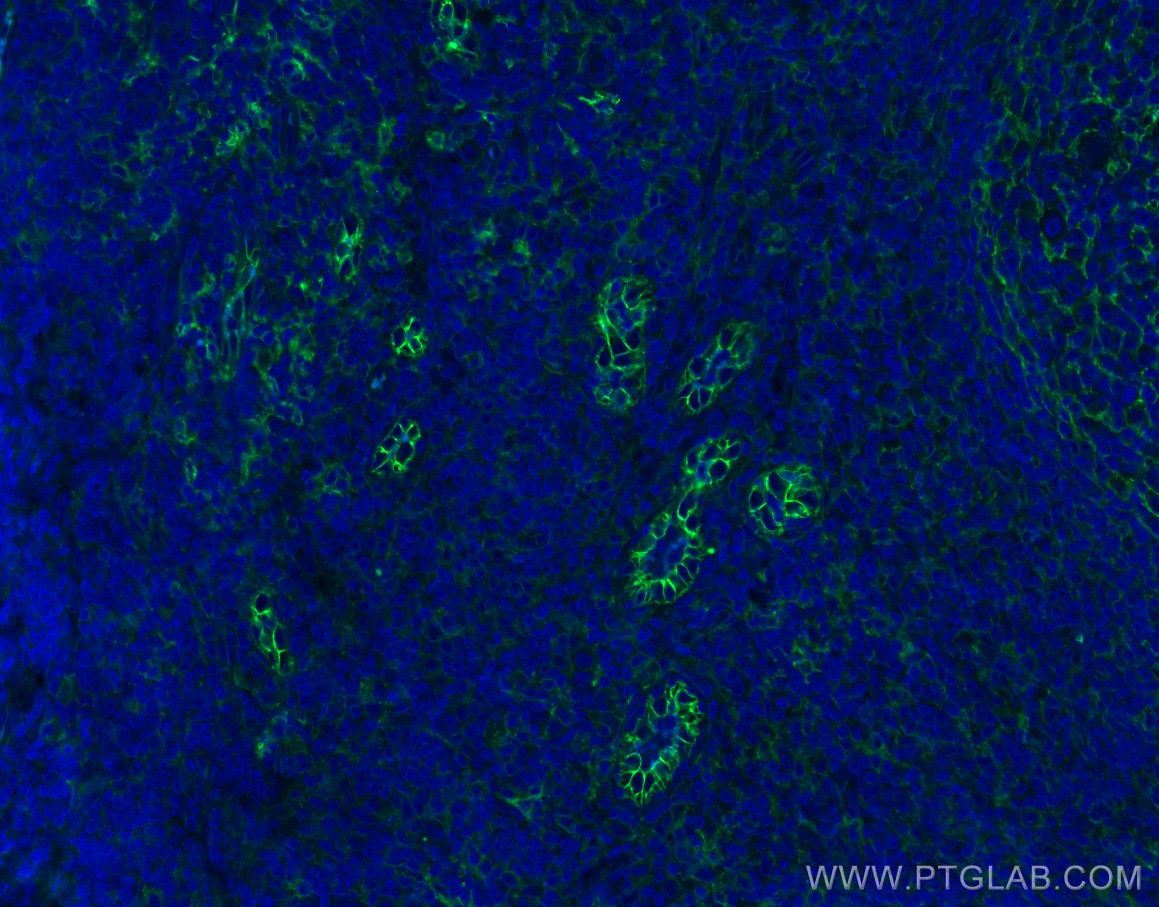
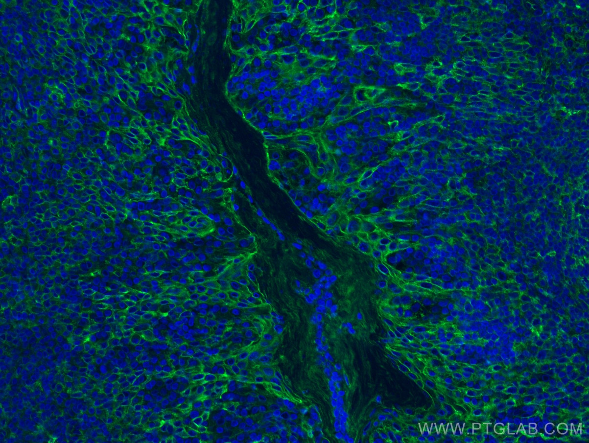
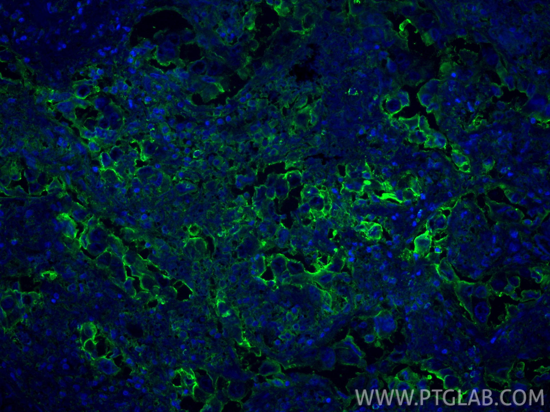
| Positive IF-P detected in | human tonsillitis tissue, human lung cancer tissue |
Recommended dilution
| Application | Dilution |
|---|---|
| Western Blot (WB) | WB : 1:5000-1:50000 |
| Immunoprecipitation (IP) | IP : 0.5-4.0 ug for 1.0-3.0 mg of total protein lysate |
| Immunohistochemistry (IHC) | IHC : 1:5000-1:20000 |
| Immunofluorescence (IF)-P | IF-P : 1:50-1:500 |
| It is recommended that this reagent should be titrated in each testing system to obtain optimal results. | |
| Sample-dependent, Check data in validation data gallery. | |
Product Information
10831-1-AP targets ICAM-1/CD54 in WB, IHC, IF-P, IP, CoIP, ChIP, ELISA, Blocking, Blocking assay applications and shows reactivity with human samples.
| Tested Reactivity | human |
| Cited Reactivity | human, pig, bovine, goat |
| Host / Isotype | Rabbit / IgG |
| Class | Polyclonal |
| Type | Antibody |
| Immunogen |
CatNo: Ag1188 Product name: Recombinant human ICAM-1 protein Source: e coli.-derived, PGEX-4T Tag: GST Domain: 181-532 aa of BC015969 Sequence: GANFSCRTELDLRPQGLELFENTSAPYQLQTFVLPATPPQLVSPRVLEVDTQGTVVCSLDGLFPVSEAQVHLALGDQRLNPTVTYGNDSFSAKASVSVTAEDEGTQRLTCAVILGNQSQETLQTVTIYSFPAPNVILTKPEVSEGTEVTVKCEAHPRAKVTLNGVPAQPLGPRAQLLLKATPEDNGRSFSCSATLEVAGQLIHKNQTRELRVLYGPRLDERDCPGNWTWPENSQQTPMCQAWGNPLPELKCLKDGTFPLPIGESVTVTRDLEGTYLCRARSTQGEVTRKVTVNVLSPRYEIVIITVVAAAVIMGTAGLSTYLYNRQRKIKKYRLQQAQKGTPMKPNTQATPP Predict reactive species |
| Full Name | intercellular adhesion molecule 1 |
| Calculated Molecular Weight | 90 kDa |
| Observed Molecular Weight | 90-100 kDa |
| GenBank Accession Number | BC015969 |
| Gene Symbol | ICAM-1 |
| Gene ID (NCBI) | 3383 |
| ENSEMBL Gene ID | ENSG00000090339 |
| RRID | AB_2264494 |
| Conjugate | Unconjugated |
| Form | Liquid |
| Purification Method | Antigen affinity purification |
| UNIPROT ID | P05362 |
| Storage Buffer | PBS with 0.02% sodium azide and 50% glycerol, pH 7.3. |
| Storage Conditions | Store at -20°C. Stable for one year after shipment. Aliquoting is unnecessary for -20oC storage. 20ul sizes contain 0.1% BSA. |
Background Information
Where is ICAM-1 expressed?
Intercellular Adhesion Molecule 1 (ICAM-1), also known as Cluster of Differentiation 54 (CD54) is a transmembrane glycoprotein constitutively expressed at low levels in endothelial cells, pericytes and on some lymphocytes and monocytes1. It is located at the cytoplasmic membrane, with a large extracellular region of mainly hydrophobic amino acids joined to a small transmembrane region and a cytoplasmic tail. It has a molecular weight of 75 to 115 kDa depending on the level of glycosylation.
What is the function of ICAM-1?
ICAM-1 is important in both innate and adaptive immune responses as an adhesion molecule. Although it is constitutively expressed, in the presence of pro-inflammatory cytokines such as TNFα the endothelial cells are activated and upregulate expression of ICAM-12. In blood vessels lined with endothelial cells, leukocytes that are rolling over the surface are able to bind to ICAM-1 and transmigrate through the endothelial barrier and into the tissue. The initial binding of the leukocytes to ICAM-1 causes a Ca2+ release that initiates endothelial cell contraction and weakening of the intercellular tight junctions3, 4. This protein can be used as an indicator of endothelial activation and of vascular inflammation.
What is the role of ICAM-1 in disease?
Beyond the role in the immune response, ICAM-1 has also been identified as the target of attachment for the human rhinovirus, the cause of the common cold. Binding of the virus to ICAM-1 causes the viral capsid to uncoat and leads to release of the genetic material5.
Hubbard, A. K. & Rothlein, R. Intercellular adhesion molecule-1 (ICAM-1) expression and cell signaling cascades. Free Radic. Biol. Med. 28, 1379-86 (2000).
Long, E. O. ICAM-1: getting a grip on leukocyte adhesion. J. Immunol. 186, 5021-3 (2011).
Lawson, C. & Wolf, S. ICAM-1 signaling in endothelial cells. (2009).
Lyck, R. & Enzmann, G. The physiological roles of ICAM-1 and ICAM-2 in neutrophil migration into tissues. Curr. Opin. Hematol. 22, 53-59 (2015).
Xing, L., Casasnovas, J. M. & Cheng, R. H. Structural analysis of human rhinovirus complexed with ICAM-1 reveals the dynamics of receptor-mediated virus uncoating. J. Virol. 77, 6101-7 (2003).
Protocols
| Product Specific Protocols | |
|---|---|
| IF protocol for ICAM-1/CD54 antibody 10831-1-AP | Download protocol |
| IHC protocol for ICAM-1/CD54 antibody 10831-1-AP | Download protocol |
| IP protocol for ICAM-1/CD54 antibody 10831-1-AP | Download protocol |
| WB protocol for ICAM-1/CD54 antibody 10831-1-AP | Download protocol |
| Standard Protocols | |
|---|---|
| Click here to view our Standard Protocols |
Publications
What published studies show
The anti-ICAM-1 antibody (Cat# 10831-1-AP) has accumulated 243 citations in journals including Nature Immunology, Nature Communications, Nature Microbiology, Cell Stem Cell, Cancer Research, Advanced Science, Science Advances, Biomaterials, and Theranostics. Research fields span atherosclerosis, endothelial dysfunction, inflammation, multiple cancers (lung, liver, gastric, breast, cervical), diabetes complications, neuroinflammation, sepsis, rheumatoid arthritis, nanomedicine, immunotherapy, blood-brain barrier biology, and vascular remodeling.
| Species | Application | Title |
|---|---|---|
Signal Transduct Target Ther Pulsed electromagnetic fields inhibit atherosclerosis by regulating pyroptosis through membrane tension-mediated mechanosensitive channels | ||
Mol Cancer Integrated multi-omics identifies a CD54(+) iCAF-ITGAL(+) macrophage niche driving immunosuppression via CXCL8-PDL1 axis in cervical cancer. | ||
Adv Mater Biomimetic Immunosuppressive Exosomes that Inhibit Cytokine Storms Contribute to the Alleviation of Sepsis. | ||
Nat Immunol Sparcl1 mitigates abdominal aortic aneurysm through inhibiting lymphangiogenesis-mediated TLS formation | ||
Bioact Mater TLR2 inhibition attenuates NET-driven inflammation and restenosis during poly-lactic acid bioresorbable scaffold degradation. | ||
Cell Stem Cell Modeling blood-brain barrier formation and cerebral cavernous malformations in human PSC-derived organoids |
Reviews
What customers say
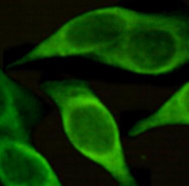
Both reviewers gave this antibody a perfect 5-star rating for immunofluorescence. Julia Derdau used it at 1:300 dilution on human paraffin-embedded colon tissue and reported being happy with the results. Uthra Rajamani used it at 1:150 on leukocytes and described it simply as "Good." Overall, users find this antibody reliable and effective for IF applications across different tissue types and cell preparations.
The reviews below have been submitted by verified Proteintech customers who received an incentive for providing their feedback.
Julia (Verified Customer) (05-07-2026) | This antibody worked in human paraffin samples. Happy with the results!
|
Uthra (Verified Customer) (10-03-2022) | Good!!!
 |


