- Phare
- Validé par KD/KO
Anticorps Polyclonal de lapin anti-CCR2a-specific
CCR2a-specific Polyclonal Antibody for WB, IHC, ELISA
Hôte / Isotype
Lapin / IgG
Réactivité testée
Humain, rat, souris
Applications
WB, IF, IHC, ELISA
Conjugaison
Non conjugué
N° de cat : 16153-1-AP
Synonymes
Galerie de données de validation
Applications testées
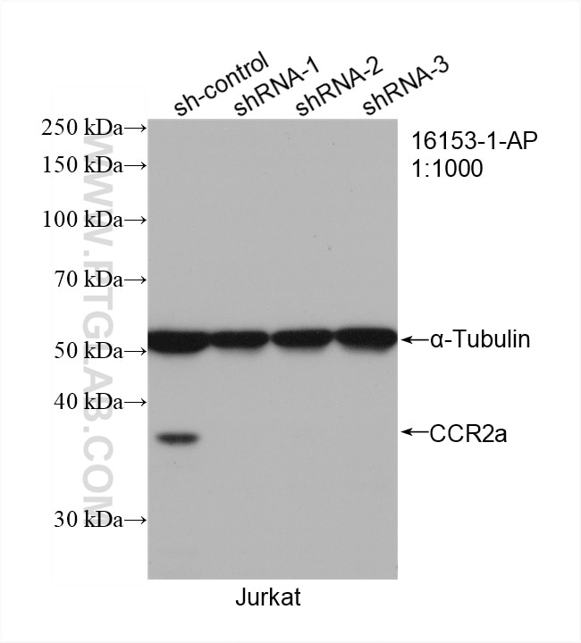
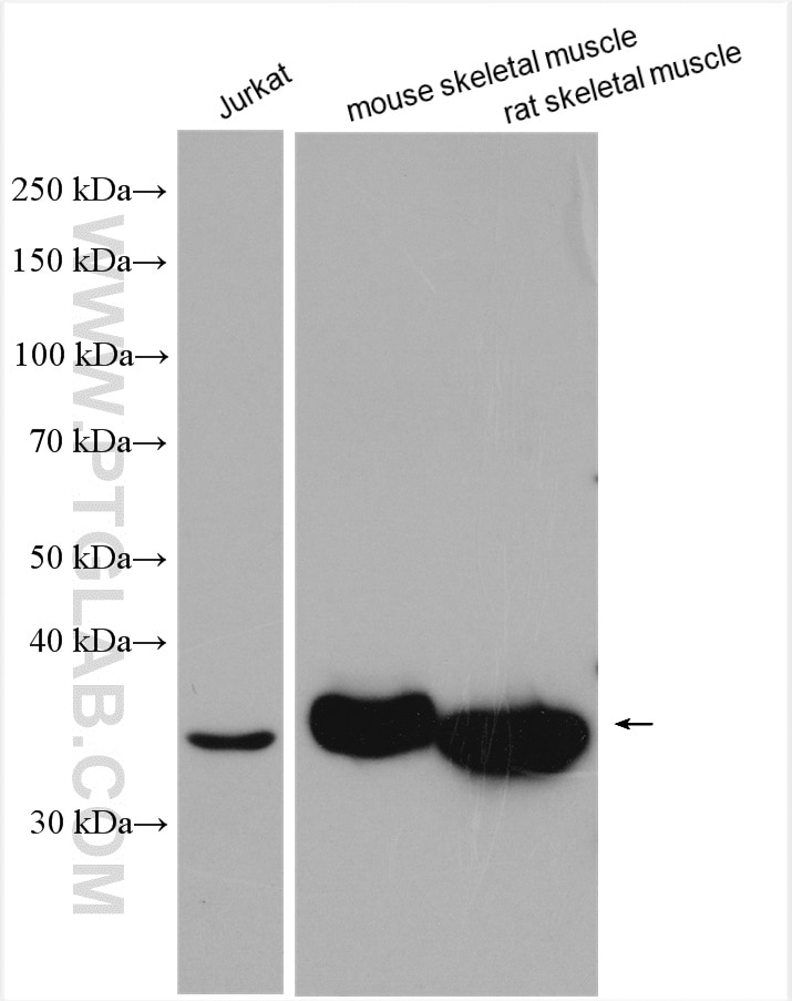
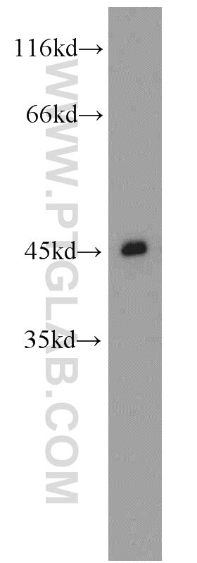
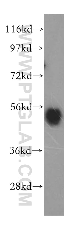
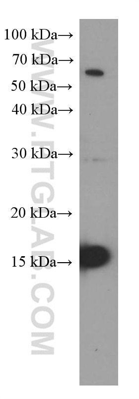
| Résultats positifs en WB | cellules Jurkat, tissu de muscle squelettique de rat, tissu de muscle squelettique de souris |
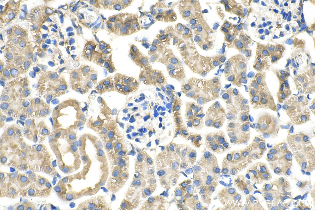
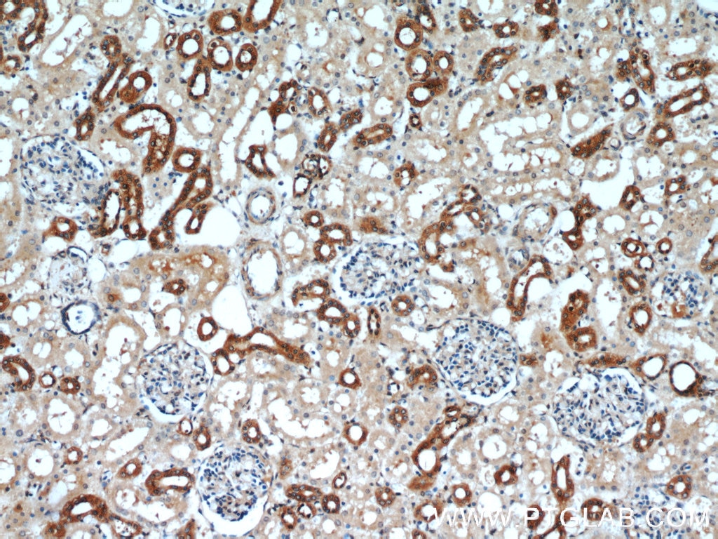
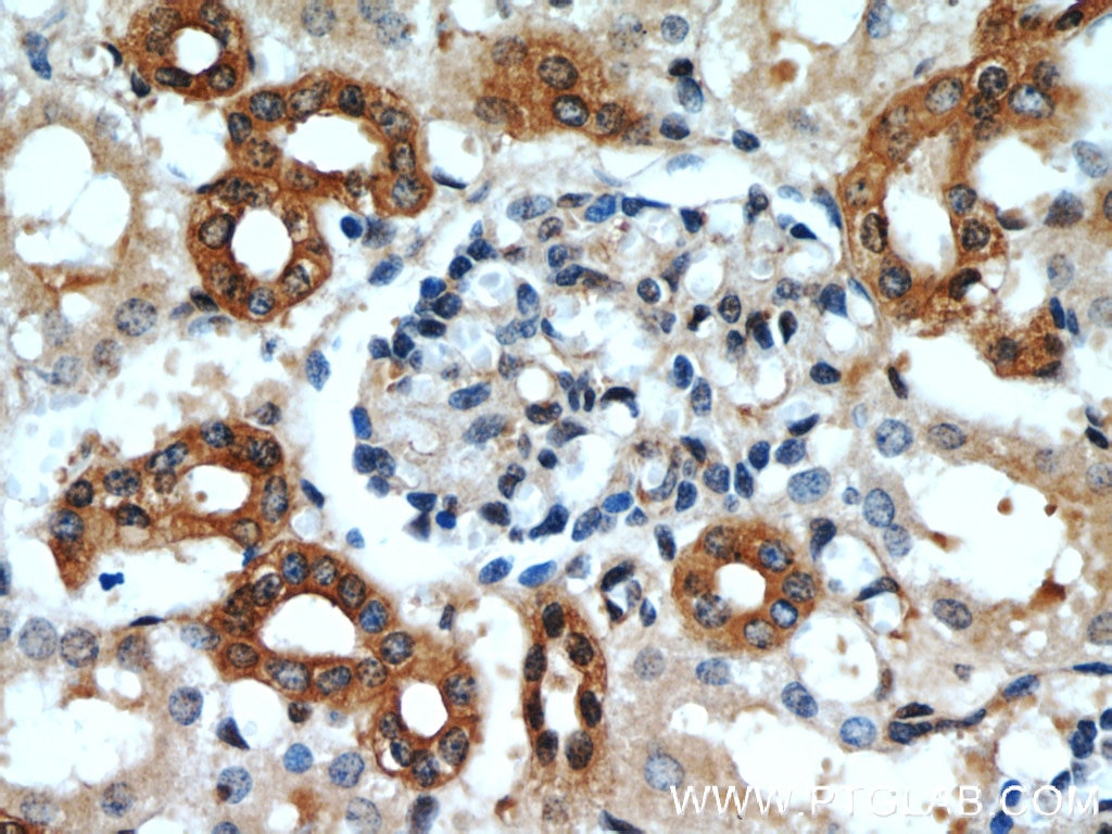
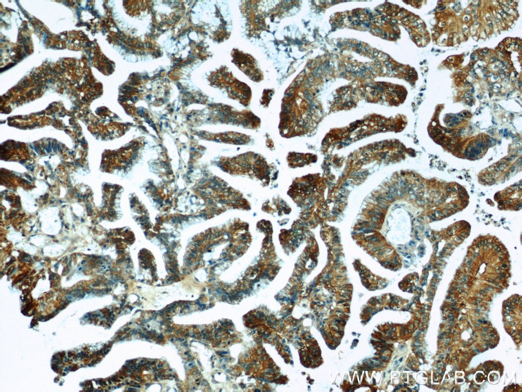
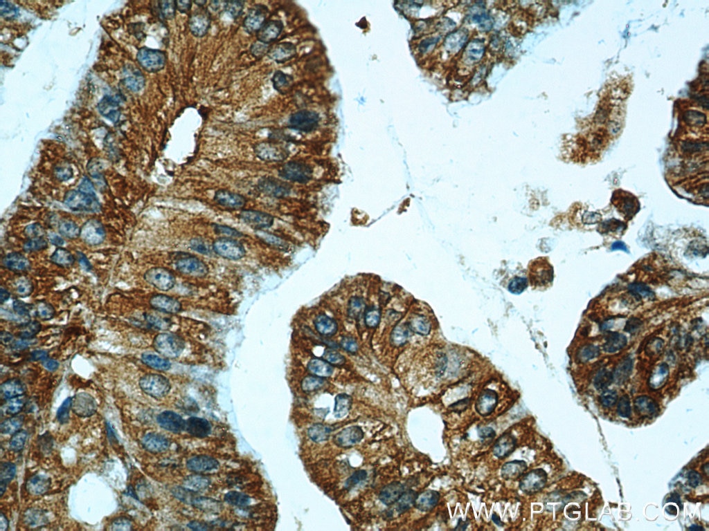
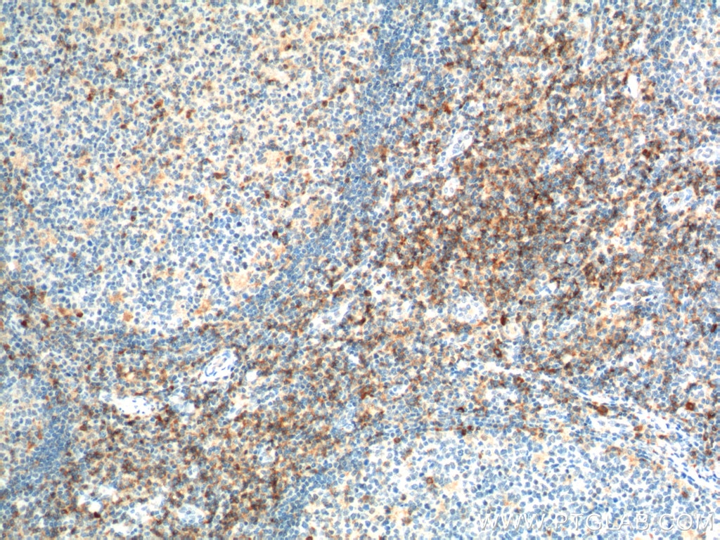
| Résultats positifs en IHC | tissu rénal de souris, tissu de tumeur ovarienne humain, tissu rénal humain il est suggéré de démasquer l'antigène avec un tampon de TE buffer pH 9.0; (*) À défaut, 'le démasquage de l'antigène peut être 'effectué avec un tampon citrate pH 6,0. |
Dilution recommandée
| Application | Dilution |
|---|---|
| Western Blot (WB) | WB : 1:1000-1:4000 |
| Immunohistochimie (IHC) | IHC : 1:50-1:500 |
| It is recommended that this reagent should be titrated in each testing system to obtain optimal results. | |
| Sample-dependent, check data in validation data gallery | |
Applications publiées
| WB | See 11 publications below |
| IHC | See 6 publications below |
| IF | See 4 publications below |
Informations sur le produit
16153-1-AP cible CCR2a-specific dans les applications de WB, IF, IHC, ELISA et montre une réactivité avec des échantillons Humain, rat, souris
| Réactivité | Humain, rat, souris |
| Réactivité citée | rat, Humain, souris |
| Hôte / Isotype | Lapin / IgG |
| Clonalité | Polyclonal |
| Type | Anticorps |
| Immunogène | Peptide |
| Nom complet | chemokine (C-C motif) receptor 2 |
| Masse moléculaire calculée | 42 kDa |
| Poids moléculaire observé | 35 kDa |
| Numéro d’acquisition GenBank | NM_000647 |
| Symbole du gène | CCR2 |
| Identification du gène (NCBI) | 729230 |
| Conjugaison | Non conjugué |
| Forme | Liquide |
| Méthode de purification | Purification par affinité contre l'antigène |
| Tampon de stockage | PBS avec azoture de sodium à 0,02 % et glycérol à 50 % pH 7,3 |
| Conditions de stockage | Stocker à -20°C. Stable pendant un an après l'expédition. L'aliquotage n'est pas nécessaire pour le stockage à -20oC Les 20ul contiennent 0,1% de BSA. |
Informations générales
CCR2 encodes a receptor for MCP1, functioning in inflammatory processes, tumor infiltration, and monocyte invasion of artery walls in atherosclerosis. There are two isoforms of CCR2, CCR2A and CCR2B. CCR2 isoforms were expressed by different cell subsets in both normal and IIM muscle. CCR2A was expressed in vessel walls and by some mononuclear cells, especially in cells involved in partial invasion in PM and IBM. CCR2B expression was observed in all satellite cells, in the muscular domain of neuromuscular junctions, and in some regenerative fibers of IIM, but not in inflammatory exudates. The sequences of the two isoforms are different from 314-374 aa. This antibody 16153-1-AP is raised against the peptide of 344-358 amino acids of CCR2a. It is specific to CCR2a.
Protocole
| Product Specific Protocols | |
|---|---|
| WB protocol for CCR2a-specific antibody 16153-1-AP | Download protocol |
| IHC protocol for CCR2a-specific antibody 16153-1-AP | Download protocol |
| Standard Protocols | |
|---|---|
| Click here to view our Standard Protocols |
Publications
| Species | Application | Title |
|---|---|---|
Rheumatology (Oxford) Interleukin-6 trans-signaling regulates monocyte chemoattractant protein-1 production in immune-mediated necrotizing myopathy | ||
Lab Invest Matrix metalloproteinase 11 (MMP11) in macrophages promotes the migration of HER2-positive breast cancer cells and monocyte recruitment through CCL2-CCR2 signaling. | ||
Oncol Rep CCL2/CCR2 axis induces hepatocellular carcinoma invasion and epithelial-mesenchymal transition in vitro through activation of the Hedgehog pathway. | ||
J Ethnopharmacol Identification of potential regulating effect of baicalin on NFκB/CCL2/CCR2 signaling pathway in rats with cerebral ischemia by antibody-based array and bioinformatics analysis | ||
Mol Immunol QKI deficiency in macrophages protects mice against JEV infection by regulating cell migration and antiviral response. | ||
J Physiol Biochem Hypoxia-induced CCL2/CCR2 axis in adipose-derived stem cells (ADSCs) promotes angiogenesis by human dermal microvascular endothelial cells (HDMECs) in flap tissues |
Avis
The reviews below have been submitted by verified Proteintech customers who received an incentive forproviding their feedback.
FH Kamal Baral (Verified Customer) (01-26-2024) | Bone marrow mesenchymal stem cells from mouse stained great at the provided dilution.
|
FH Kamal (Verified Customer) (01-26-2024) | Bands appeared at 35 kDa. Antibody works well when diluted in 1X TBST.
|
FH Alexandru (Verified Customer) (11-13-2023) | Great stainig results on paraffin-embedded tissue!
|
