- Phare
- Validé par KD/KO
Anticorps Polyclonal de lapin anti-MUC1/CA15-3 C-terminal
MUC1/CA15-3 C-terminal Polyclonal Antibody for WB, IHC, ELISA
Hôte / Isotype
Lapin / IgG
Réactivité testée
Humain et plus (1)
Applications
WB, IF, IHC, ELISA
Conjugaison
Non conjugué
N° de cat : 23614-1-AP
Synonymes
Galerie de données de validation
Applications testées
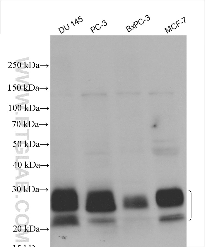
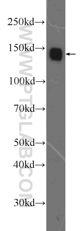
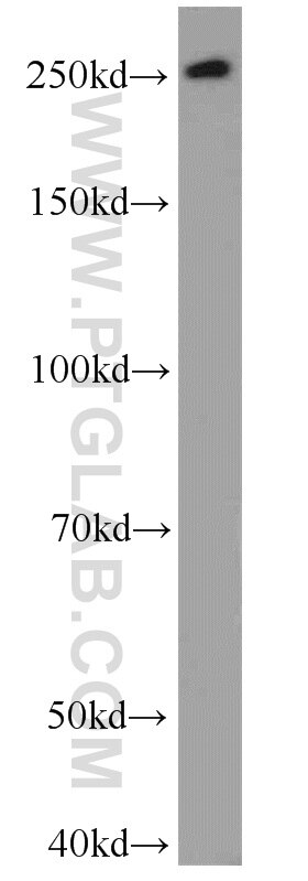
| Résultats positifs en WB | cellules DU 145, cellules BxPC-3, cellules MCF-7, cellules PC-3 |
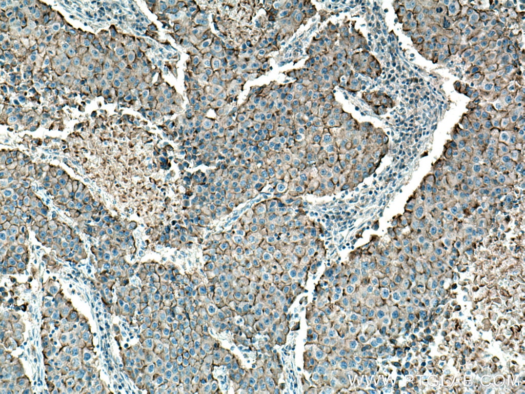
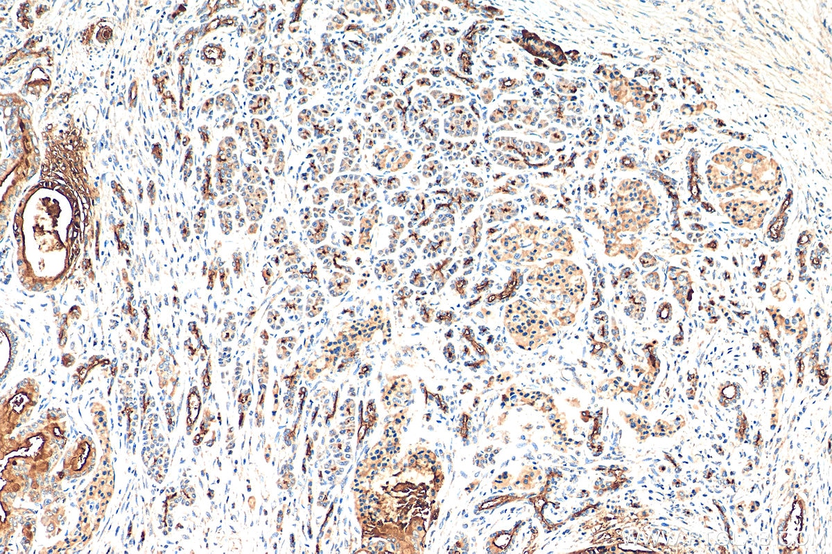
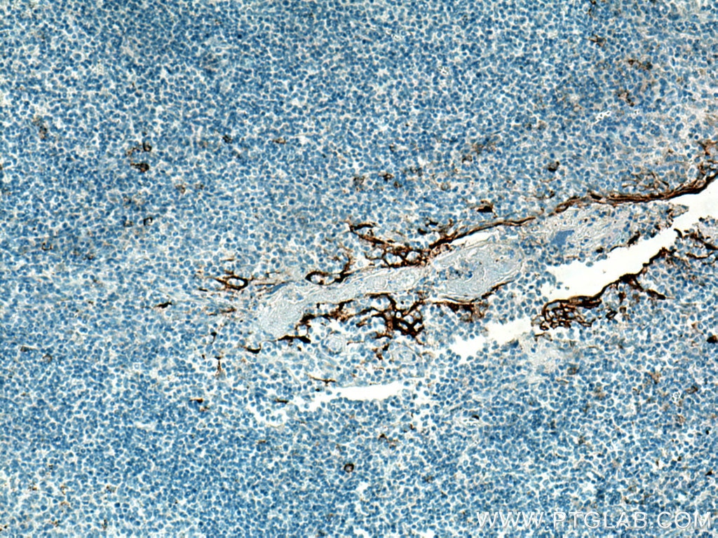
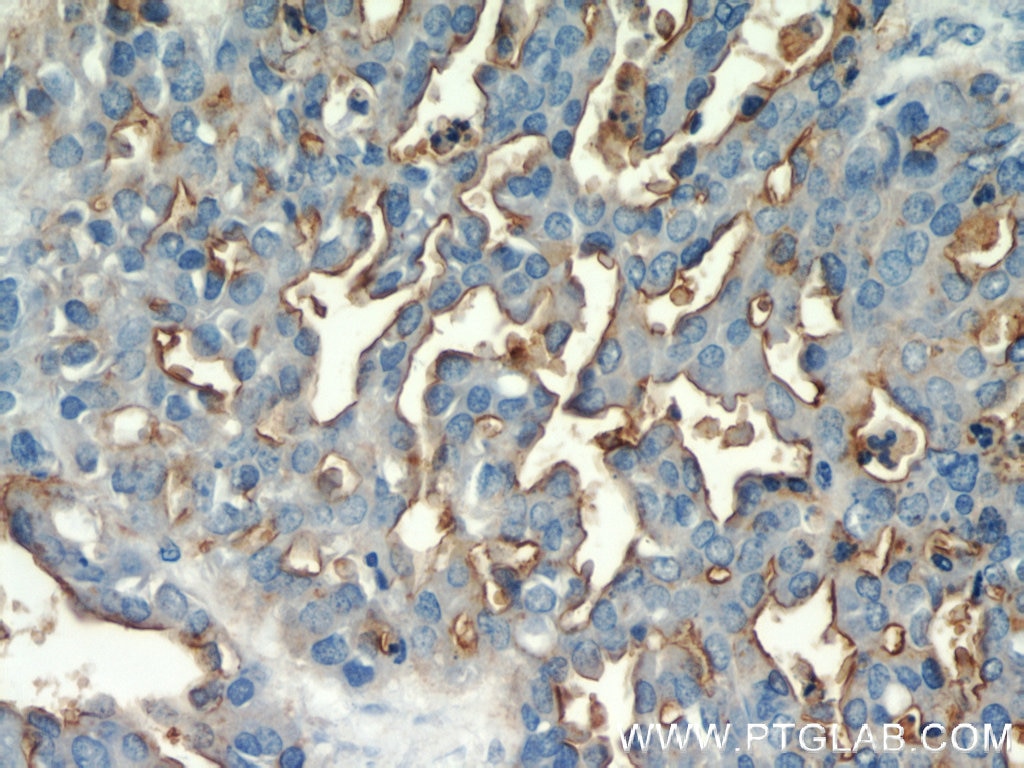
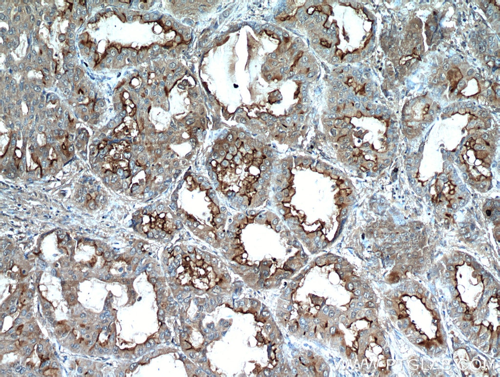
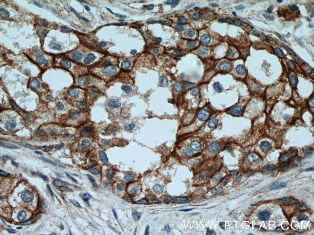
| Résultats positifs en IHC | tissu de cancer du pancréas humain, tissu d'amygdalite humain, tissu de cancer du sein humain, tissu de tumeur ovarienne humain il est suggéré de démasquer l'antigène avec un tampon de TE buffer pH 9.0; (*) À défaut, 'le démasquage de l'antigène peut être 'effectué avec un tampon citrate pH 6,0. |
Dilution recommandée
| Application | Dilution |
|---|---|
| Western Blot (WB) | WB : 1:500-1:2000 |
| Immunohistochimie (IHC) | IHC : 1:50-1:500 |
| It is recommended that this reagent should be titrated in each testing system to obtain optimal results. | |
| Sample-dependent, check data in validation data gallery | |
Applications publiées
| KD/KO | See 1 publications below |
| WB | See 9 publications below |
| IHC | See 4 publications below |
| IF | See 1 publications below |
| FC | See 1 publications below |
Informations sur le produit
23614-1-AP cible MUC1/CA15-3 C-terminal dans les applications de WB, IF, IHC, ELISA et montre une réactivité avec des échantillons Humain
| Réactivité | Humain |
| Réactivité citée | Humain, souris |
| Hôte / Isotype | Lapin / IgG |
| Clonalité | Polyclonal |
| Type | Anticorps |
| Immunogène | MUC1/CA15-3 C-terminal Protéine recombinante Ag20366 |
| Nom complet | mucin 1, cell surface associated |
| Masse moléculaire calculée | 1264 aa, 123 kDa |
| Poids moléculaire observé | 23-28 kDa |
| Numéro d’acquisition GenBank | BC120975 |
| Symbole du gène | MUC1/CA15-3 |
| Identification du gène (NCBI) | 4582 |
| Conjugaison | Non conjugué |
| Forme | Liquide |
| Méthode de purification | Purification par affinité contre l'antigène |
| Tampon de stockage | PBS avec azoture de sodium à 0,02 % et glycérol à 50 % pH 7,3 |
| Conditions de stockage | Stocker à -20°C. Stable pendant un an après l'expédition. L'aliquotage n'est pas nécessaire pour le stockage à -20oC Les 20ul contiennent 0,1% de BSA. |
Informations générales
MUC1 is a type I transmembrane glycoprotein expressed by various epithelial cells of the female reproductive tract, lung, breast, kidney, stomach, and pancreas. MUC1 is transcribed as a large precursor gene product, and upon translation, is cleaved in the endoplasmic reticulum, yielding two subunits: the large extracellular N-terminal subunit (MUC1-N, about 120-200 kDa) and the small cytoplasmic C-terminal subunit (MUC1-C, about 23-30 kDa). Among the known mucins, MUC1 is best studied and plays crucial roles in regulating many cellular properties, including cell proliferation, apoptosis, adhesion, and invasion. MUC1 is overexpressed in a wide range of human epithelial malignancies. This antibody was raised against the C-terminal region of human MUC1, thus it recognizes the 23-30 kDa cytoplasmic MUC1.
Protocole
| Product Specific Protocols | |
|---|---|
| WB protocol for MUC1/CA15-3 C-terminal antibody 23614-1-AP | Download protocol |
| IHC protocol for MUC1/CA15-3 C-terminal antibody 23614-1-AP | Download protocol |
| FC protocol for MUC1/CA15-3 C-terminal antibody 23614-1-AP | Download protocol |
| Standard Protocols | |
|---|---|
| Click here to view our Standard Protocols |
Publications
| Species | Application | Title |
|---|---|---|
ACS Sens Rolling Circle Amplification-Assisted Flow Cytometry Approach for Simultaneous Profiling of Exosomal Surface Proteins. | ||
Cell Cycle LINC02535/miR-30a-5p/GALNT3 axis contributes to lung adenocarcinoma progression via the NF- κ B signaling pathway. | ||
Pathol Res Pract MUC1 promotes cancer stemness and predicts poor prognosis in osteosarcoma
| ||
Cell Death Discov Silencing of lncRNA MALAT1 facilitates erastin-induced ferroptosis in endometriosis through miR-145-5p/MUC1 signaling. | ||
Cell Death Discov Loss of QKI in macrophage aggravates inflammatory bowel disease through amplified ROS signaling and microbiota disproportion. | ||
Front Oncol A ferroptosis-related gene signature associated with immune landscape and therapeutic response in osteosarcoma |
