

Tested Applications
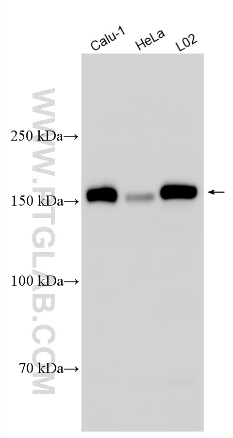
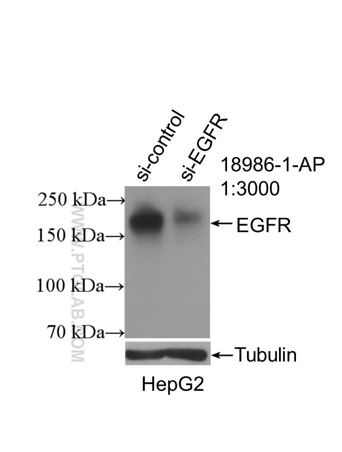
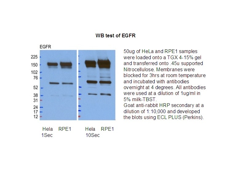
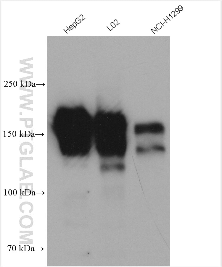
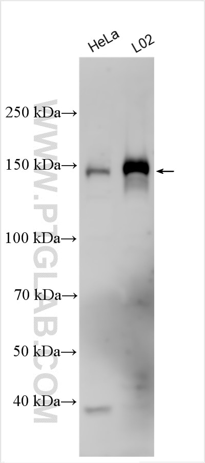
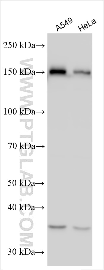
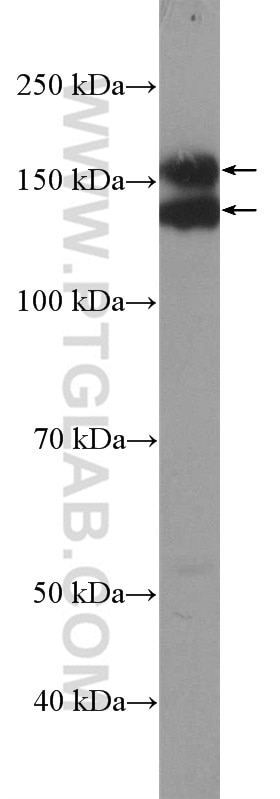
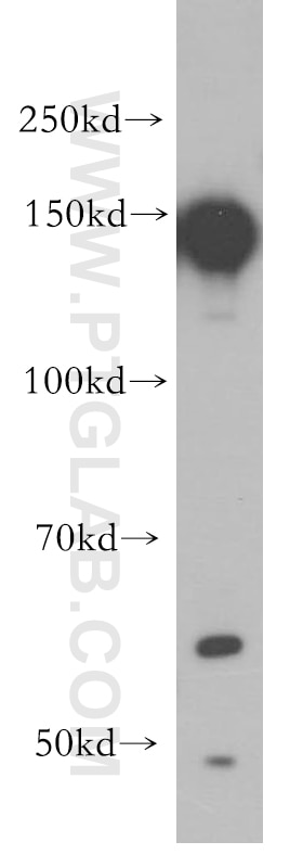
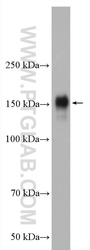
| Positive WB detected in | Calu-1 cells, A431 cells, A549 cells, HeLa cells, HeLa/RPE1 cells, HepG2 cells, MCF-7 cells, NCI-H1299 cells, L02 cells, NCL-H1299 cells |
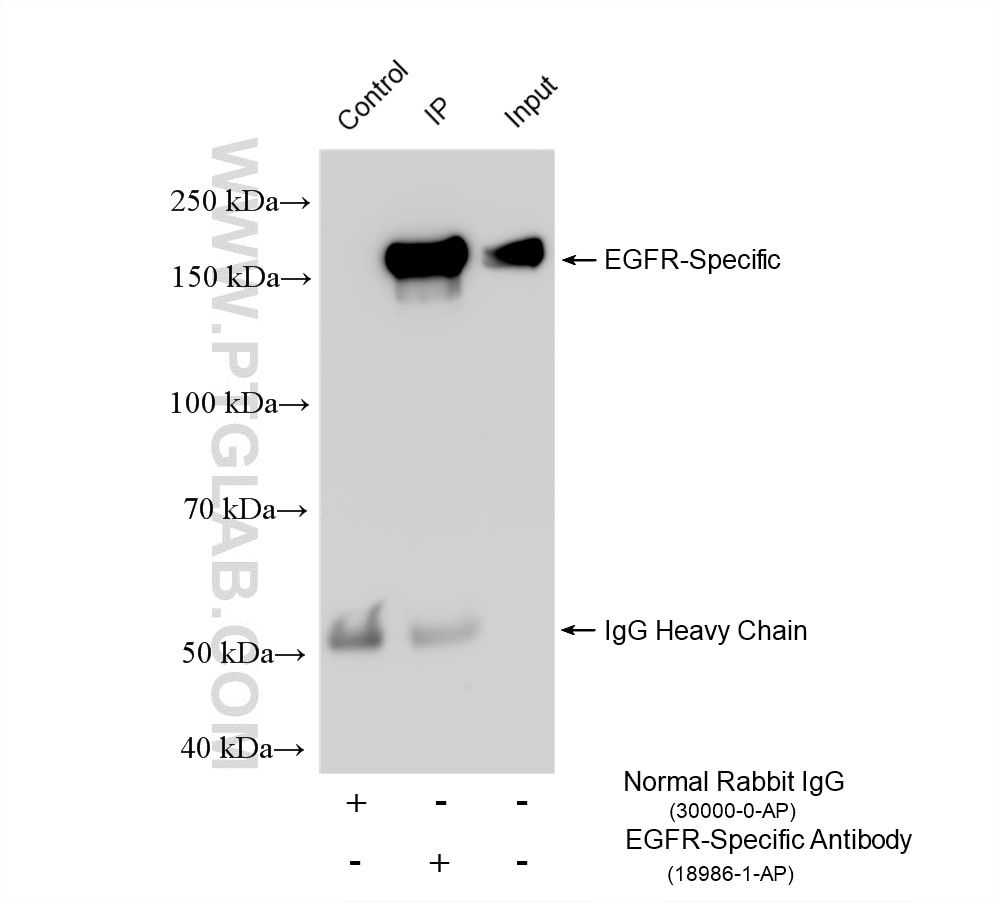
| Positive IP detected in | L02 cells |
Recommended dilution
| Application | Dilution |
|---|---|
| Western Blot (WB) | WB : 1:1000-1:8000 |
| Immunoprecipitation (IP) | IP : 0.5-4.0 ug for 1.0-3.0 mg of total protein lysate |
| It is recommended that this reagent should be titrated in each testing system to obtain optimal results. | |
| Sample-dependent, Check data in validation data gallery. | |
Product Information
18986-1-AP targets EGFR-Specific in WB, IHC, IF, IP, CoIP, ChIP, ELISA applications and shows reactivity with human samples.
| Tested Reactivity | human |
| Cited Reactivity | human, mouse, rat, pig, bovine, goat, sheep, sus scrofa (pig) |
| Host / Isotype | Rabbit / IgG |
| Class | Polyclonal |
| Type | Antibody |
| Immunogen |
Peptide Predict reactive species |
| Full Name | epidermal growth factor receptor (erythroblastic leukemia viral (v-erb-b) oncogene homolog, avian) |
| Calculated Molecular Weight | 134 kDa |
| Observed Molecular Weight | 145-165 kDa |
| GenBank Accession Number | NM_005228 |
| Gene Symbol | EGFR |
| Gene ID (NCBI) | 1956 |
| RRID | AB_10596476 |
| Conjugate | Unconjugated |
| Form | Liquid |
| Purification Method | Antigen affinity purification |
| UNIPROT ID | P00533 |
| Storage Buffer | PBS with 0.02% sodium azide and 50% glycerol, pH 7.3. |
| Storage Conditions | Store at -20°C. Stable for one year after shipment. Aliquoting is unnecessary for -20oC storage. 20ul sizes contain 0.1% BSA. |
Background Information
ERBB1, also named as EGFR, is a receptor for EGF, but also for other members of the EGF family, as TGF-alpha, amphiregulin, betacellulin, heparin-binding EGF-like growth factor, GP30 and vaccinia virus growth factor. ERBB1 is involved in the control of cell growth and differentiation. Phosphorylates MUC1 in breast cancer cells and increases the interaction of MUC1 with C-SRC and CTNNB1/beta-catenin. Isoform 2/truncated isoform of ERBB1 may act as an antagonist. EGFR is associated with lung cancer. The antibody is specific to isoform1.
Protocols
| Product Specific Protocols | |
|---|---|
| IP protocol for EGFR-Specific antibody 18986-1-AP | Download protocol |
| WB protocol for EGFR-Specific antibody 18986-1-AP | Download protocol |
| Standard Protocols | |
|---|---|
| Click here to view our Standard Protocols |
Publications
What published studies show
EGFR-Specific Polyclonal Antibody (Cat# 18986-1-AP) has 231 citations across high-impact journals including Nature Communications, Cell Research, Immunity, Cancer Research, Autophagy, Science Advances, and PNAS. Research fields span oncology (lung, breast, colorectal, liver, glioma), EGFR signaling and drug resistance, EMT, autophagy, immunotherapy, nanomedicine, cardiovascular disease, reproductive biology, and pharmacology. Primary applications are Western blot, IHC, IF, IP, CoIP, and ChIP.
| Species | Application | Title |
|---|---|---|
Mol Cancer Micropeptide MIAC inhibits the tumor progression by interacting with AQP2 and inhibiting EREG/EGFR signaling in renal cell carcinoma | ||
Cell Res In vivo self-assembled small RNAs as a new generation of RNAi therapeutics.
| ||
Immunity The cGAS-STING pathway activates transcription factor TFEB to stimulate lysosome biogenesis and pathogen clearance | ||
Exploration (Beijing) Targeting MGAT4A-Mediated N-Glycosylation as a Therapeutic Strategy to Inhibit Glioblastoma Stem Cell Invasion. | ||
Adv Mater Dual Single-Atom Engineered NADH Oxidase-Mimic Boosts In Situ NAD(+) Biotransformation for Targeted Myocardial Ischemia/Reperfusion Protection. | ||
Cancer Commun (Lond) Blockage of EGFR/AKT and mevalonate pathways synergize the antitumor effect of temozolomide by reprogramming energy metabolism in glioblastoma |
Reviews
What customers say
Users report this EGFR antibody performs well across Western blot, immunofluorescence, and IHC, with an average rating of 4.4/5. Recommended dilutions range from 1:200 (IF/IHC) to 1:1000 (WB). It works on human, mouse, and rat samples including cell lines and tissues. A few users noted non-specific bands at lower molecular weights on WB, and one reported difficulty with the phosphorylated form. Overall, reviewers find it reliable with good signal-to-noise, though some suggest optimizing dilution for stronger results.
The reviews below have been submitted by verified Proteintech customers who received an incentive for providing their feedback.
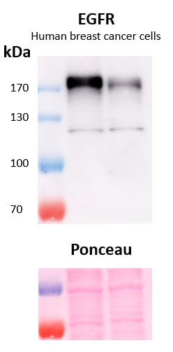
Marina (Verified Customer) (02-13-2023) | A stronger band can be observed slighly above 170 kDa and a fainter band below 130 kDa. Ponceau red is used as total protein loading control. Primary antibody dilution 1:500, overnight incubation at 4ºC. Exposure time 3 minutes.
 |
Clarisse (Verified Customer) (12-12-2022) | This antibody works very well on tissue fixed overnight at 4 degrees C with 2% PFA. No antigen retrieval required.
|
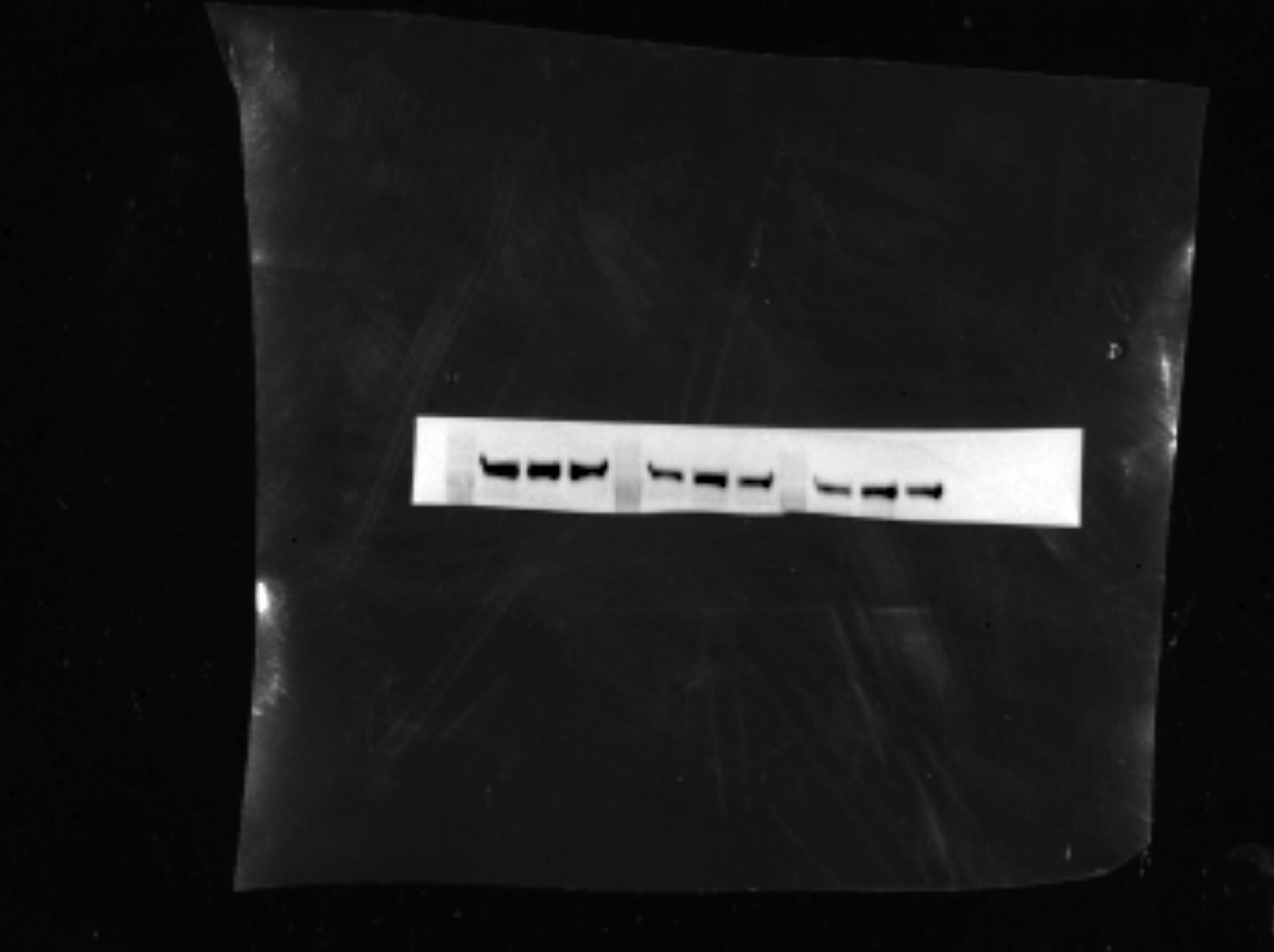
Morgan (Verified Customer) (10-26-2021) | I had luck with this antibody as seen from the image attached. It took a little bit of time to develop though. Maybe using more than 1:500 would be better. Unfortunately I couldn't get the phosphorylated version (p-EGF) to work though. I used OV90 (ovarian cancer) cells
 |
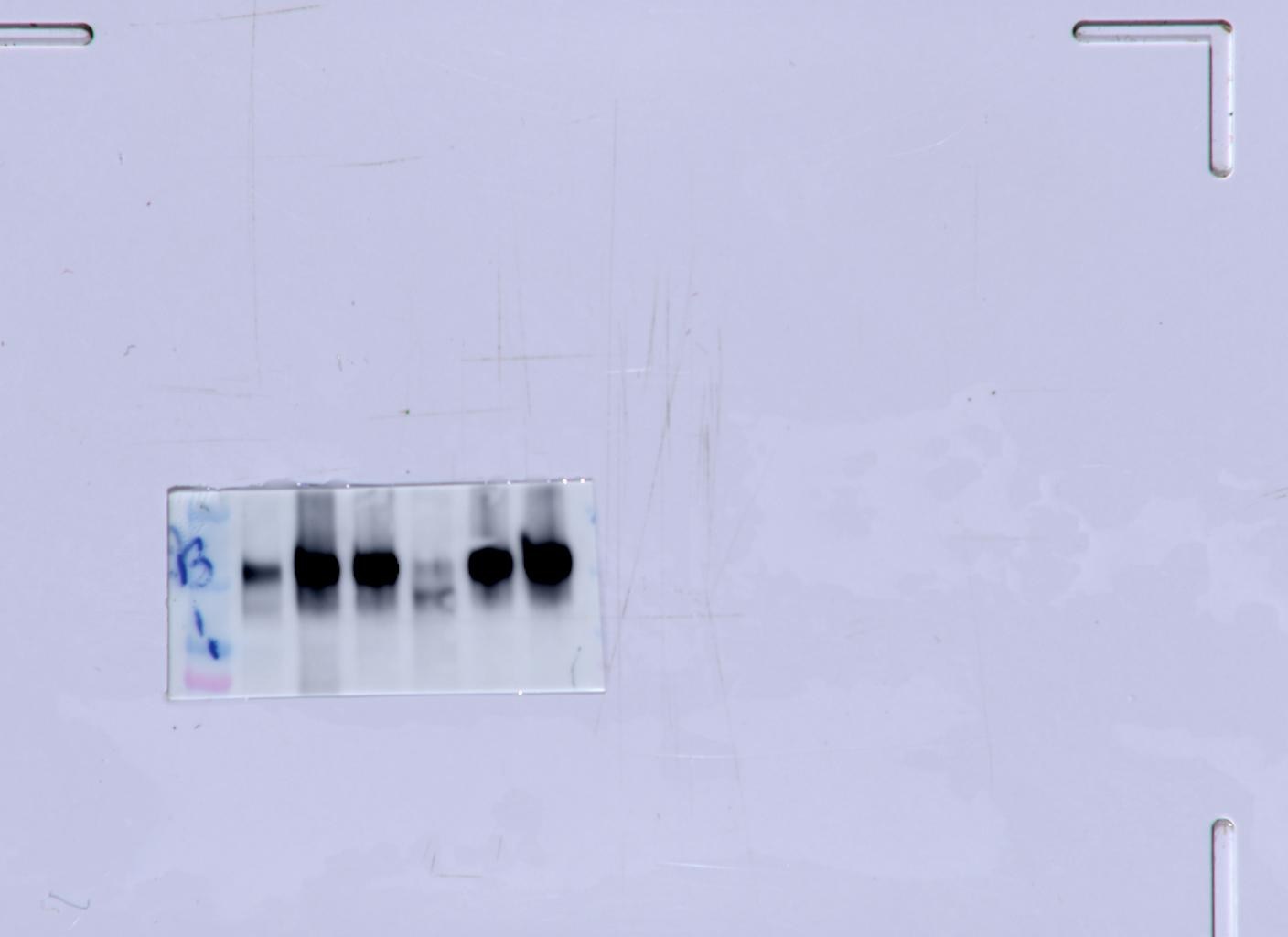
K (Verified Customer) (12-20-2020) | worked well for WB (1:1000)IF (1:200)and IHC (1:200)
 |
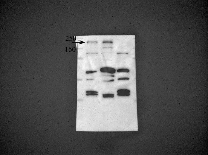
XB (Verified Customer) (10-29-2020) | It is OK to use it to probe EGFR (about 180 kD), but there are some unspecific bands at the lower part of the gel.
 |


