Filter:
 at dilution of 1:10000 incubated at room temperature for 1.5 hours.")
 at dilution of 1:10000 incubated at room temperature for 1.5 hours.")
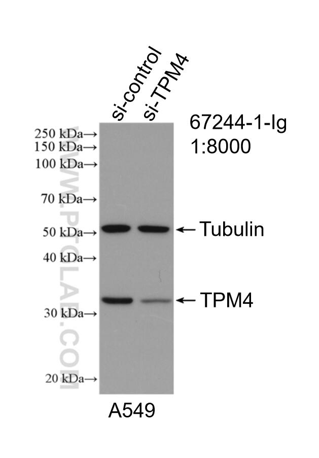
 with sh-Control and sh-TPM4 transfected A549 cells.")
 at dilution of 1:3000 incubated at room temperature for 1.5 hours.")
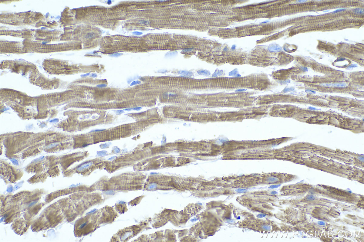
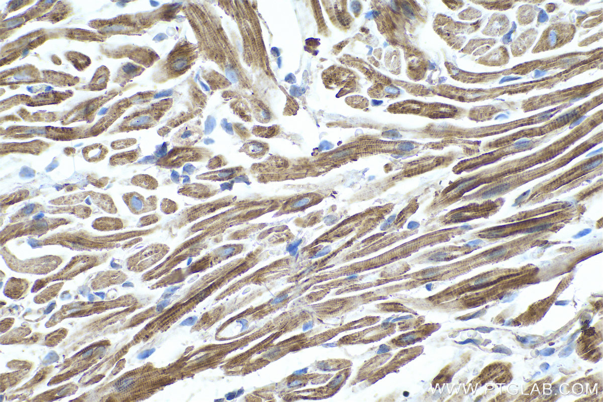
 at dilution of 1:2000 (under 40x lens). Heat mediated antigen retrieval with Tris-EDTA buffer (pH 9.0).")
 at dilution of 1:2000 (under 40x lens). Heat mediated antigen retrieval with Tris-EDTA buffer (pH 9.0).")
 fixed MCF-7 cells using TPM4 antibody (67244-1-Ig, Clone: 2E12D12 ) at dilution of 1:400 and CoraLite®488-Conjugated Goat Anti-Mouse IgG(H+L).")
Tested Applications
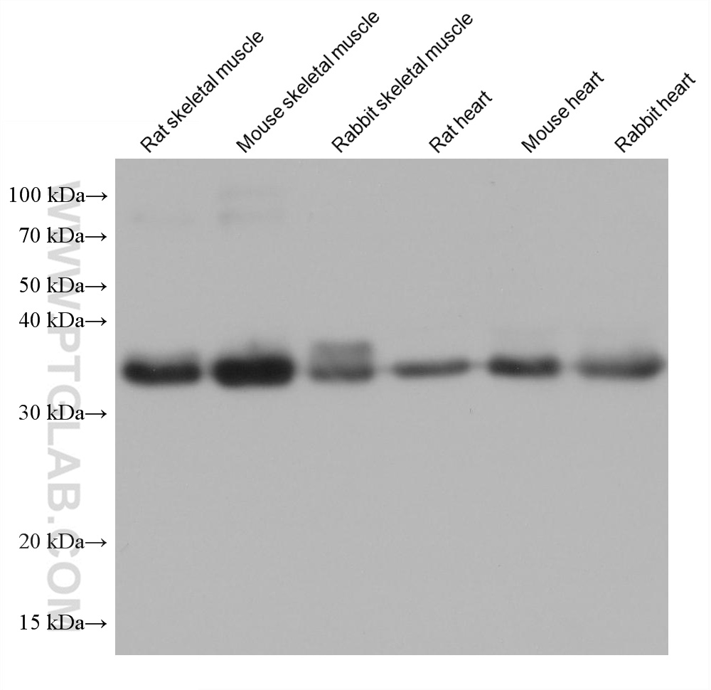
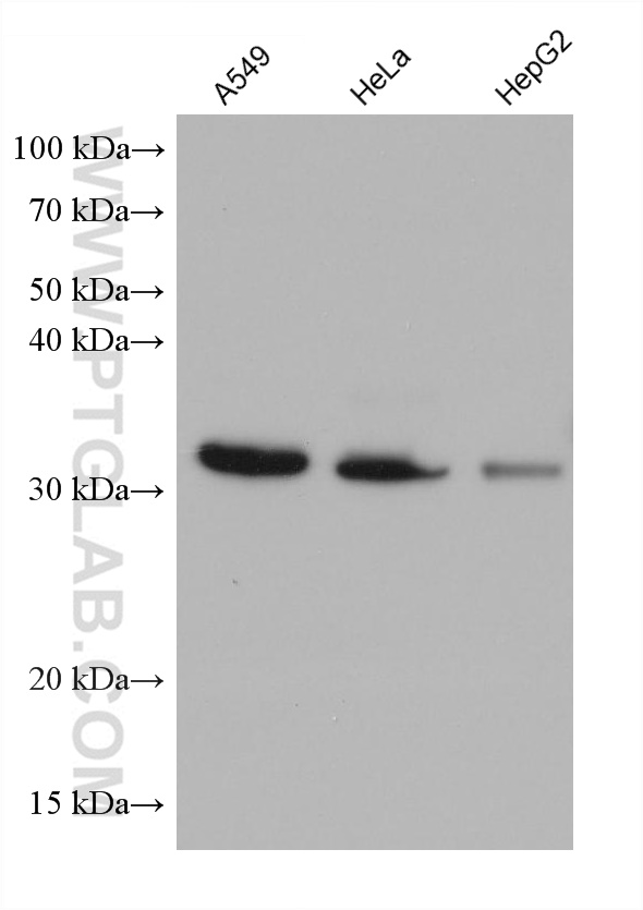
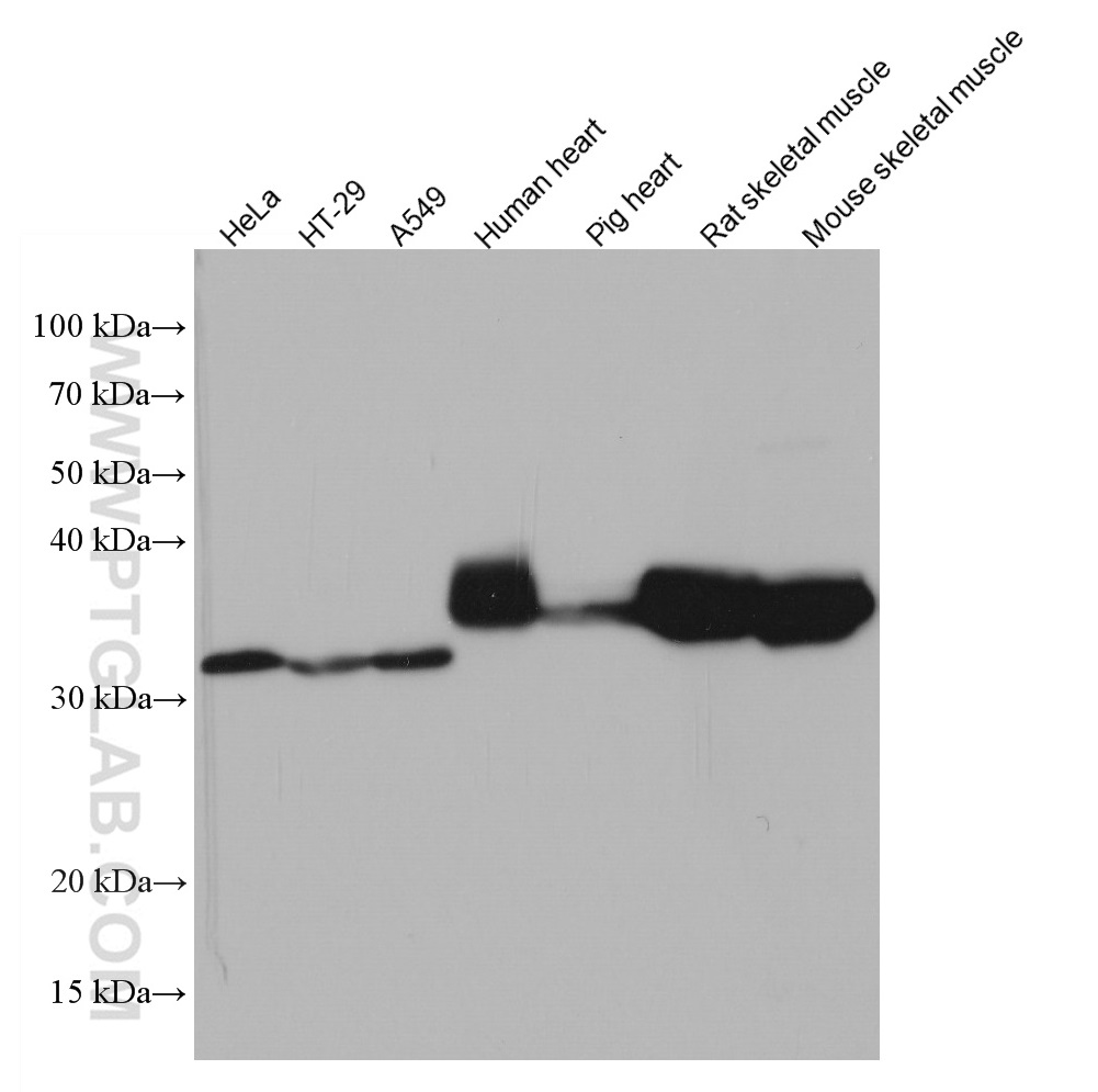
| Positive WB detected in | rat skeletal muscle tissue, A549 cells, HeLa cells, HepG2 cells, mouse skeletal muscle tissue, rabbit skeletal muscle tissue, rat heart tissue, mouse heart tissue, rabbit heart tissue, HT-29 cells, human heart tissue, pig heart tissue |
| Positive IHC detected in | mouse heart tissue, rat heart tissue Note: suggested antigen retrieval with TE buffer pH 9.0; (*) Alternatively, antigen retrieval may be performed with citrate buffer pH 6.0 |
| Positive IF/ICC detected in | MCF-7 cells |
Recommended dilution
| Application | Dilution |
|---|---|
| Western Blot (WB) | WB : 1:5000-1:50000 |
| Immunohistochemistry (IHC) | IHC : 1:1000-1:4000 |
| Immunofluorescence (IF)/ICC | IF/ICC : 1:200-1:800 |
| It is recommended that this reagent should be titrated in each testing system to obtain optimal results. | |
| Sample-dependent, Check data in validation data gallery. | |
Published Applications
| WB | See 2 publications below |
| IF | See 1 publications below |
Product Information
67244-1-Ig targets TPM4 in WB, IHC, IF/ICC, ELISA applications and shows reactivity with human, mouse, rat, pig, rabbit samples.
| Tested Reactivity | human, mouse, rat, pig, rabbit |
| Cited Reactivity | human, mouse |
| Host / Isotype | Mouse / IgG1 |
| Class | Monoclonal |
| Type | Antibody |
| Immunogen | TPM4 fusion protein Ag6947 Predict reactive species |
| Full Name | tropomyosin 4 |
| Calculated Molecular Weight | 248 aa, 29 kDa |
| Observed Molecular Weight | 32-35 kDa |
| GenBank Accession Number | BC037576 |
| Gene Symbol | TPM4 |
| Gene ID (NCBI) | 7171 |
| ENSEMBL Gene ID | ENSG00000167460 |
| RRID | AB_2882523 |
| Conjugate | Unconjugated |
| Form | Liquid |
| Purification Method | Protein A purification |
| UNIPROT ID | P67936 |
| Storage Buffer | PBS with 0.02% sodium azide and 50% glycerol pH 7.3. |
| Storage Conditions | Store at -20°C. Stable for one year after shipment. Aliquoting is unnecessary for -20oC storage. 20ul sizes contain 0.1% BSA. |
Background Information
TPM4 is a member of tropomyosins (TPMs), a multi-gene family of actin-binding proteins present in all eukaryotic cells. In muscle, TPMs are responsible for mediating contraction via regulation of the actin-myosin interaction. As to non-muscle cells, its proposed role is to stabilize the actin filaments by modulating the interaction with proteins that are responsible for the regulation of actin dynamics. The form of TPM4 in muscle has a higher molecular weight than the form found in non-muscle cells.
Protocols
| Product Specific Protocols | |
|---|---|
| WB protocol for TPM4 antibody 67244-1-Ig | Download protocol |
| IHC protocol for TPM4 antibody 67244-1-Ig | Download protocol |
| IF protocol for TPM4 antibody 67244-1-Ig | Download protocol |
| Standard Protocols | |
|---|---|
| Click here to view our Standard Protocols |
Publications
| Species | Application | Title |
|---|---|---|
Rheumatology (Oxford) Association of Anti-TPM4 autoantibodies with vasculopathic cutaneous manifestations in juvenile dermatomyositis | ||
PLoS One Proteomic changes in the hippocampus of large mammals after total-body low dose radiation | ||
Cell Discov TPM4 condensates glycolytic enzymes and facilitates actin reorganization under hyperosmotic stress |