Various lysates were subjected to SDS PAGE followed by western blot with 66105-1-Ig (PGM1 antibody) at dilution of 1:5000 incubated at room temperature for 1.5 hours.
Various lysates were subjected to SDS PAGE followed by western blot with 66105-1-Ig (PGM1 antibody) at dilution of 1:5000 incubated at room temperature for 1.5 hours.
IHC staining of human colon using 66105-1-Ig
Immunohistochemical analysis of paraffin-embedded human colon tissue slide using 66105-1-Ig (PGM1 Antibody) at dilution of 1:200 (under 10x lens).
The Proteintech guarantee covers Proteintech antibodies in any species and any application, including those not listed on the datasheet. If the antibody doesn’t perform, you can receive a hassle-free refund or credit note.
human colon tissue, human liver cancer tissue Note: suggested antigen retrieval with TE buffer pH 9.0; (*) Alternatively, antigen retrieval may be performed with citrate buffer pH 6.0
Positive IF/ICC detected in
HepG2 cells
Recommended dilution
Application
Dilution
Western Blot (WB)
WB : 1:2000-1:10000
Immunohistochemistry (IHC)
IHC : 1:50-1:500
Immunofluorescence (IF)/ICC
IF/ICC : 1:500-1:2000
It is recommended that this reagent should be titrated in each testing system to obtain optimal results.
Sample-dependent, Check data in validation data gallery.
Product Information
66105-1-Ig targets PGM1 in WB, IHC, IF/ICC, ELISA applications and shows reactivity with human, mouse, rat, rabbit, pig samples.
PBS with 0.02% sodium azide and 50% glycerol , pH 7.3
Storage Conditions
Store at -20°C. Stable for one year after shipment. Aliquoting is unnecessary for -20oC storage. 20ul sizes contain 0.1% BSA.
Background Information
PGM1(Phosphoglucomutase-1) is also named as glucose phosphomutase 1 and belongs to the phosphohexose mutase family. It catalyzes the transfer of phosphate between the 1 and 6 positions of glucose. In most cell types, PGM1 isozymes predominate, representing about 90% of total PGM activity. One exception is red cells, where PGM2 is a major isozyme(PMID:8257433). Defects in PGM1 are the cause of glycogen storage disease type 14 (GSD14)(PMID:19625727). It has 2 isoforms produced by alternative splicing.
Various lysates were subjected to SDS PAGE followed by western blot with 66105-1-Ig (PGM1 antibody) at dilution of 1:5000 incubated at room temperature for 1.5 hours.
IHC Figures
IHC staining of human colon using 66105-1-Ig
Immunohistochemical analysis of paraffin-embedded human colon tissue slide using 66105-1-Ig (PGM1 Antibody) at dilution of 1:200 (under 10x lens).
IHC staining of human colon using 66105-1-Ig
Immunohistochemical analysis of paraffin-embedded human colon tissue slide using 66105-1-Ig (PGM1 Antibody) at dilution of 1:200 (under 40x lens).
IHC staining of human liver cancer using 66105-1-Ig
Immunohistochemical analysis of paraffin-embedded human liver cancer tissue slide using 66105-1-Ig (PGM1 Antibody) at dilution of 1:100.
IHC staining of human liver cancer using 66105-1-Ig
Immunohistochemical analysis of paraffin-embedded human liver cancer tissue slide using 66105-1-Ig (PGM1 Antibody) at dilution of 1:100.
IF/ICC Figures
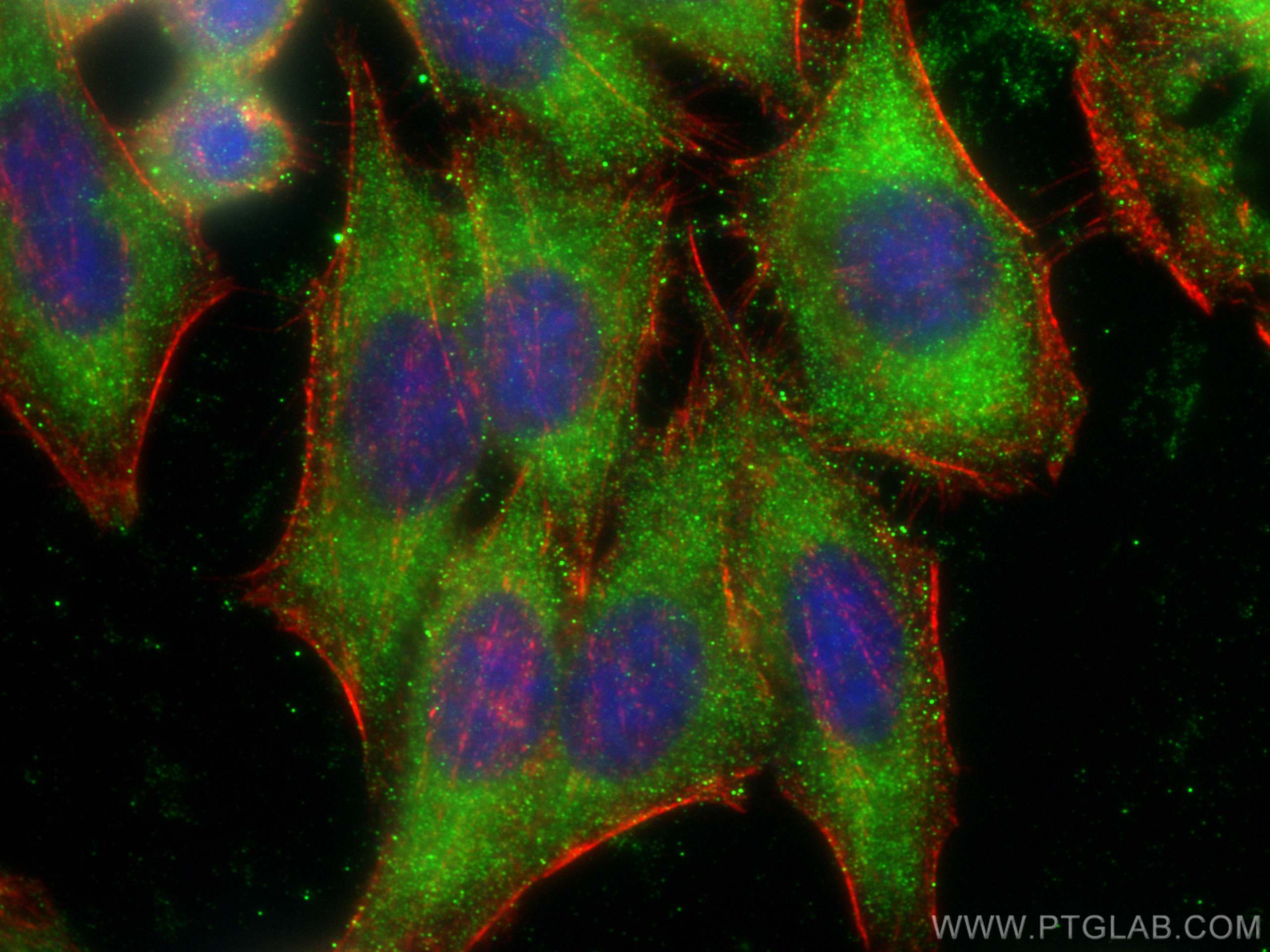
IF Staining of HepG2 using 66105-1-Ig
Immunofluorescent analysis of (-20°C Ethanol) fixed HepG2 cells using PGM1 antibody (66105-1-Ig, Clone: 2G5A8 ) at dilution of 1:1000 and CoraLite®488-Conjugated Goat Anti-Mouse IgG(H+L), CL594-phalloidin (red).
The species listed in Tested Reactivity are in-house verified and applicable species. For unlisted species, please refer to the homology analysis of the immunogen sequence and related species. For rabbit polyclonal antibodies, homology >70% is recommended. For mouse monoclonal antibodies and rabbit recombinant antibodies, homology >90% is recommended. Generally, the higher the homology, the greater the applicability. However, there will be certain differences in protein expression in different species, tissues or cells. Therefore, the homology analysis results are for reference only and do not serve as a guarantee.
At Proteintech, we pride ourselves on our antibody quality, customer service and transparency. As such, we are comparing our antibodies with other vendors, enabling easy identification and comparisons of key data to help you choose the suitable antibody for your needs.
We have selected the top cited antibodies from these vendors for you to compare.
 at dilution of 1:5000 incubated at room temperature for 1.5 hours.")
 at dilution of 1:200 (under 10x lens).")
 at dilution of 1:200 (under 40x lens).")
 at dilution of 1:100.")
 at dilution of 1:100.")
 fixed HepG2 cells using PGM1 antibody (66105-1-Ig, Clone: 2G5A8 ) at dilution of 1:1000 and CoraLite®488-Conjugated Goat Anti-Mouse IgG(H+L), CL594-phalloidin (red).")