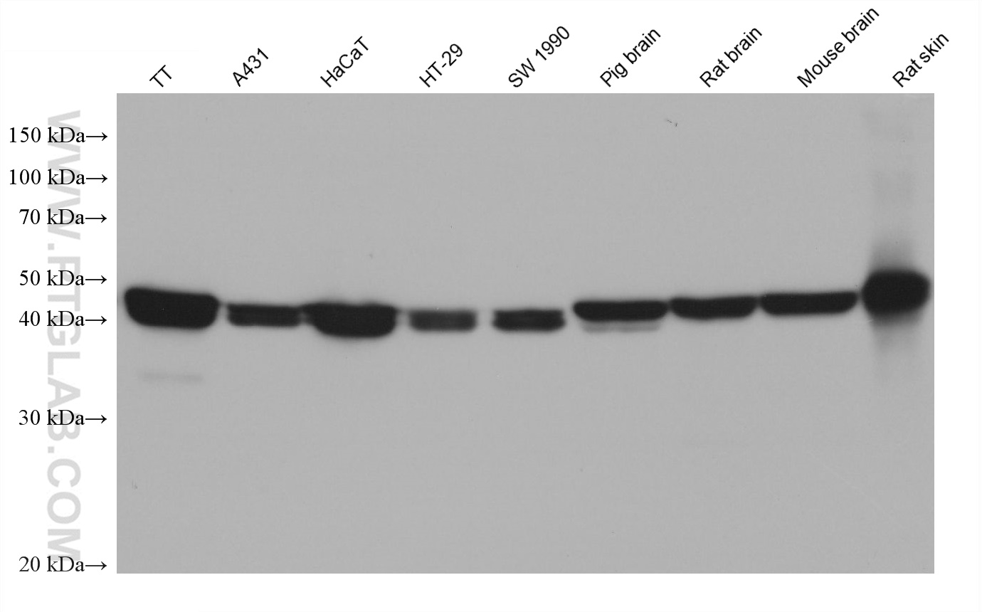
Various lysates were subjected to SDS PAGE followed by western blot with 67427-1-Ig (KLK11 antibody) at dilution of 1:20000 incubated at room temperature for 1.5 hours.
Various lysates were subjected to SDS PAGE followed by western blot with 67427-1-Ig (KLK11 antibody) at dilution of 1:20000 incubated at room temperature for 1.5 hours.
WB analysis using 67427-1-Ig
Various lysates were subjected to SDS PAGE followed by western blot with 67427-1-Ig (KLK11 antibody) at dilution of 1:3000 incubated at room temperature for 1.5 hours.
Various lysates were subjected to SDS PAGE followed by western blot with 67427-1-Ig (KLK11 antibody) at dilution of 1:3000 incubated at room temperature for 1.5 hours.
IHC staining of human breast cancer using 67427-1-Ig
Immunohistochemical analysis of paraffin-embedded human breast cancer tissue slide using 67427-1-Ig (KLK11 antibody) at dilution of 1:1000 (under 40x lens). Heat mediated antigen retrieval with Tris-EDTA buffer (pH 9.0).
Immunohistochemical analysis of paraffin-embedded human breast cancer tissue slide using 67427-1-Ig (KLK11 antibody) at dilution of 1:1000 (under 40x lens). Heat mediated antigen retrieval with Tris-EDTA buffer (pH 9.0).
IHC staining of human breast cancer using 67427-1-Ig
Immunohistochemical analysis of paraffin-embedded human breast cancer tissue slide using 67427-1-Ig (KLK11 antibody) at dilution of 1:1000 (under 10x lens). Heat mediated antigen retrieval with Tris-EDTA buffer (pH 9.0).
Immunohistochemical analysis of paraffin-embedded human breast cancer tissue slide using 67427-1-Ig (KLK11 antibody) at dilution of 1:1000 (under 10x lens). Heat mediated antigen retrieval with Tris-EDTA buffer (pH 9.0).
IHC staining of human prostate cancer using 67427-1-Ig
Immunohistochemical analysis of paraffin-embedded human prostate cancer tissue slide using 67427-1-Ig (KLK11 antibody) at dilution of 1:1000 (under 10x lens). Heat mediated antigen retrieval with Tris-EDTA buffer (pH 9.0).
Immunohistochemical analysis of paraffin-embedded human prostate cancer tissue slide using 67427-1-Ig (KLK11 antibody) at dilution of 1:1000 (under 10x lens). Heat mediated antigen retrieval with Tris-EDTA buffer (pH 9.0).
IHC staining of human prostate cancer using 67427-1-Ig
Immunohistochemical analysis of paraffin-embedded human prostate cancer tissue slide using 67427-1-Ig (KLK11 antibody) at dilution of 1:1000 (under 40x lens). Heat mediated antigen retrieval with Tris-EDTA buffer (pH 9.0).
Immunohistochemical analysis of paraffin-embedded human prostate cancer tissue slide using 67427-1-Ig (KLK11 antibody) at dilution of 1:1000 (under 40x lens). Heat mediated antigen retrieval with Tris-EDTA buffer (pH 9.0).
IF Staining of human breast cancer using 67427-1-Ig
Immunofluorescent analysis of (4% PFA) fixed human breast cancer tissue using KLK11 antibody (67427-1-Ig, Clone: 1E2B4 ) at dilution of 1:400 and CoraLite®488-Conjugated Goat Anti-Mouse IgG(H+L).
Immunofluorescent analysis of (4% PFA) fixed human breast cancer tissue using KLK11 antibody (67427-1-Ig, Clone: 1E2B4 ) at dilution of 1:400 and CoraLite®488-Conjugated Goat Anti-Mouse IgG(H+L).
The Proteintech guarantee covers Proteintech antibodies in any species and any application, including those not listed on the datasheet. If the antibody doesn’t perform, you can receive a hassle-free refund or credit note.
human breast cancer tissue, human prostate cancer tissue Note: suggested antigen retrieval with TE buffer pH 9.0; (*) Alternatively, antigen retrieval may be performed with citrate buffer pH 6.0
Positive IF-P detected in
human breast cancer tissue
Recommended dilution
Application
Dilution
Western Blot (WB)
WB : 1:5000-1:50000
Immunohistochemistry (IHC)
IHC : 1:500-1:2000
Immunofluorescence (IF)-P
IF-P : 1:200-1:800
It is recommended that this reagent should be titrated in each testing system to obtain optimal results.
Sample-dependent, Check data in validation data gallery.
PBS with 0.02% sodium azide and 50% glycerol pH 7.3.
Storage Conditions
Store at -20°C. Stable for one year after shipment. Aliquoting is unnecessary for -20oC storage. 20ul sizes contain 0.1% BSA.
Background Information
KLK11(Kallikrein-11) is also named as PRSS20, TLSP and belongs to the peptidase S1 family and Kallikrein subfamily. It is a multifunctinal protease cleaving several substrates for kallikrein and trypsin and involved in normal physiology processes in bronchus. This full length protein has a signal peptide, a propeptide and four glycosylation sites. It has 4 isoforms produced by alternative splicing with the molecular weight of 31 kDa, 27 kDa,34 kDa and 30 kDa.
Various lysates were subjected to SDS PAGE followed by western blot with 67427-1-Ig (KLK11 antibody) at dilution of 1:20000 incubated at room temperature for 1.5 hours.
WB analysis using 67427-1-Ig
Various lysates were subjected to SDS PAGE followed by western blot with 67427-1-Ig (KLK11 antibody) at dilution of 1:3000 incubated at room temperature for 1.5 hours.
WB analysis using 67427-1-Ig
Various lysates were subjected to SDS PAGE followed by western blot with 67427-1-Ig (KLK11 antibody) at dilution of 1:3000 incubated at room temperature for 1.5 hours.
IHC Figures
IHC staining of human breast cancer using 67427-1-Ig
Immunohistochemical analysis of paraffin-embedded human breast cancer tissue slide using 67427-1-Ig (KLK11 antibody) at dilution of 1:1000 (under 40x lens). Heat mediated antigen retrieval with Tris-EDTA buffer (pH 9.0).
IHC staining of human breast cancer using 67427-1-Ig
Immunohistochemical analysis of paraffin-embedded human breast cancer tissue slide using 67427-1-Ig (KLK11 antibody) at dilution of 1:1000 (under 10x lens). Heat mediated antigen retrieval with Tris-EDTA buffer (pH 9.0).
IHC staining of human prostate cancer using 67427-1-Ig
Immunohistochemical analysis of paraffin-embedded human prostate cancer tissue slide using 67427-1-Ig (KLK11 antibody) at dilution of 1:1000 (under 10x lens). Heat mediated antigen retrieval with Tris-EDTA buffer (pH 9.0).
IHC staining of human prostate cancer using 67427-1-Ig
Immunohistochemical analysis of paraffin-embedded human prostate cancer tissue slide using 67427-1-Ig (KLK11 antibody) at dilution of 1:1000 (under 40x lens). Heat mediated antigen retrieval with Tris-EDTA buffer (pH 9.0).
IF-P Figures
IF Staining of human breast cancer using 67427-1-Ig
Immunofluorescent analysis of (4% PFA) fixed human breast cancer tissue using KLK11 antibody (67427-1-Ig, Clone: 1E2B4 ) at dilution of 1:400 and CoraLite®488-Conjugated Goat Anti-Mouse IgG(H+L).
IF Staining of human breast cancer using 67427-1-Ig
Immunofluorescent analysis of (4% PFA) fixed human breast cancer tissue using KLK11 antibody (67427-1-Ig, Clone: 1E2B4 ) at dilution of 1:400 and CoraLite®488-Conjugated Goat Anti-Mouse IgG(H+L).
The species listed in Tested Reactivity are in-house verified and applicable species. For unlisted species, please refer to the homology analysis of the immunogen sequence and related species. For rabbit polyclonal antibodies, homology >70% is recommended. For mouse monoclonal antibodies and rabbit recombinant antibodies, homology >90% is recommended. Generally, the higher the homology, the greater the applicability. However, there will be certain differences in protein expression in different species, tissues or cells. Therefore, the homology analysis results are for reference only and do not serve as a guarantee.
At Proteintech, we pride ourselves on our antibody quality, customer service and transparency. As such, we are comparing our antibodies with other vendors, enabling easy identification and comparisons of key data to help you choose the suitable antibody for your needs.
We have selected the top cited antibodies from these vendors for you to compare.
 at dilution of 1:20000 incubated at room temperature for 1.5 hours.")
 at dilution of 1:3000 incubated at room temperature for 1.5 hours.")
 at dilution of 1:3000 incubated at room temperature for 1.5 hours.")
 at dilution of 1:1000 (under 40x lens). Heat mediated antigen retrieval with Tris-EDTA buffer (pH 9.0).")
 at dilution of 1:1000 (under 10x lens). Heat mediated antigen retrieval with Tris-EDTA buffer (pH 9.0).")
 at dilution of 1:1000 (under 10x lens). Heat mediated antigen retrieval with Tris-EDTA buffer (pH 9.0).")
 at dilution of 1:1000 (under 40x lens). Heat mediated antigen retrieval with Tris-EDTA buffer (pH 9.0).")
 fixed human breast cancer tissue using KLK11 antibody (67427-1-Ig, Clone: 1E2B4 ) at dilution of 1:400 and CoraLite®488-Conjugated Goat Anti-Mouse IgG(H+L).")
 fixed human breast cancer tissue using KLK11 antibody (67427-1-Ig, Clone: 1E2B4 ) at dilution of 1:400 and CoraLite®488-Conjugated Goat Anti-Mouse IgG(H+L).")