at dilution of 1:20000 and incubated at room temperature for 1.5 hours.")
 at dilution of 1:20000 and incubated at room temperature for 1.5 hours.")
 at dilution of 1:20000 and incubated at room temperature for 1.5 hours.")
 at dilution of 1:4000 incubated at room temperature for 1.5 hours.")
 at dilution of 1:20000 and incubated at room temperature for 1.5 hours.")
. Heat mediated antigen retrieval with Tris-EDTA buffer (pH 9.0).")
. Heat mediated antigen retrieval with Tris-EDTA buffer (pH 9.0).")
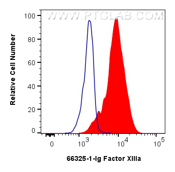
 and CoraLite®488-Conjugated Goat Anti-Mouse IgG(H+L) (SA00013-1)(red), or 0.25 ug Mouse IgG2b isotype control Mouse McAb (66360-3-Ig, Clone: 11B8C4) (blue). Cells were fixed with 4% PFA and permeabilized with Flow Cytometry Perm Buffer.")
Tested Applications
| Positive WB detected in | pig colon tissue, human placenta tissue, human peripheral blood platelets, rat colon tissue, rabbit large intestine tissue, pig liver tissue |
| Positive IHC detected in | human liver cancer tissue Note: suggested antigen retrieval with TE buffer pH 9.0; (*) Alternatively, antigen retrieval may be performed with citrate buffer pH 6.0 |
| Positive FC (Intra) detected in | U-937 cells |
Recommended dilution
| Application | Dilution |
|---|---|
| Western Blot (WB) | WB : 1:5000-1:50000 |
| Immunohistochemistry (IHC) | IHC : 1:100-1:1000 |
| Flow Cytometry (FC) (INTRA) | FC (INTRA) : 0.25 ug per 10^6 cells in a 100 µl suspension |
| It is recommended that this reagent should be titrated in each testing system to obtain optimal results. | |
| Sample-dependent, Check data in validation data gallery. | |
Published Applications
| WB | See 1 publications below |
| IHC | See 1 publications below |
Product Information
66325-1-Ig targets Factor XIIIa in WB, IHC, ELISA applications and shows reactivity with human, mouse, rat, pig, rabbit samples.
| Tested Reactivity | human, mouse, rat, pig, rabbit |
| Cited Reactivity | human |
| Host / Isotype | Mouse / IgG2b |
| Class | Monoclonal |
| Type | Antibody |
| Immunogen |
CatNo: Ag11105 Product name: Recombinant human F13A1 protein Source: e coli.-derived, PET28a Tag: 6*His Domain: 383-732 aa of BC027963 Sequence: RPDLPVGFGGWQAVDSTPQENSDGMYRCGPASVQAIKHGHVCFQFDAPFVFAEVNSDLIYITAKKDGTHVVENVDATHIGKLIVTKQIGGDGMMDITDTYKFQEGQEEERLALETALMYGAKKPLNTEGVMKSRSNVDMDFEVENAVLGKDFKLSITFRNNSHNRYTITAYLSANITFYTGVPKAEFKKETFDVTLEPLSFKKEAVLIQAGEYMGQLLEQASLHFFVTARINETRDVLAKQKSTVLTIPEIIIKVRGTQVVGSDMTVIVEFTNPLKETLRNVWVHLDGPGVTRPMKKMFREIRPNSTVQWEEVCRPWVSGHRKLIASMSSDSLRHVYGELDVQIQRRPSM Predict reactive species |
| Full Name | coagulation factor XIII, A1 polypeptide |
| Calculated Molecular Weight | 732 aa, 83 kDa |
| Observed Molecular Weight | 75-80 kDa |
| GenBank Accession Number | BC027963 |
| Gene Symbol | Factor XIIIa |
| Gene ID (NCBI) | 2162 |
| RRID | AB_2881706 |
| Conjugate | Unconjugated |
| Form | Liquid |
| Purification Method | Protein A purification |
| UNIPROT ID | P00488 |
| Storage Buffer | PBS with 0.02% sodium azide and 50% glycerol, pH 7.3. |
| Storage Conditions | Store at -20°C. Stable for one year after shipment. Aliquoting is unnecessary for -20oC storage. 20ul sizes contain 0.1% BSA. |
Background Information
Factor XIIIa is a blood proenzyme that catalyzes intermolecular cross-linking of fibrinogen and is involved in clotting cascade. It has been identified in various tissues including placenta, uterus, and prostate. Anti- Factor XIIIa has been found to be useful in differentiating between dermatofibroma (90% positive) and desmoplastic malignant melanoma (negative). Factor XIIIa positivity is also seen in capillary hemagioblastoma, hemangioendothelioma, hemangiopericytoma, xanthogranuloma, xanthoma, hepatocellular carcinoma, glomus tumor , and meningioma.
Protocols
| Product Specific Protocols | |
|---|---|
| FC protocol for Factor XIIIa antibody 66325-1-Ig | Download protocol |
| IHC protocol for Factor XIIIa antibody 66325-1-Ig | Download protocol |
| WB protocol for Factor XIIIa antibody 66325-1-Ig | Download protocol |
| Standard Protocols | |
|---|---|
| Click here to view our Standard Protocols |