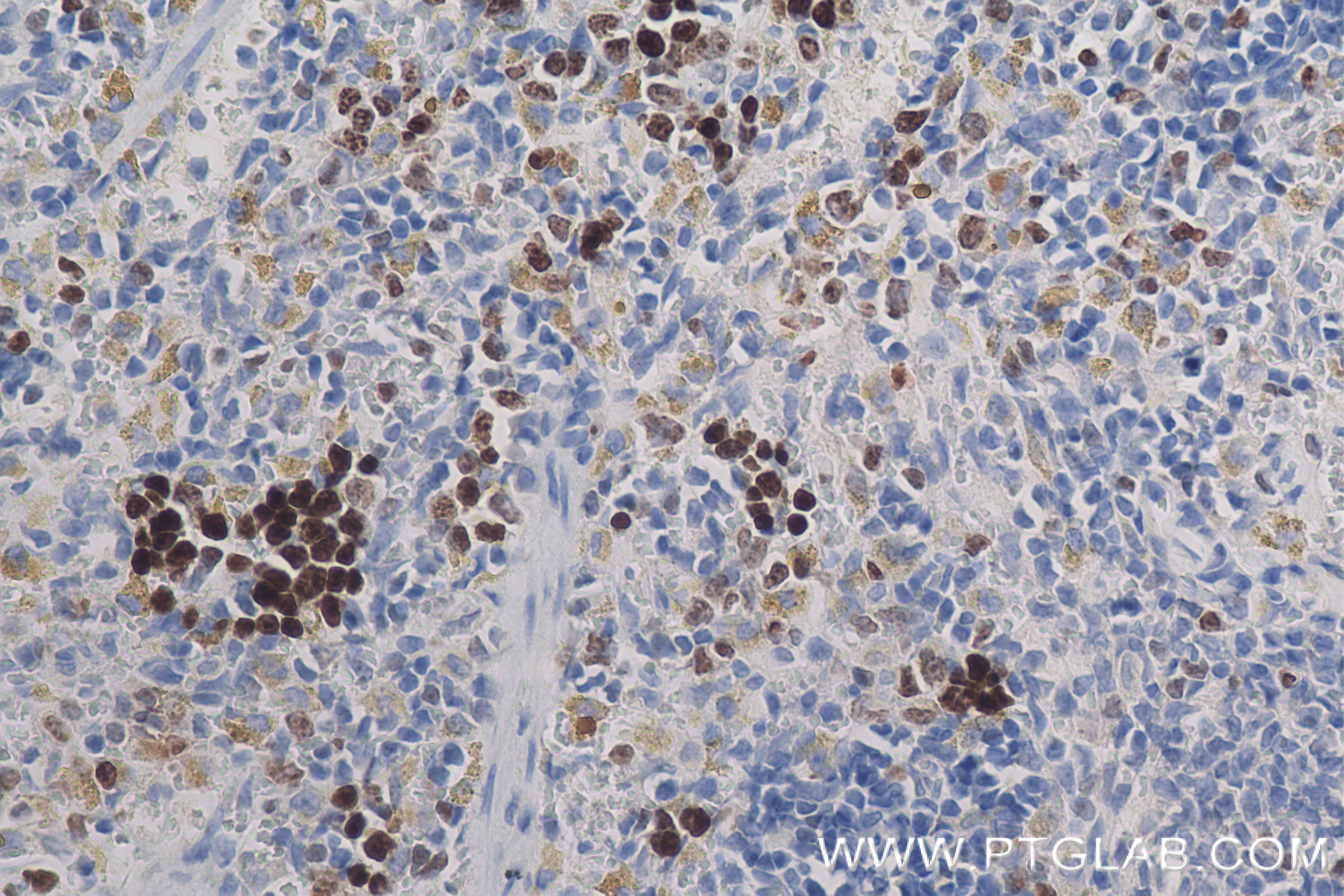
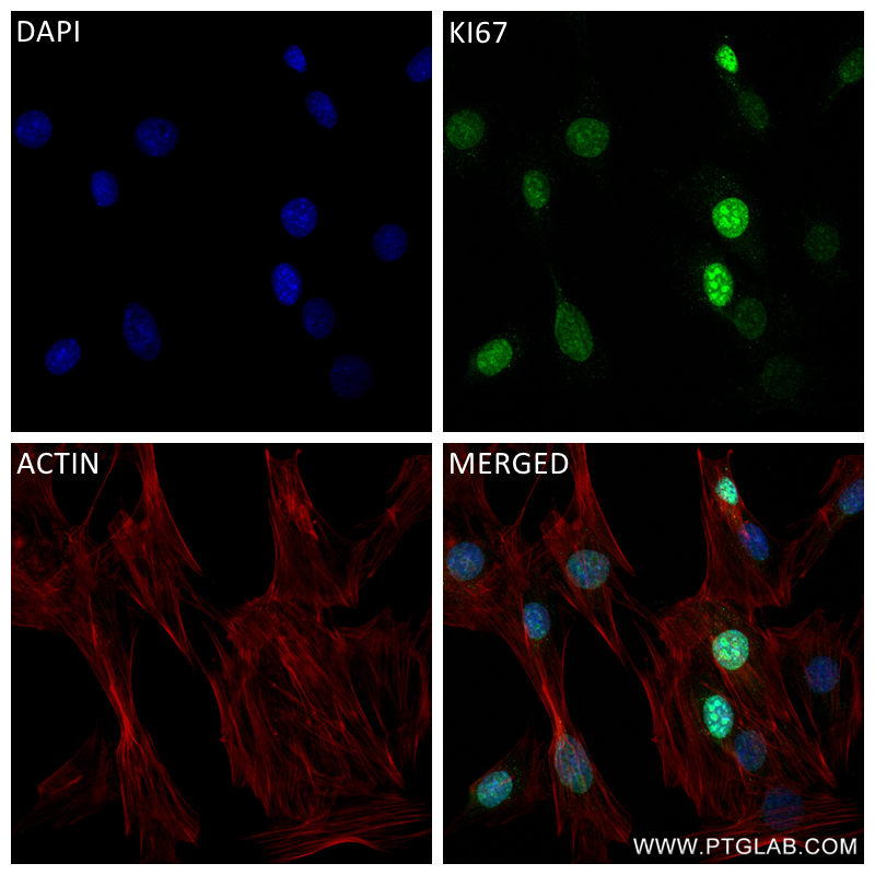
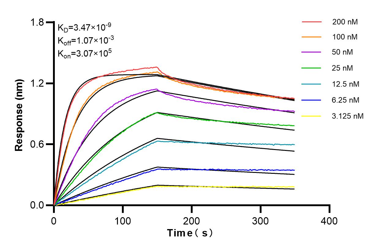
Mouse Ki-67 Recombinant antibody, PBS Only (Detector)
Ki-67 Uni-rAbTM Recombinant Antibody for WB, IHC, IF/ICC, Cytometric bead array, Indirect ELISA
Host / Isotype
Rabbit / IgG
Reactivity
mouse, rat
Applications
WB, IHC, IF/ICC, Cytometric bead array, Indirect ELISA
Conjugate
Unconjugated
CloneNo.
241768E2
Cat no : 84432-1-PBS
Synonyms
Validation Data Gallery
Product Information
84432-1-PBS targets Ki-67 as part of a matched antibody pair:
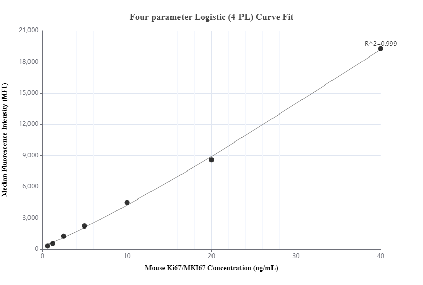
MP01277-1: 84432-2-PBS capture and 84432-1-PBS detection (validated in Cytometric bead array)
Unconjugated rabbit recombinant monoclonal antibody in PBS only (BSA and azide free) storage buffer at a concentration of 1 mg/mL, ready for conjugation. Created using Proteintech’s proprietary in-house recombinant technology. Recombinant production enables unrivalled batch-to-batch consistency, easy scale-up, and future security of supply.
This conjugation ready format makes antibodies ideal for use in many applications including: ELISAs, multiplex assays requiring matched pairs, mass cytometry, and multiplex imaging applications.Antibody use should be optimized by the end user for each application and assay.
| Tested Reactivity | mouse, rat |
| Host / Isotype | Rabbit / IgG |
| Class | Recombinant |
| Type | Antibody |
| Immunogen | FusionProtein |
| Full Name | antigen identified by monoclonal antibody Ki 67 |
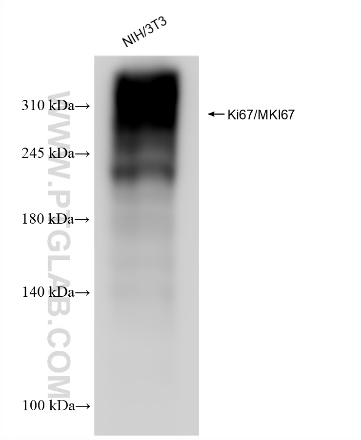
| Calculated Molecular Weight | 351kDa |
| Observed Molecular Weight | 350 kDa |
| GenBank Accession Number | NM_001081117.2 |
| Gene Symbol | ki67 |
| Gene ID (NCBI) | 17345 |
| Conjugate | Unconjugated |
| Form | Liquid |
| Purification Method | Protein A purification |
| Storage Buffer | PBS Only |
| Storage Conditions | Store at -80°C. |
Background Information
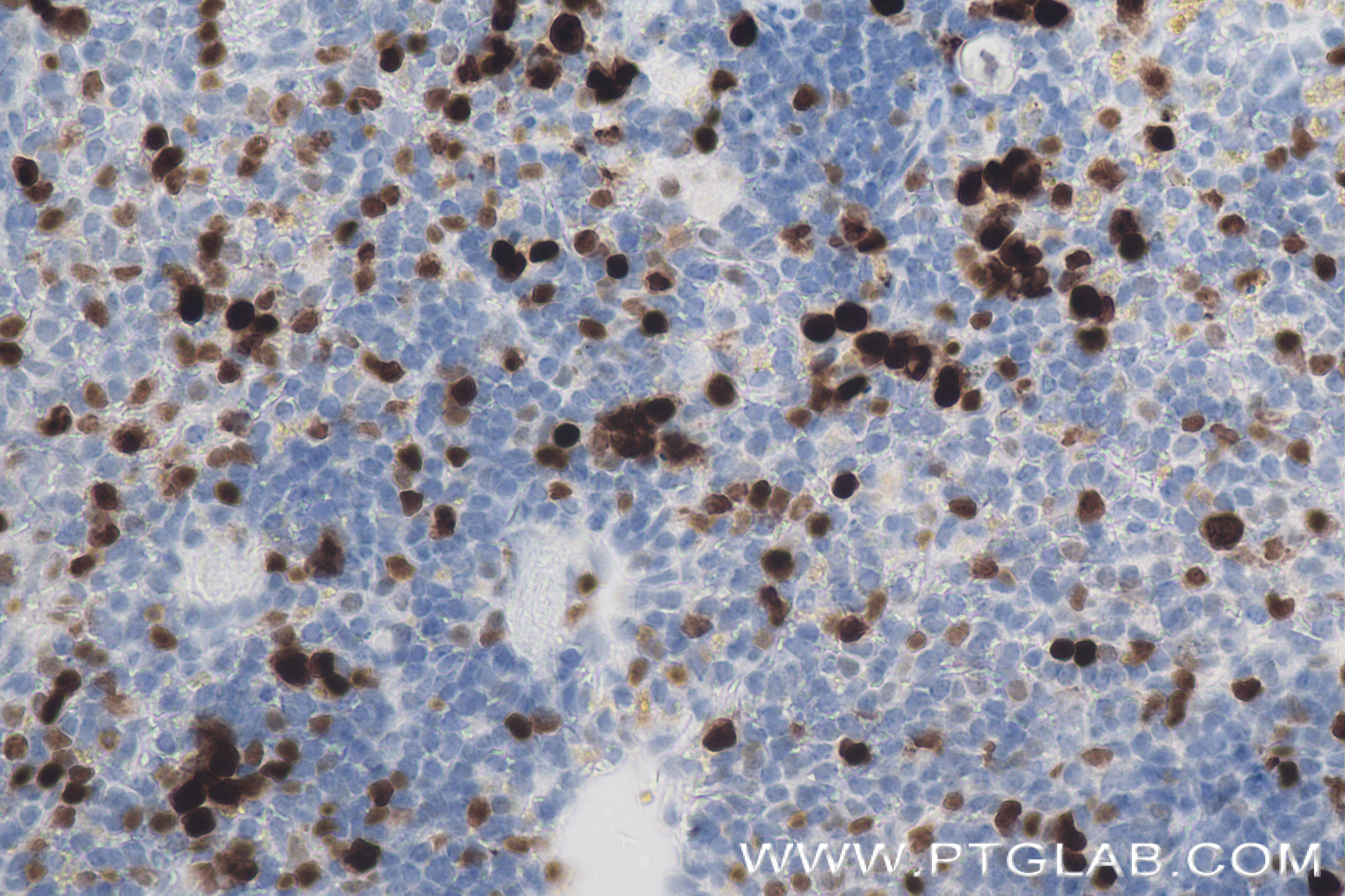
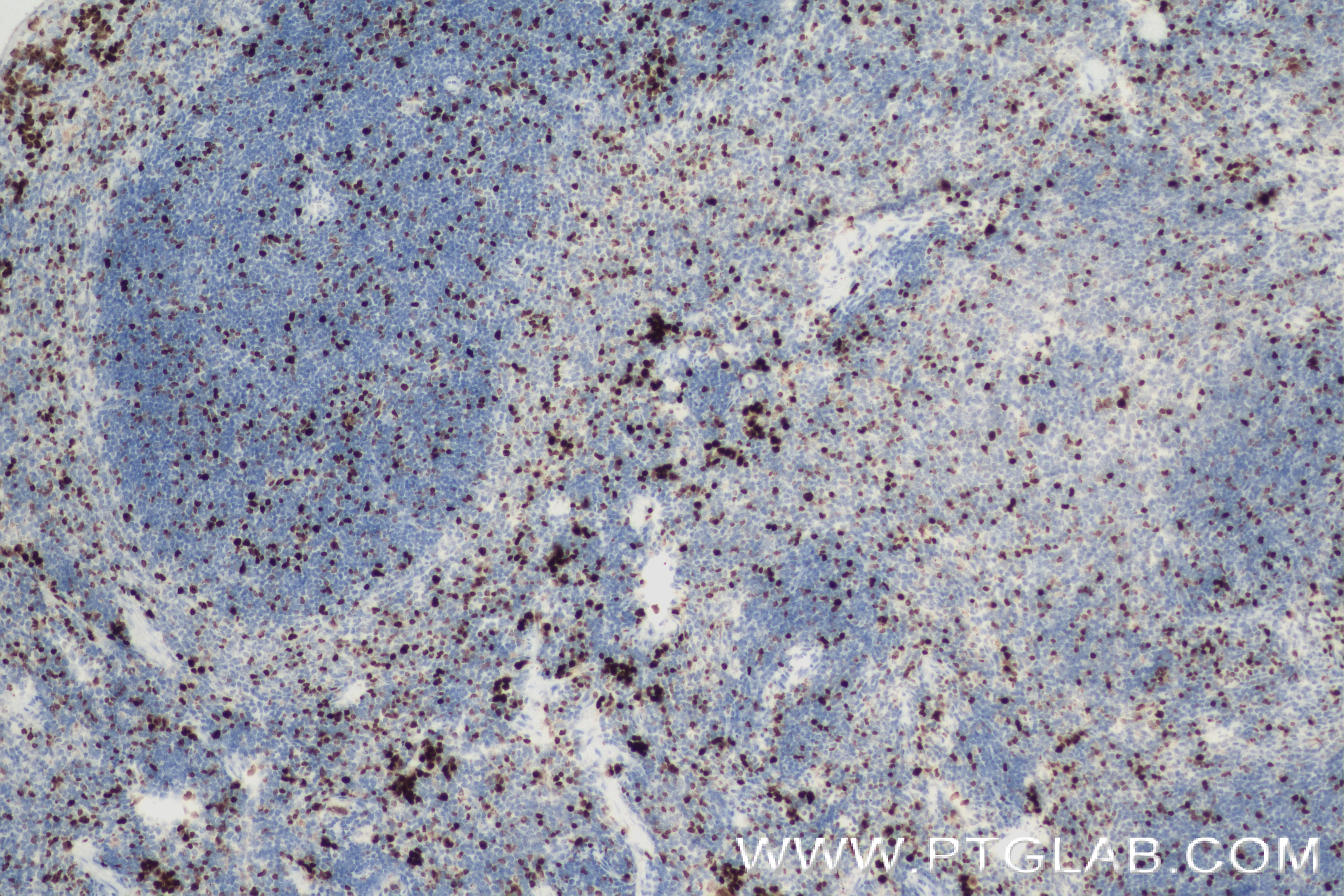
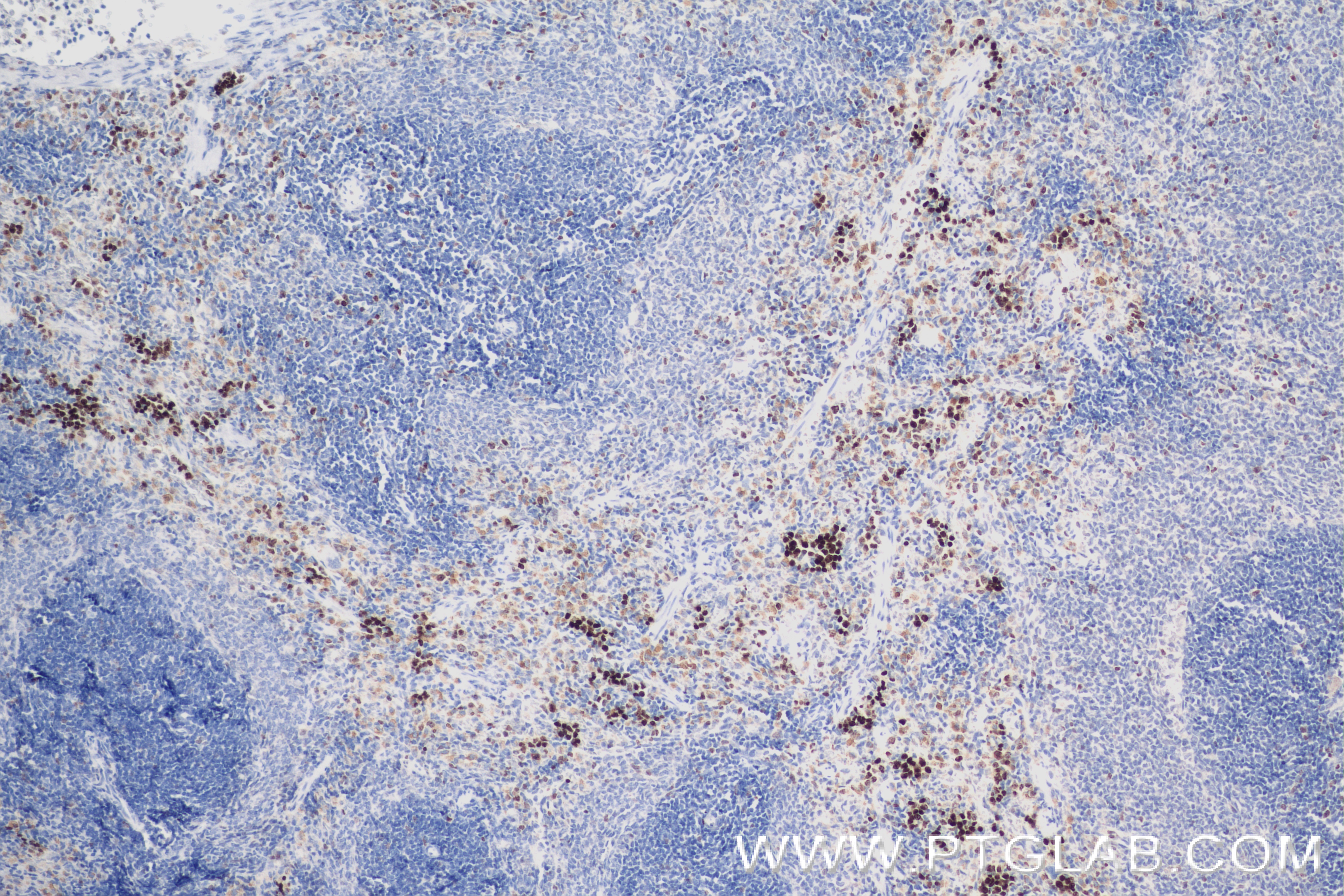
The Ki-67 protein (also known as MKI67) is a cellular marker for proliferation. Ki67 is present during all active phases of the cell cycle (G1, S, G2 and M), but is absent in resting cells (G0). Cellular content of Ki-67 protein markedly increases during cell progression through S phase of the cell cycle. Therefore, the nuclear expression of Ki67 can be evaluated to assess tumor proliferation by immunohistochemistry.

