Various lysates were subjected to SDS PAGE followed by western blot with 68100-1-Ig (p115, USO1 antibody) at dilution of 1:20000 incubated at room temperature for 1.5 hours. The membrane was stripped and reblotted with Lamin B1 Monoclonal antibody (66095-1-Ig) as loading control.
Various lysates were subjected to SDS PAGE followed by western blot with 68100-1-Ig (p115, USO1 antibody) at dilution of 1:20000 incubated at room temperature for 1.5 hours. The membrane was stripped and reblotted with Lamin B1 Monoclonal antibody (66095-1-Ig) as loading control.
WB analysis of HeLa using 68100-1-Ig
WB result of p115, USO1 antibody (68100-1-Ig; 1:20000; incubated at room temperature for 1.5 hours) with sh-Control and sh-p115, USO1 transfected HeLa cells.
WB result of p115, USO1 antibody (68100-1-Ig; 1:20000; incubated at room temperature for 1.5 hours) with sh-Control and sh-p115, USO1 transfected HeLa cells.
IHC staining of human liver cancer using 68100-1-Ig
Immunohistochemical analysis of paraffin-embedded human liver cancer tissue slide using 68100-1-Ig (p115, USO1 antibody) at dilution of 1:1000 (under 10x lens). Heat mediated antigen retrieval with Tris-EDTA buffer (pH 9.0).
Immunohistochemical analysis of paraffin-embedded human liver cancer tissue slide using 68100-1-Ig (p115, USO1 antibody) at dilution of 1:1000 (under 10x lens). Heat mediated antigen retrieval with Tris-EDTA buffer (pH 9.0).
IHC staining of human liver cancer using 68100-1-Ig
Immunohistochemical analysis of paraffin-embedded human liver cancer tissue slide using 68100-1-Ig (p115, USO1 antibody) at dilution of 1:1000 (under 40x lens). Heat mediated antigen retrieval with Tris-EDTA buffer (pH 9.0).
Immunohistochemical analysis of paraffin-embedded human liver cancer tissue slide using 68100-1-Ig (p115, USO1 antibody) at dilution of 1:1000 (under 40x lens). Heat mediated antigen retrieval with Tris-EDTA buffer (pH 9.0).
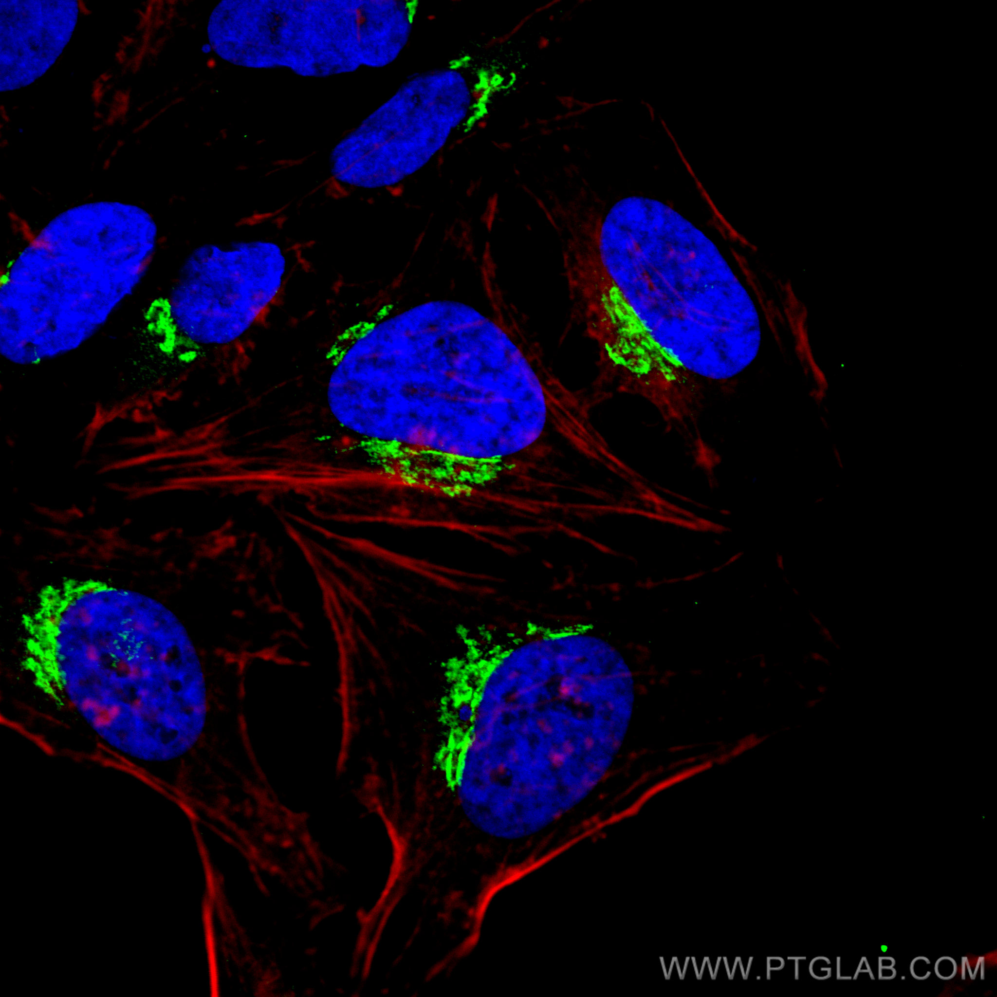
IF Staining of HeLa using 68100-1-Ig
Immunofluorescent analysis of (4% PFA) fixed HeLa cells using p115, USO1 antibody (68100-1-Ig, Clone: 3B7D8 ) at dilution of 1:500 and CoraLite®488-Conjugated AffiniPure Goat Anti-Mouse IgG(H+L), CL594-Phalloidin (red).
Immunofluorescent analysis of (4% PFA) fixed HeLa cells using p115, USO1 antibody (68100-1-Ig, Clone: 3B7D8 ) at dilution of 1:500 and CoraLite®488-Conjugated AffiniPure Goat Anti-Mouse IgG(H+L), CL594-Phalloidin (red).
IF Staining of HeLa using 68100-1-Ig
Immunofluorescent analysis of (4% PFA) fixed HeLa cells using p115, USO1 antibody (68100-1-Ig, Clone: 3B7D8 ) at dilution of 1:2000 and CoraLite®488-Conjugated AffiniPure Goat Anti-Mouse IgG(H+L), CL594-Phalloidin (red).
Immunofluorescent analysis of (4% PFA) fixed HeLa cells using p115, USO1 antibody (68100-1-Ig, Clone: 3B7D8 ) at dilution of 1:2000 and CoraLite®488-Conjugated AffiniPure Goat Anti-Mouse IgG(H+L), CL594-Phalloidin (red).
The Proteintech guarantee covers Proteintech antibodies in any species and any application, including those not listed on the datasheet. If the antibody doesn’t perform, you can receive a hassle-free refund or credit note.
human liver cancer tissue Note: suggested antigen retrieval with TE buffer pH 9.0; (*) Alternatively, antigen retrieval may be performed with citrate buffer pH 6.0
Positive IF/ICC detected in
HeLa cells
Recommended dilution
Application
Dilution
Western Blot (WB)
WB : 1:5000-1:50000
Immunohistochemistry (IHC)
IHC : 1:500-1:2000
Immunofluorescence (IF)/ICC
IF/ICC : 1:250-1:1000
It is recommended that this reagent should be titrated in each testing system to obtain optimal results.
Sample-dependent, Check data in validation data gallery.
Product Information
68100-1-Ig targets p115, USO1 in WB, IHC, IF/ICC, ELISA applications and shows reactivity with human, mouse, rat samples.
PBS with 0.02% sodium azide and 50% glycerol, pH 7.3.
Storage Conditions
Store at -20°C. Stable for one year after shipment. Aliquoting is unnecessary for -20oC storage. 20ul sizes contain 0.1% BSA.
Background Information
p115, also known as USO1, TAP (transcytosis-associated protein) or VDP (vesicle docking protein) is a general vesicular transport factor and plays an important role at different steps of vesicular transport. It is a 962-residue peripheral membrane protein which recycles between the cytosol and the Golgi apparatus during interphase (PMID: 9478999). p115 forms stable homodimers (PMID: 19247479). Rab1 recruits p115 to coat protein complex II (COPII) vesicles during budding from the endoplasmic reticulum, where p115 interacts directly with a select set of SNARE proteins (PMID: 10903204). p115 is required for intra-Golgi transport, and also functions in endoplasmic reticulum to Golgi trafficking, Golgi biogenesis and exocytotic transport (PMID: 19247479).
Various lysates were subjected to SDS PAGE followed by western blot with 68100-1-Ig (p115, USO1 antibody) at dilution of 1:20000 incubated at room temperature for 1.5 hours. The membrane was stripped and reblotted with Lamin B1 Monoclonal antibody (66095-1-Ig) as loading control.
WB analysis of HeLa using 68100-1-Ig
WB result of p115, USO1 antibody (68100-1-Ig; 1:20000; incubated at room temperature for 1.5 hours) with sh-Control and sh-p115, USO1 transfected HeLa cells.
IHC Figures
IHC staining of human liver cancer using 68100-1-Ig
Immunohistochemical analysis of paraffin-embedded human liver cancer tissue slide using 68100-1-Ig (p115, USO1 antibody) at dilution of 1:1000 (under 10x lens). Heat mediated antigen retrieval with Tris-EDTA buffer (pH 9.0).
IHC staining of human liver cancer using 68100-1-Ig
Immunohistochemical analysis of paraffin-embedded human liver cancer tissue slide using 68100-1-Ig (p115, USO1 antibody) at dilution of 1:1000 (under 40x lens). Heat mediated antigen retrieval with Tris-EDTA buffer (pH 9.0).
IF/ICC Figures
IF Staining of HeLa using 68100-1-Ig
Immunofluorescent analysis of (4% PFA) fixed HeLa cells using p115, USO1 antibody (68100-1-Ig, Clone: 3B7D8 ) at dilution of 1:500 and CoraLite®488-Conjugated AffiniPure Goat Anti-Mouse IgG(H+L), CL594-Phalloidin (red).
IF Staining of HeLa using 68100-1-Ig
Immunofluorescent analysis of (4% PFA) fixed HeLa cells using p115, USO1 antibody (68100-1-Ig, Clone: 3B7D8 ) at dilution of 1:2000 and CoraLite®488-Conjugated AffiniPure Goat Anti-Mouse IgG(H+L), CL594-Phalloidin (red).
The species listed in Tested Reactivity are in-house verified and applicable species. For unlisted species, please refer to the homology analysis of the immunogen sequence and related species. For rabbit polyclonal antibodies, homology >70% is recommended. For mouse monoclonal antibodies and rabbit recombinant antibodies, homology >90% is recommended. Generally, the higher the homology, the greater the applicability. However, there will be certain differences in protein expression in different species, tissues or cells. Therefore, the homology analysis results are for reference only and do not serve as a guarantee.
At Proteintech, we pride ourselves on our antibody quality, customer service and transparency. As such, we are comparing our antibodies with other vendors, enabling easy identification and comparisons of key data to help you choose the suitable antibody for your needs.
We have selected the top cited antibodies from these vendors for you to compare.
 at dilution of 1:20000 incubated at room temperature for 1.5 hours. The membrane was stripped and reblotted with Lamin B1 Monoclonal antibody (66095-1-Ig) as loading control.")
 with sh-Control and sh-p115, USO1 transfected HeLa cells.")
 at dilution of 1:1000 (under 10x lens). Heat mediated antigen retrieval with Tris-EDTA buffer (pH 9.0).")
 at dilution of 1:1000 (under 40x lens). Heat mediated antigen retrieval with Tris-EDTA buffer (pH 9.0).")
 fixed HeLa cells using p115, USO1 antibody (68100-1-Ig, Clone: 3B7D8 ) at dilution of 1:500 and CoraLite®488-Conjugated AffiniPure Goat Anti-Mouse IgG(H+L), CL594-Phalloidin (red).")
 fixed HeLa cells using p115, USO1 antibody (68100-1-Ig, Clone: 3B7D8 ) at dilution of 1:2000 and CoraLite®488-Conjugated AffiniPure Goat Anti-Mouse IgG(H+L), CL594-Phalloidin (red).")