Tested Applications
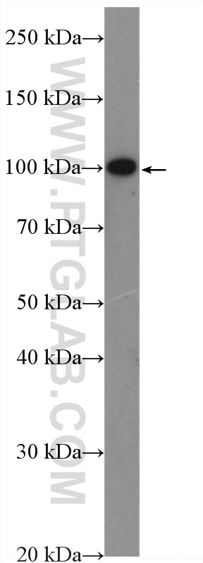
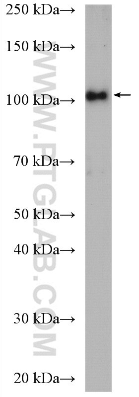
| Positive WB detected in | SH-SY5Y cells, HEK-293 cells |
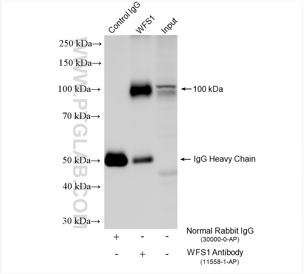
| Positive IP detected in | SH-SY5Y cells |
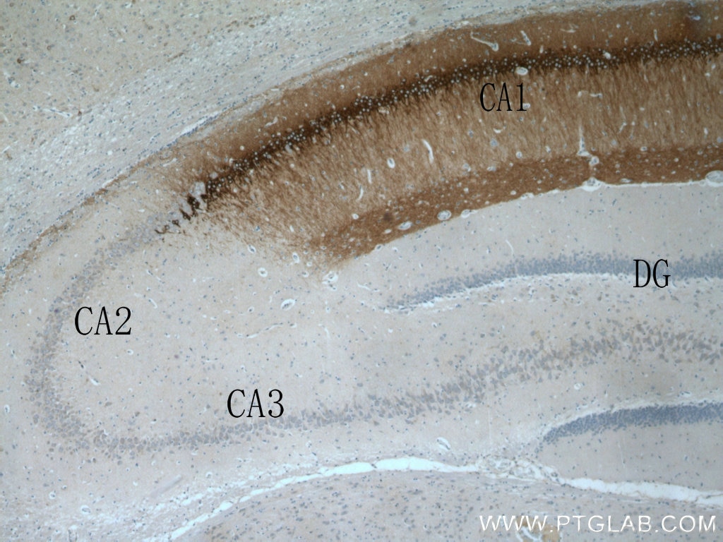
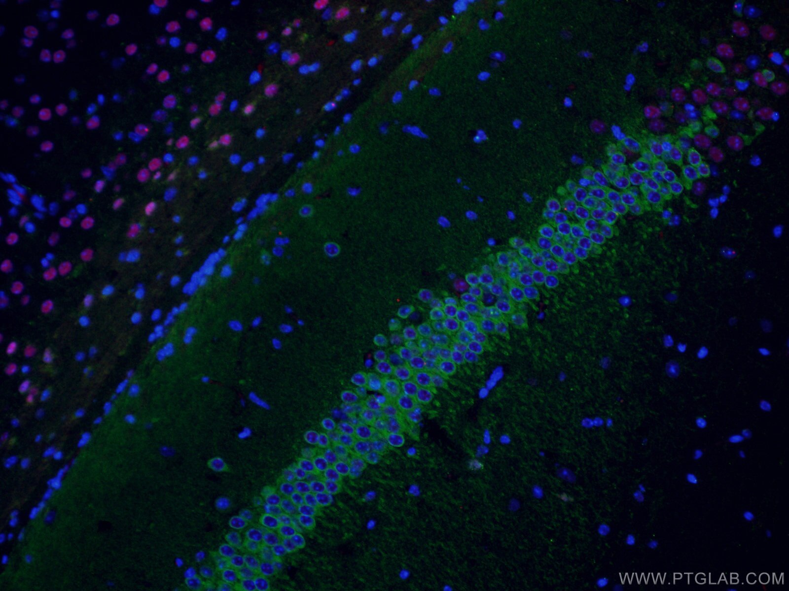
| Positive IHC detected in | rat brain tissue Note: suggested antigen retrieval with TE buffer pH 9.0; (*) Alternatively, antigen retrieval may be performed with citrate buffer pH 6.0 |
Recommended dilution
| Application | Dilution |
|---|---|
| Western Blot (WB) | WB : 1:500-1:1000 |
| Immunoprecipitation (IP) | IP : 0.5-4.0 ug for 1.0-3.0 mg of total protein lysate |
| Immunohistochemistry (IHC) | IHC : 1:200-1:800 |
| It is recommended that this reagent should be titrated in each testing system to obtain optimal results. | |
| Sample-dependent, Check data in validation data gallery. | |
Published Applications
| KD/KO | See 3 publications below |
| WB | See 12 publications below |
| IHC | See 19 publications below |
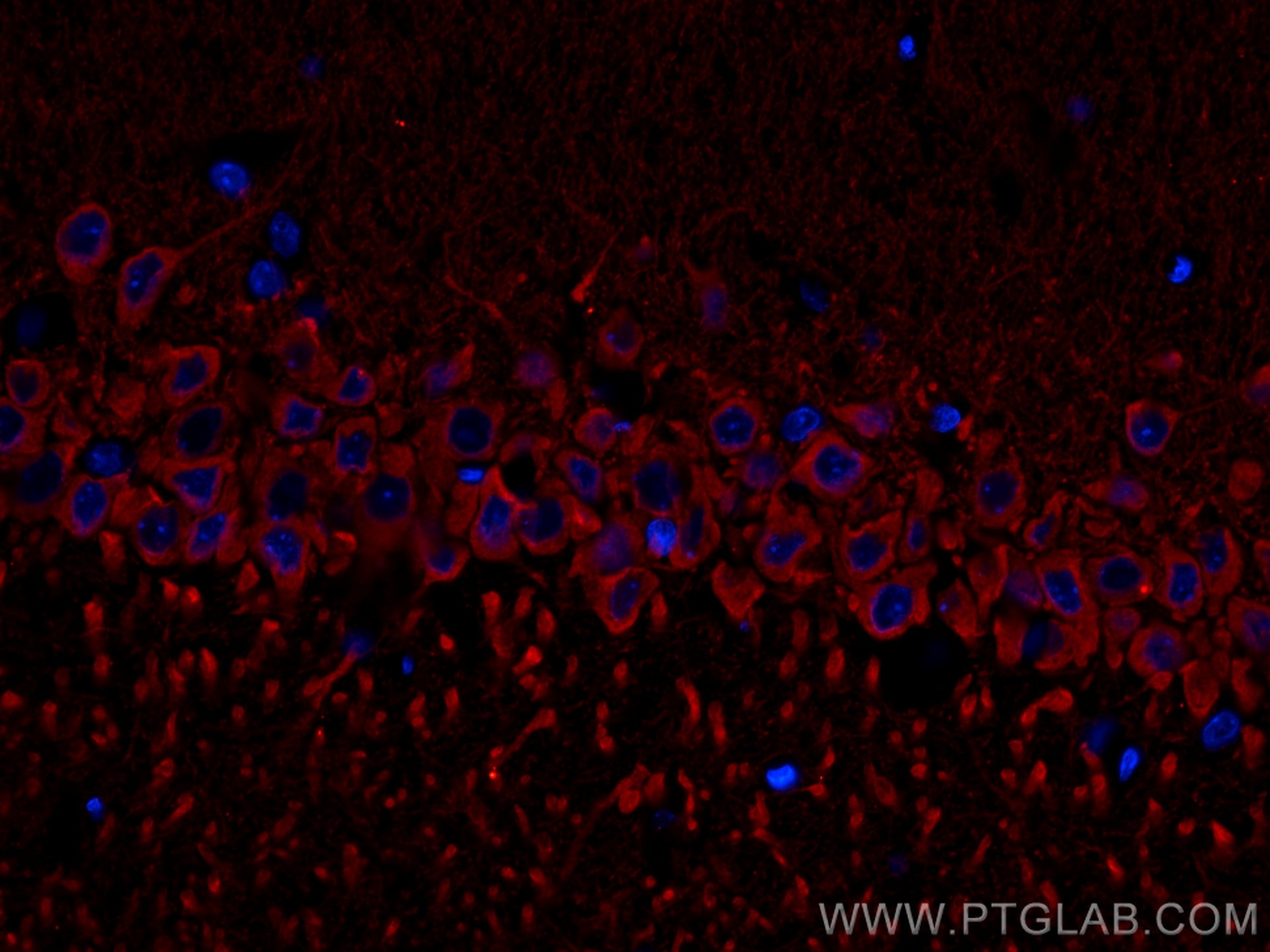
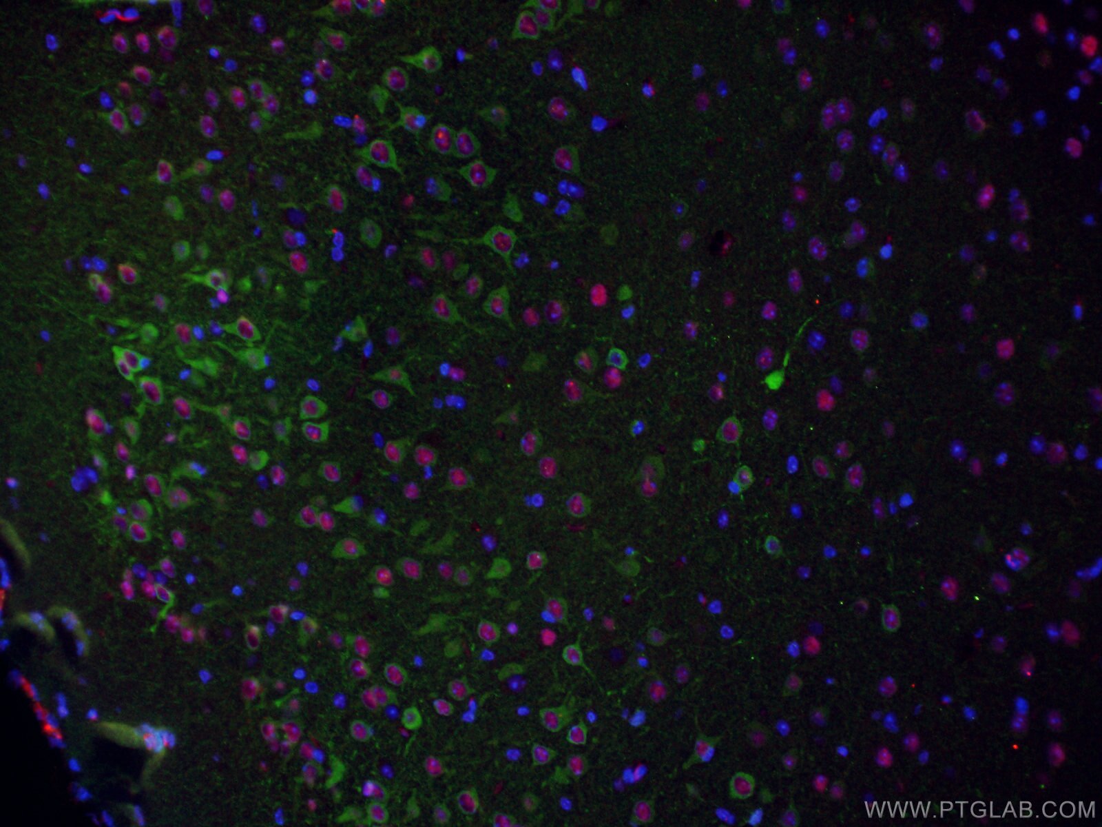
| IF | See 41 publications below |
| CoIP | See 1 publications below |
Product Information
11558-1-AP targets WFS1 in WB, IHC, IF-P, IP, coIP, ELISA applications and shows reactivity with human, mouse, rat samples.
| Tested Reactivity | human, mouse, rat |
| Cited Reactivity | human, mouse, rat |
| Host / Isotype | Rabbit / IgG |
| Class | Polyclonal |
| Type | Antibody |
| Immunogen |
CatNo: Ag2114 Product name: Recombinant human WFS1 protein Source: e coli.-derived, PGEX-4T Tag: GST Domain: 1-314 aa of BC030130 Sequence: MDSNTAPLGPSCPQPPPAPQPQARSRLNATASLEQERSERPRAPGPQAGPGPGVRDAAAPAEPQAQHTRSRERADGTGPTKGDMEIPFEEVLERAKAGDPKAQTEVGKHYLQLAGDTDEELNSCTAVDWLVLAAKQGRREAVKLLRRCLADRRGITSENEREVRQLSSETDLERAVRKAALVMYWKLNPKKKKQVAVAELLENVGQVNEHDGGAQPGPVPKSLQKQRRMLERLVSSESKNYIALDDFVEITKKYAKGVIPSSLFLQDDEDDDELAGKSPEDLPLRLKVVKYPLHAIMEIKEYLIDMASRAGMHW Predict reactive species |
| Full Name | Wolfram syndrome 1 (wolframin) |
| Calculated Molecular Weight | 890 aa, 100 kDa |
| Observed Molecular Weight | 100 kDa |
| GenBank Accession Number | BC030130 |
| Gene Symbol | WFS1 |
| Gene ID (NCBI) | 7466 |
| RRID | AB_2216046 |
| Conjugate | Unconjugated |
| Form | Liquid |
| Purification Method | Antigen affinity purification |
| UNIPROT ID | O76024 |
| Storage Buffer | PBS with 0.02% sodium azide and 50% glycerol, pH 7.3. |
| Storage Conditions | Store at -20°C. Stable for one year after shipment. Aliquoting is unnecessary for -20oC storage. 20ul sizes contain 0.1% BSA. |
Background Information
Wolfram syndrome protein (WFS1), also called wolframin, is a transmembrane protein, which is located primarily in the endoplasmic reticulum and its expression is induced in response to ER stress, partially through transcriptional activation. ER localization suggests that WFS1 protein has physiological functions in membrane trafficking, secretion, processing and/or regulation of ER calcium homeostasis. It is ubiquitously expressed with highest levels in brain, pancreas, heart, and insulinoma beta-cell lines. Mutations of the WFS1 gene are responsible for two hereditary diseases, autosomal recessive Wolfram syndrome and autosomal dominant low frequency sensorineural hearing loss.
Publications
| Species | Application | Title |
|---|---|---|
Cell A Hippocampus-Accumbens Tripartite Neuronal Motif Guides Appetitive Memory in Space. | ||
Reviews
The reviews below have been submitted by verified Proteintech customers who received an incentive for providing their feedback.
FH James (Verified Customer) (09-05-2025) | Specific to protein with minimal bleeding. Still has a strong affinity for target protein with accurate and clear images after microscopy.
|
FH Lana (Verified Customer) (05-29-2020) | SDS-PAGE: 15 ug/ul RIPA lysate of whole brain tissue or mitochondrial fraction. 4-12% Bis-tris gradient gel.Transfer: Immobilon-FL transfer membranes (Millipore) O/N at 30V, 4CBlocking: SEA Block Blocking Buffer 1hPrimary Ab: O/N incubation at 4C, 1:1000Secondary Ab: IRDye 800CW Goat anti-Rabbit, 1:15000Lines of WB: 1 – protein ladder, 2 – whole brain tissue lysate, 3 – mitochondrial fraction lysate.
 |

