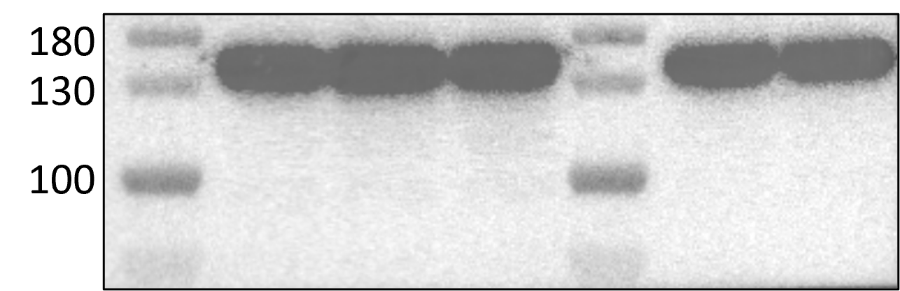
at dilution of 1:20000 incubated at room temperature for 1.5 hours.")
 at dilution of 1:20000 incubated at room temperature for 1.5 hours.")
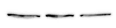
 at dilution of 1:10000 incubated at room temperature for 1.5 hours.")
 at dilution of 1:5000 incubated at room temperature for 1.5 hours.")
 at dilution of 1:400 (under 10x lens). Heat mediated antigen retrieval with Tris-EDTA buffer (pH 9.0).")
 at dilution of 1:400 (under 40x lens). Heat mediated antigen retrieval with Tris-EDTA buffer (pH 9.0).")
 at dilution of 1:200 (under 10x lens). Heat mediated antigen retrieval with Tris-EDTA buffer (pH 9.0).")
 at dilution of 1:200 (under 40x lens). Heat mediated antigen retrieval with Tris-EDTA buffer (pH 9.0).")
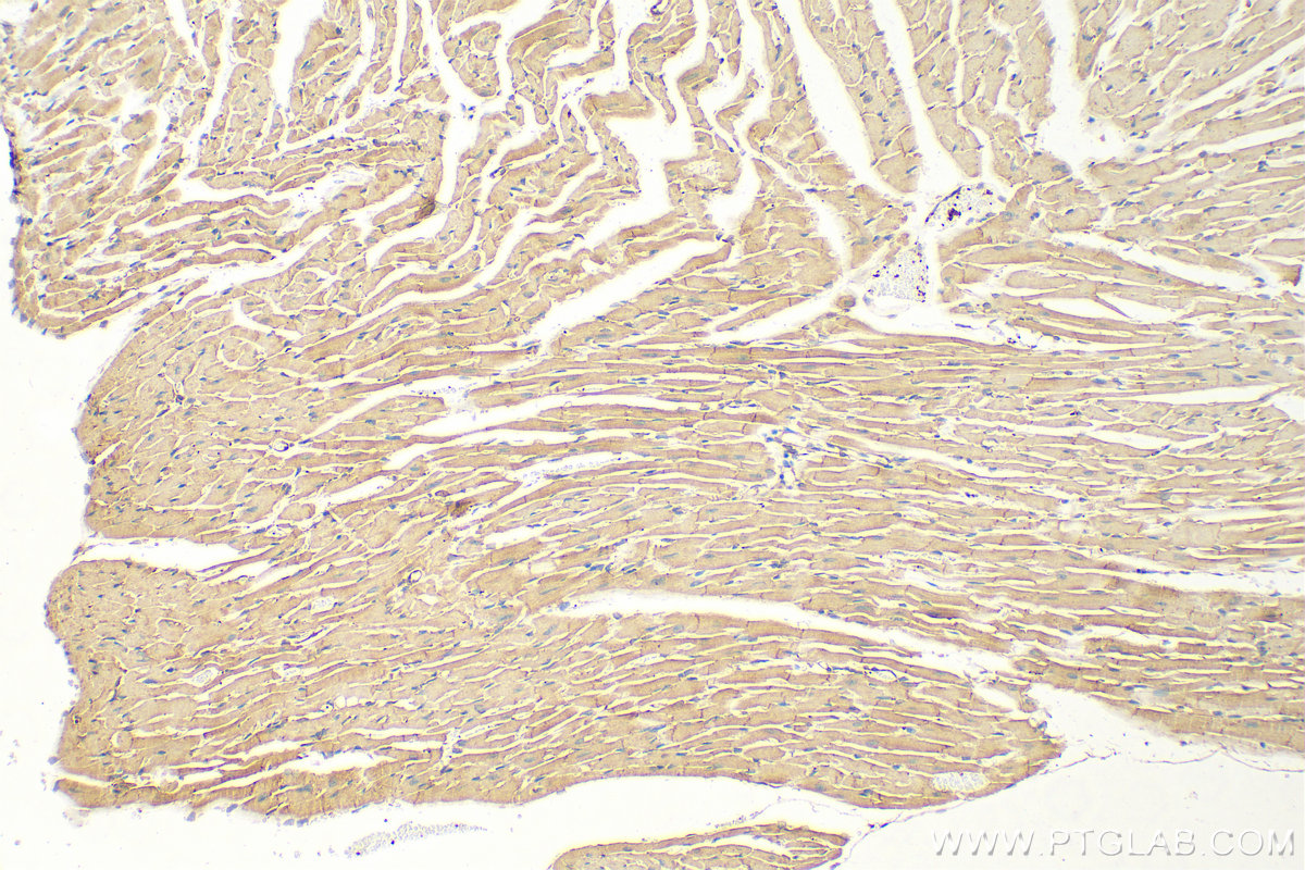
 at dilution of 1:200 (under 10x lens). Heat mediated antigen retrieval with Tris-EDTA buffer (pH 9.0).")
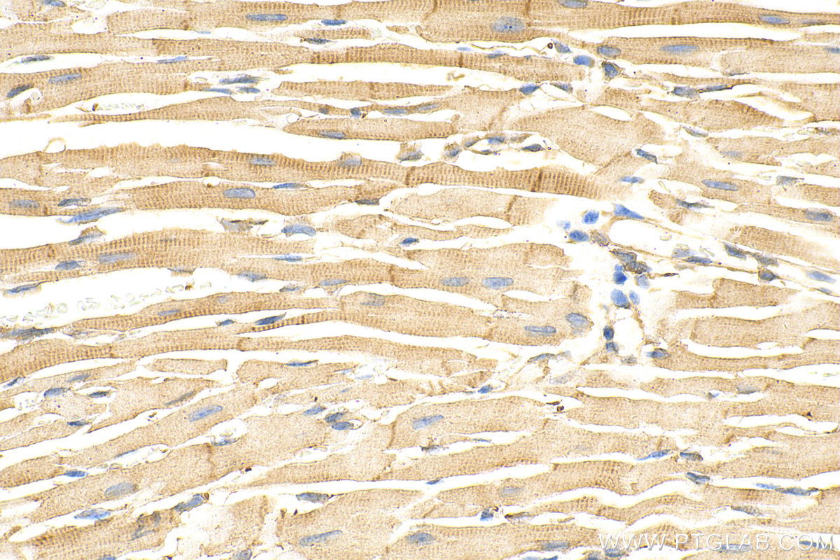
 at dilution of 1:200 (under 40x lens). Heat mediated antigen retrieval with Tris-EDTA buffer (pH 9.0).")
 fixed HeLa cells using 66305-1-Ig(Vinculin antibody) at dilution of 1:200 and Alexa Fluor 488-conjugated AffiniPure Goat Anti-Mouse IgG(H+L).")
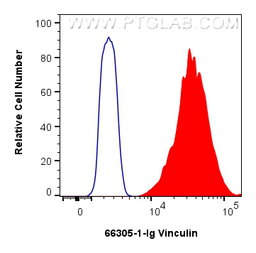
 and CoraLite®488-Conjugated Goat Anti-Mouse IgG(H+L) (SA00013-1)(red), or 0.25 ug Mouse IgG1 isotype control Mouse McAb (66360-1-Ig, Clone: 1F8D3) (blue). Cells were fixed with 4% PFA and permeabilized with Flow Cytometry Perm Buffer.")
"Vinculin Antibodies" Comparison
View side-by-side comparison of Vinculin antibodies from other vendors to find the one that best suits your research needs.
Tested Applications
| Positive WB detected in | HepG2 cells, pig heart tissue, U-251 cells, LNCaP cells, U-87 MG cells, C6 cells, HEK-293 cells, HeLa cells, HuH-7 cells, L02 cells, HSC-T6 cells, NIH/3T3 cells |
| Positive IHC detected in | human breast cancer tissue, human prostate cancer tissue, mouse heart tissue Note: suggested antigen retrieval with TE buffer pH 9.0; (*) Alternatively, antigen retrieval may be performed with citrate buffer pH 6.0 |
| Positive IF/ICC detected in | HeLa cells |
| Positive FC (Intra) detected in | HeLa cells |
Recommended dilution
| Application | Dilution |
|---|---|
| Western Blot (WB) | WB : 1:5000-1:50000 |
| Immunohistochemistry (IHC) | IHC : 1:50-1:500 |
| Immunofluorescence (IF)/ICC | IF/ICC : 1:50-1:500 |
| Flow Cytometry (FC) (INTRA) | FC (INTRA) : 0.25 ug per 10^6 cells in a 100 µl suspension |
| It is recommended that this reagent should be titrated in each testing system to obtain optimal results. | |
| Sample-dependent, Check data in validation data gallery. | |
Published Applications
| WB | See 171 publications below |
| IHC | See 2 publications below |
| IF | See 35 publications below |
| CoIP | See 1 publications below |
Product Information
66305-1-Ig targets Vinculin in WB, IHC, IF/ICC, FC (Intra), CoIP, ELISA applications and shows reactivity with human, mouse, rat, pig samples.
| Tested Reactivity | human, mouse, rat, pig |
| Cited Reactivity | human, mouse, rat, pig, rabbit, zebrafish, hamster |
| Host / Isotype | Mouse / IgG1 |
| Class | Monoclonal |
| Type | Antibody |
| Immunogen |
CatNo: Ag24946 Product name: Recombinant human Vinculin protein Source: e coli.-derived, PET30a Tag: 6*His Domain: 777-1066 aa of BC039174 Sequence: PKFREAVKAASDELSKTISPMVMDAKAVAGNISDPGLQKSFLDSGYRILGAVAKVREAFQPQEPDFPPPPPDLEQLRLTDELAPPKPPLPEGEVPPPRPPPPEEKDEEFPEQKAGEVINQPMMMAARQLHDEARKWSSKGNDIIAAAKRMALLMAEMSRLVRGGSGTKRALIQCAKDIAKASDEVTRLAKEVAKQCTDKRIRTNLLQVCERIPTISTQLKILSTVKATMLGRTNISDEESEQATEMLVHNAQNLMQSVKETVREAEAASIKIRTDAGFTLRWVRKTPWYQ Predict reactive species |
| Full Name | vinculin |
| Calculated Molecular Weight | 1133 aa, 124 kDa |
| Observed Molecular Weight | 117 kDa |
| GenBank Accession Number | BC039174 |
| Gene Symbol | Vinculin |
| Gene ID (NCBI) | 7414 |
| RRID | AB_2810300 |
| Conjugate | Unconjugated |
| Form | Liquid |
| Purification Method | Protein G purification |
| UNIPROT ID | P18206 |
| Storage Buffer | PBS with 0.02% sodium azide and 50% glycerol, pH 7.3. |
| Storage Conditions | Store at -20°C. Stable for one year after shipment. Aliquoting is unnecessary for -20oC storage. 20ul sizes contain 0.1% BSA. |
Background Information
Vinculin belongs to the vinculin/alpha-catenin family. It is an actin filament (F-actin)-binding protein which involved in cell-matrix adhesion and cell-cell adhesion. Vinculin regulates cell-surface E-cadherin expression and potentiates mechanosensing by the E-cadherin complex. It may also play important roles in cell morphology and locomotion. Vinculin is a 117-kDa, 1,066-amino-acid protein which is ubiquitously expressed. Its splice variant, metavinculin (124 kDa), is muscle-specific.
Publications
| Species | Application | Title |
|---|---|---|
Crit Care Recombinant ACE2 protein protects against acute lung injury induced by SARS-CoV-2 spike RBD protein. | ||
Adv Sci (Weinh) RBMS1 Coordinates with the m6 A Reader YTHDF1 to Promote NSCLC Metastasis through Stimulating S100P Translation | ||
Cell Rep Med Development of an orally bioavailable CDK12/13 degrader and induction of synthetic lethality with AKT pathway inhibition | ||
Dev Cell HMOX1-LDHB interaction promotes ferroptosis by inducing mitochondrial dysfunction in foamy macrophages during advanced atherosclerosis |
Reviews
The reviews below have been submitted by verified Proteintech customers who received an incentive for providing their feedback.
FH Zubair Ahmed (Verified Customer) (11-07-2025) | I think antibody is good
|
FH Monifa (Verified Customer) (08-31-2025) | I used vinculin as an alternative loading control to beta actin. After adjusting my transfer buffer to help vinculin transfer to my membrane, I can visualize the protein very well (with chemiluminescence).
|
FH Aditya (Verified Customer) (01-31-2025) | very clean bands, much better than the abcam antibody
|
FH Morgane (Verified Customer) (01-09-2025) | Very good loading control with larger molecular size
 |
FH Daniel (Verified Customer) (10-24-2024) | The antibody works really well and it gives a very clean Western blot.
|
FH Lisa (Verified Customer) (04-29-2024) | Works super well!
|
FH Parijat (Verified Customer) (08-21-2023) | Works well as loading control
|
FH Udesh (Verified Customer) (08-16-2023) | Worked well in WB at 1:3000 and IF at 1:100
|
FH Mohamad (Verified Customer) (07-03-2023) | Very good antibody
|
FH Priya (Verified Customer) (01-17-2023) | I have used for human cardiomyocytes, mouse skin and liver tissues
|
FH Macarena (Verified Customer) (10-07-2022) | excellent results.
|
FH Jonas (Verified Customer) (07-29-2022) | This is my go to loading control stain to validate evenly loaded lanes Binding could be stronger but by using 1:500 dilution, a reliable staining can be achieved
 |
FH Charlotte (Verified Customer) (07-26-2022) | Very good antibody. Very specific, super fast to reveal. Here we see Gli1 (160 kDa) because it is a mouse antibody too.
 |
FH AKIMASA (Verified Customer) (04-28-2021) | I could get the good quality band!
|
FH Chun (Verified Customer) (09-07-2020) | This is a fairly good antibody for immunoblotting.
|
FH Huai-Chin (Verified Customer) (06-08-2019) | Serving as a loading control, this antibody is not that good compare to other. Still work to some extent.
|