with sh-Control and sh-p115, USO1 transfected HeLa cells.")
 at dilution of 1:4000 incubated at room temperature for 1.5 hours.")
 at dilution of 1:4000 incubated at room temperature for 1.5 hours.")
 at dilution of 1:4000 incubated at room temperature for 1.5 hours.")
 at dilution of 1:4000 incubated at room temperature for 1.5 hours.")
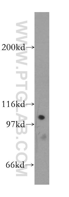
 at dilution of 1:1500 incubated at room temperature for 1.5 hours.")
 with mouse brain tissue lysate 7000ug.")
 at dilution of 1:200 (under 10x lens). Heat mediated antigen retrieval with Tris-EDTA buffer (pH 9.0).")
 at dilution of 1:200 (under 40x lens). Heat mediated antigen retrieval with Tris-EDTA buffer (pH 9.0).")
 fixed HeLa cells using p115, USO1 antibody (13509-1-AP) at dilution of 1:1000 and CoraLite®488-Conjugated Goat Anti-Rabbit IgG(H+L).")
 fixed HeLa cells using 13509-1-AP (p115, USO1 antibody) at dilution of 1:100 and Alexa Fluor 488-conjugated Goat Anti-Rabbit IgG(H+L).")
 fixed HeLa cells using 13509-1-AP (p115, USO1 antibody) at dilution of 1:50 and Alexa Fluor 488-conjugated Goat Anti-Rabbit IgG(H+L).")
 fixed HeLa cells using p115, USO1 antibody (13509-1-AP) at dilution of 1:200 and CoraLite®488-Conjugated Goat Anti-Rabbit IgG(H+L).")
Tested Applications
| Positive WB detected in | HEK-293 cells, mouse testis tissue, mouse thymus tissue, HeLa cells, human brain tissue, SH-SY5Y cells, HepG2 cells |
| Positive IP detected in | mouse brain tissue |
| Positive IHC detected in | human gliomas tissue Note: suggested antigen retrieval with TE buffer pH 9.0; (*) Alternatively, antigen retrieval may be performed with citrate buffer pH 6.0 |
| Positive IF/ICC detected in | HeLa cells |
Recommended dilution
| Application | Dilution |
|---|---|
| Western Blot (WB) | WB : 1:1000-1:8000 |
| Immunoprecipitation (IP) | IP : 0.5-4.0 ug for 1.0-3.0 mg of total protein lysate |
| Immunohistochemistry (IHC) | IHC : 1:50-1:500 |
| Immunofluorescence (IF)/ICC | IF/ICC : 1:500-1:2000 |
| It is recommended that this reagent should be titrated in each testing system to obtain optimal results. | |
| Sample-dependent, Check data in validation data gallery. | |
Published Applications
| KD/KO | See 2 publications below |
| WB | See 17 publications below |
| IF | See 18 publications below |
| CoIP | See 1 publications below |
Product Information
13509-1-AP targets p115, USO1 in WB, IHC, IF/ICC, IP, CoIP, ELISA applications and shows reactivity with human, mouse, rat samples.
| Tested Reactivity | human, mouse, rat |
| Cited Reactivity | human, mouse |
| Host / Isotype | Rabbit / IgG |
| Class | Polyclonal |
| Type | Antibody |
| Immunogen |
CatNo: Ag4431 Product name: Recombinant human p115, USO1 protein Source: e coli.-derived, PGEX-4T Tag: GST Domain: 660-961 aa of BC032654 Sequence: EQDLQLEELRQQVSTLKCQNEQLQTAVTQQVSQIQQHKDQYNLLKIQLGKDNQHQGSYSEGAQMNGIQPEEIGRLREEIEELKRNQELLQSQLTEKDSMIENMKSSQTSGTNEQSSAIVSARDSEQVAELKQELATLKSQLNSQSVEITKLQTEKQELLQKTEAFAKSVEVQGETETIIATKTTDVEGRLSALLQETKELKNEIKALSEERTAIKEQLDSSNSTIAILQTEKDKLELEITDSKKEQDDLLVLLADQDQKILSLKNKLKDLGHPVEEEDELESGDQEDEDDESEDPGKDLDHI Predict reactive species |
| Full Name | USO1 homolog, vesicle docking protein (yeast) |
| Calculated Molecular Weight | 962 aa, 108 kDa |
| Observed Molecular Weight | 115 kDa |
| GenBank Accession Number | BC032654 |
| Gene Symbol | USO1 |
| Gene ID (NCBI) | 8615 |
| RRID | AB_2257094 |
| Conjugate | Unconjugated |
| Form | Liquid |
| Purification Method | Antigen affinity purification |
| UNIPROT ID | O60763 |
| Storage Buffer | PBS with 0.02% sodium azide and 50% glycerol, pH 7.3. |
| Storage Conditions | Store at -20°C. Stable for one year after shipment. Aliquoting is unnecessary for -20oC storage. 20ul sizes contain 0.1% BSA. |
Background Information
p115, also known as USO1, TAP (transcytosis-associated protein) or VDP (vesicle docking protein) is a general vesicular transport factor and plays an important role at different steps of vesicular transport. It is a 962-residue peripheral membrane protein which recycles between the cytosol and the Golgi apparatus during interphase (PMID: 9478999). p115 forms stable homodimers (PMID: 19247479). Rab1 recruits p115 to coat protein complex II (COPII) vesicles during budding from the endoplasmic reticulum, where p115 interacts directly with a select set of SNARE proteins (PMID: 10903204). p115 is required for intra-Golgi transport, and also functions in endoplasmic reticulum to Golgi trafficking, Golgi biogenesis and exocytotic transport (PMID: 19247479).
Protocols
| Product Specific Protocols | |
|---|---|
| IF protocol for p115, USO1 antibody 13509-1-AP | Download protocol |
| IHC protocol for p115, USO1 antibody 13509-1-AP | Download protocol |
| IP protocol for p115, USO1 antibody 13509-1-AP | Download protocol |
| WB protocol for p115, USO1 antibody 13509-1-AP | Download protocol |
| Standard Protocols | |
|---|---|
| Click here to view our Standard Protocols |
Publications
| Species | Application | Title |
|---|---|---|
Mol Cell An mTORC1-GRASP55 signaling axis controls unconventional secretion to reshape the extracellular proteome upon stress. | ||
J Cell Biol Rapid degradation of GRASP55 and GRASP65 reveals their immediate impact on the Golgi structure. | ||