Tested Applications
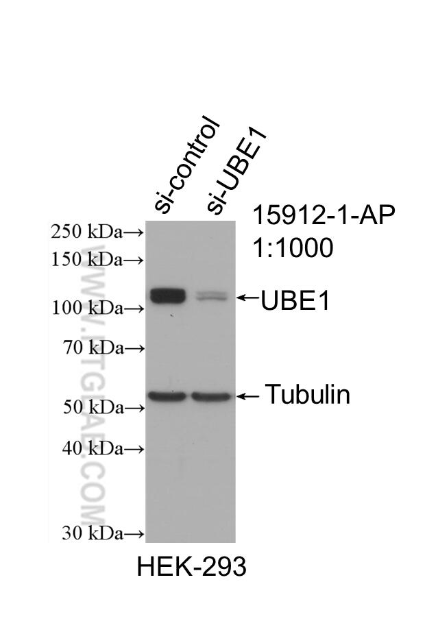
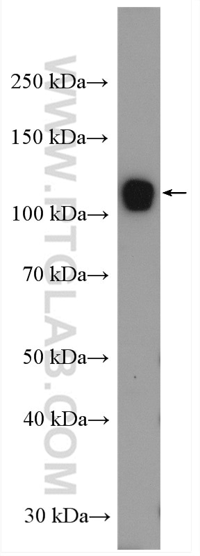
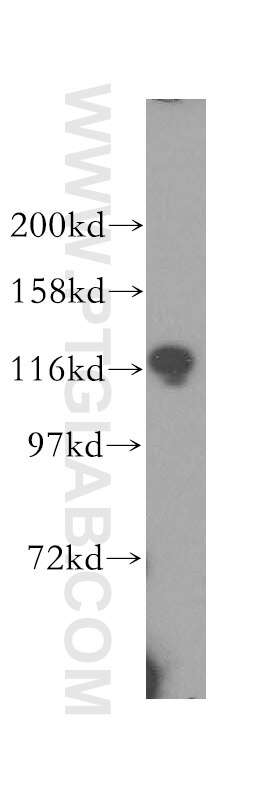
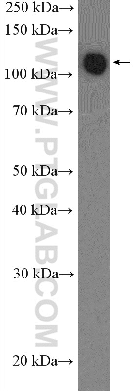
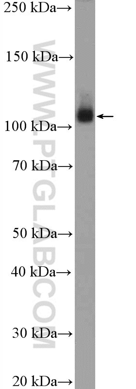
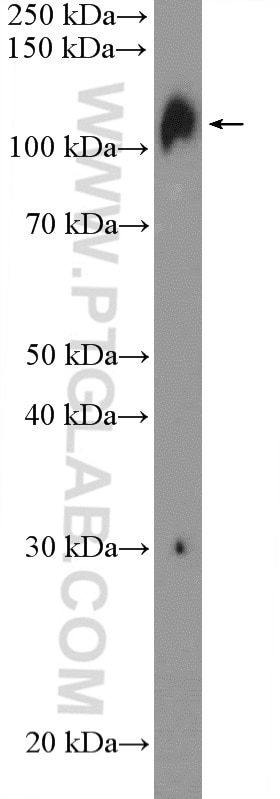
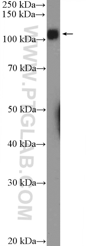
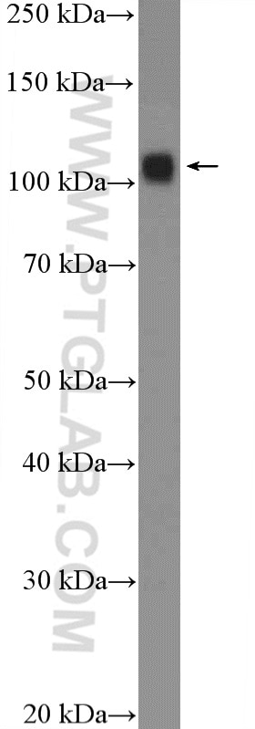
| Positive WB detected in | HL-60 cells, HeLa cells, HEK-293 cells, mouse spleen tissue, rat spleen tissue, mouse colon tissue |
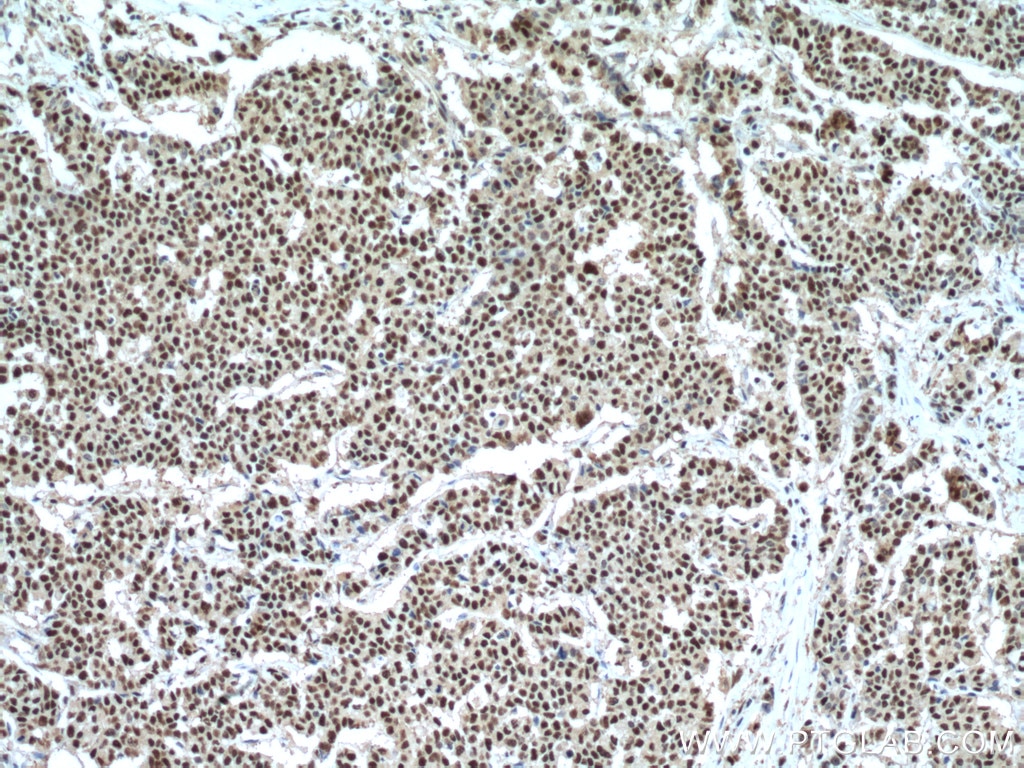
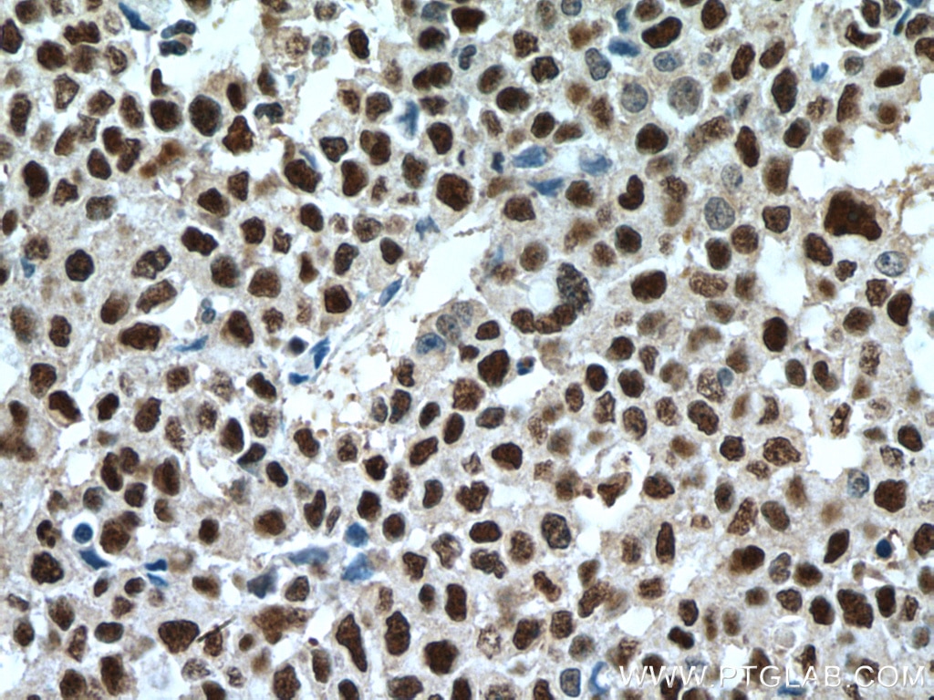
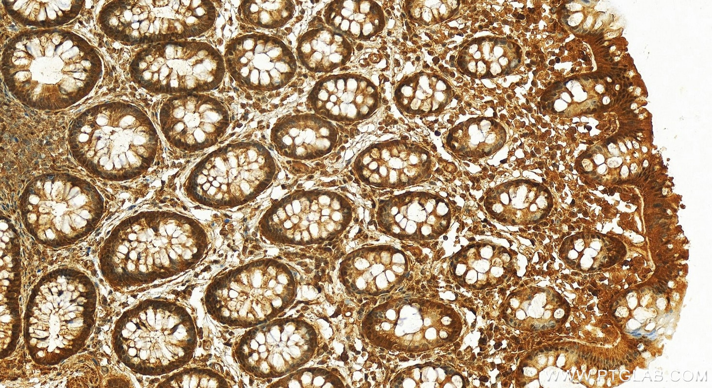
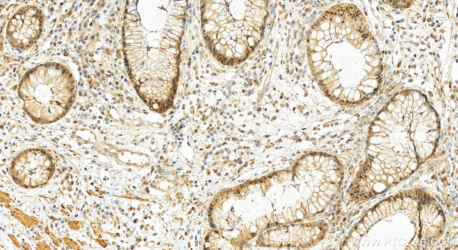
| Positive IHC detected in | human colon cancer tissue, human normal colon Note: suggested antigen retrieval with TE buffer pH 9.0; (*) Alternatively, antigen retrieval may be performed with citrate buffer pH 6.0 |
Recommended dilution
| Application | Dilution |
|---|---|
| Western Blot (WB) | WB : 1:500-1:3000 |
| Immunohistochemistry (IHC) | IHC : 1:200-1:1500 |
| It is recommended that this reagent should be titrated in each testing system to obtain optimal results. | |
| Sample-dependent, Check data in validation data gallery. | |
Published Applications
| KD/KO | See 1 publications below |
| WB | See 9 publications below |
| IHC | See 4 publications below |
Product Information
15912-1-AP targets UBE1 in WB, IHC, ELISA applications and shows reactivity with human, mouse, rat samples.
| Tested Reactivity | human, mouse, rat |
| Cited Reactivity | human, mouse |
| Host / Isotype | Rabbit / IgG |
| Class | Polyclonal |
| Type | Antibody |
| Immunogen | UBE1 fusion protein Ag8703 Predict reactive species |
| Full Name | ubiquitin-like modifier activating enzyme 1 |
| Calculated Molecular Weight | 1058 aa, 118 kDa |
| Observed Molecular Weight | 114-118 kDa |
| GenBank Accession Number | BC013041 |
| Gene Symbol | UBA1 |
| Gene ID (NCBI) | 7317 |
| RRID | AB_2211462 |
| Conjugate | Unconjugated |
| Form | Liquid |
| Purification Method | Antigen affinity purification |
| UNIPROT ID | P22314 |
| Storage Buffer | PBS with 0.02% sodium azide and 50% glycerol , pH 7.3 |
| Storage Conditions | Store at -20°C. Stable for one year after shipment. Aliquoting is unnecessary for -20oC storage. 20ul sizes contain 0.1% BSA. |
Background Information
UBE1(Ubiquitin-activating enzyme E1) is also named as A1S9T, UBE1 and belongs to the ubiquitin-activating E1 family. It catalyzes the first step in ubiquitin conjugation to mark cellular proteins for degradation and initiates the activation and conjugation of ubiquitin-like proteins. Defects in UBE1 are the cause of spinal muscular atrophy X-linked type 2 (SMAX2). UBE1 has two isoforms with the molecular mass of 118 and 114 kDa.
Protocols
| Product Specific Protocols | |
|---|---|
| WB protocol for UBE1 antibody 15912-1-AP | Download protocol |
| IHC protocol for UBE1 antibody 15912-1-AP | Download protocol |
| Standard Protocols | |
|---|---|
| Click here to view our Standard Protocols |
Publications
| Species | Application | Title |
|---|---|---|
Cancer Discov The UBA1-STUB1 axis mediates cancer immune escape and resistance to checkpoint blockade | ||
Nat Commun DNA replication initiation factor RECQ4 possesses a role in antagonizing DNA replication initiation | ||
ACS Nano Graphene Oxide Causes Disordered Zonation Due to Differential Intralobular Localization in the Liver. | ||
Cancer Biol Med CSN6 promotes tumorigenesis of gastric cancer by ubiquitin-independent proteasomal degradation of p16INK4a. | ||
Am J Cancer Res The E3 ubiquitin ligase RBCK1 promotes the invasion and metastasis of hepatocellular carcinoma by destroying the PPARγ/PGC1α complex. | ||
Front Oncol Ubiquitin-Like Modifier Activating Enzyme 1 as a Novel Diagnostic and Prognostic Indicator That Correlates With Ferroptosis and the Malignant Phenotypes of Liver Cancer Cells. |
Reviews
The reviews below have been submitted by verified Proteintech customers who received an incentive for providing their feedback.
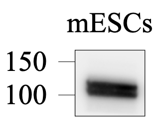
FH Tsimafei (Verified Customer) (11-19-2024) | Antibody detect a band of the expected molecular weight in mESCs
 |
