Tested Applications
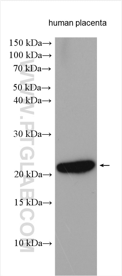
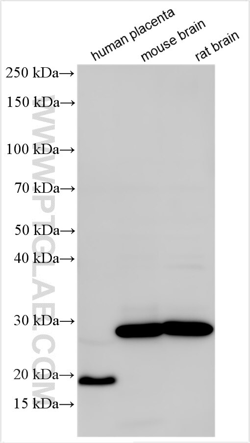
| Positive WB detected in | human placenta tissue, mouse brain tissue, rat brain tissue |
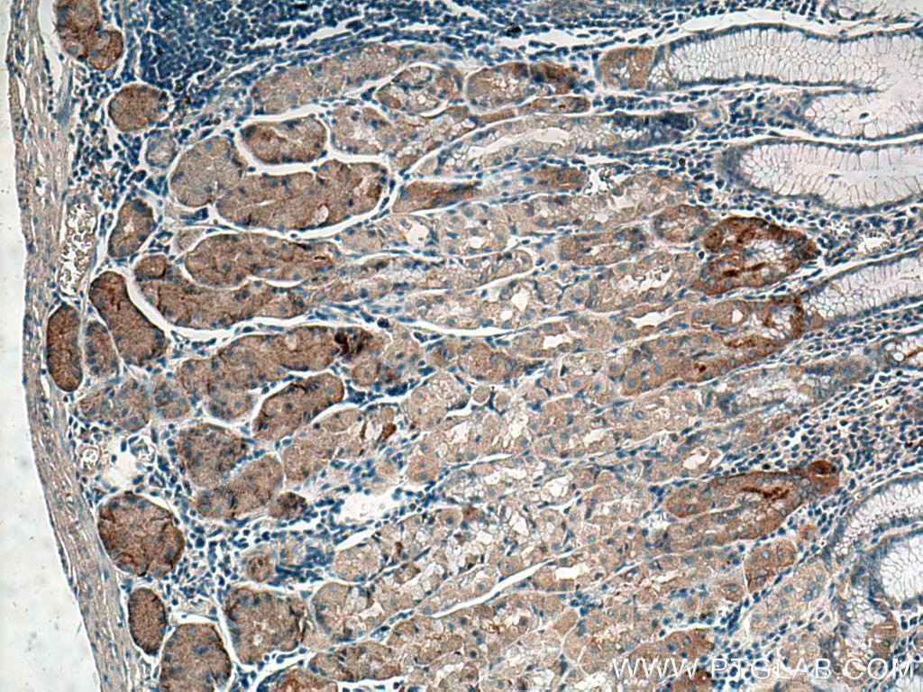
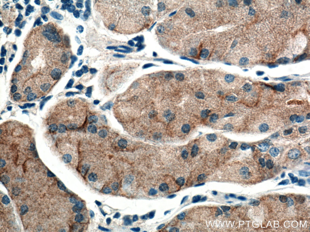
| Positive IHC detected in | human stomach tissue Note: suggested antigen retrieval with TE buffer pH 9.0; (*) Alternatively, antigen retrieval may be performed with citrate buffer pH 6.0 |
Recommended dilution
| Application | Dilution |
|---|---|
| Western Blot (WB) | WB : 1:1000-1:4000 |
| Immunohistochemistry (IHC) | IHC : 1:200-1:800 |
| It is recommended that this reagent should be titrated in each testing system to obtain optimal results. | |
| Sample-dependent, Check data in validation data gallery. | |
Published Applications
| KD/KO | See 2 publications below |
| WB | See 6 publications below |
| IHC | See 9 publications below |
Product Information
10858-1-AP targets Timp-3 in WB, IHC, ELISA applications and shows reactivity with human, mouse, rat samples.
| Tested Reactivity | human, mouse, rat |
| Cited Reactivity | human, mouse, rat |
| Host / Isotype | Rabbit / IgG |
| Class | Polyclonal |
| Type | Antibody |
| Immunogen |
CatNo: Ag1253 Product name: Recombinant human Timp-3 protein Source: e coli.-derived, PGEX-4T Tag: GST Domain: 1-211 aa of BC014277 Sequence: MTPWLGLIVLLGSWSLGDWGAEACTCSPSHPQDAFCNSDIVIRAKVVGKKLVKEGPFGTLVYTIKQMKMYRGFTKMPHVQYIHTEASESLCGLKLEVNKYQYLLTGRVYDGKMYTGLCNFVERWDQLTLSQRKGLNYRYHLGCNCKIKSCYYLPCFVTSKNECLWTDMLSNFGYPGYQSKHYACIRQKGGYCSWYRGWAPPDKSIINATDP Predict reactive species |
| Full Name | TIMP metallopeptidase inhibitor 3 |
| Calculated Molecular Weight | 24 kDa |
| Observed Molecular Weight | 20-30 kDa |
| GenBank Accession Number | BC014277 |
| Gene Symbol | TIMP3 |
| Gene ID (NCBI) | 7078 |
| RRID | AB_2204973 |
| Conjugate | Unconjugated |
| Form | Liquid |
| Purification Method | Antigen affinity purification |
| UNIPROT ID | P35625 |
| Storage Buffer | PBS with 0.02% sodium azide and 50% glycerol, pH 7.3. |
| Storage Conditions | Store at -20°C. Stable for one year after shipment. Aliquoting is unnecessary for -20oC storage. 20ul sizes contain 0.1% BSA. |
Background Information
Timp-3 (TIMP metallopeptidase inhibitor 3), also known as SFD. It is expected to be located in extracellular space, and the protein is enriched in the placenta tissue and fat tissue. The proteins encoded by this gene family are inhibitors of the matrix metalloproteinases, a group of peptidases involved in degradation of the extracellular matrix (ECM). Expression of this gene is induced in response to mitogenic stimulation, and this netrin domain-containing protein is localized to the ECM. Mutations in this gene have been associated with the autosomal dominant disorder Sorsby's fundus dystrophy. TIMP-3 is expressed as an unglycosylated 24 kDa and glycosylated 29 kDa protein with inhibitory activity against interstitial collagenase, stromelysin-1, and gelatinases A and B (PMID:11827795).
Protocols
| Product Specific Protocols | |
|---|---|
| IHC protocol for Timp-3 antibody 10858-1-AP | Download protocol |
| WB protocol for Timp-3 antibody 10858-1-AP | Download protocol |
| Standard Protocols | |
|---|---|
| Click here to view our Standard Protocols |
Publications
| Species | Application | Title |
|---|---|---|
Theranostics miR-17-3p Contributes to Exercise-Induced Cardiac Growth and Protects against Myocardial Ischemia-Reperfusion Injury. | ||
Oxid Med Cell Longev Trehalose Protects Keratinocytes against Ultraviolet B Radiation by Activating Autophagy via Regulating TIMP3 and ATG9A. | ||
Int Immunopharmacol Sublytic C5b-9 induces TIMP3 expression by glomerular mesangial cells via TRAF6-dependent KLF5 K63-linked ubiquitination in rat Thy-1 nephritis | ||
Sci Rep AMPK deficiency in chondrocytes accelerated the progression of instability-induced and ageing-associated osteoarthritis in adult mice. | ||
Cell Prolif Aspirin inhibits inflammation and scar formation in the injury tendon healing through regulating JNK/STAT-3 signalling pathway. | ||
Biomolecules Tau Protein Modulates Perineuronal Extracellular Matrix Expression in the TauP301L-acan Mouse Model. |


