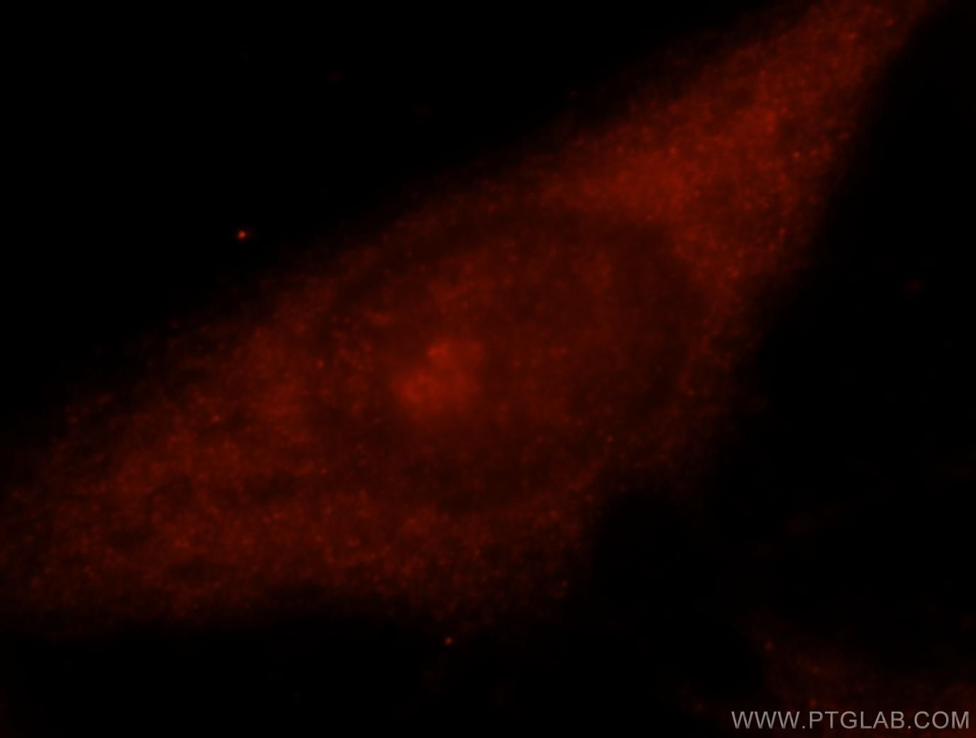
Tested Applications
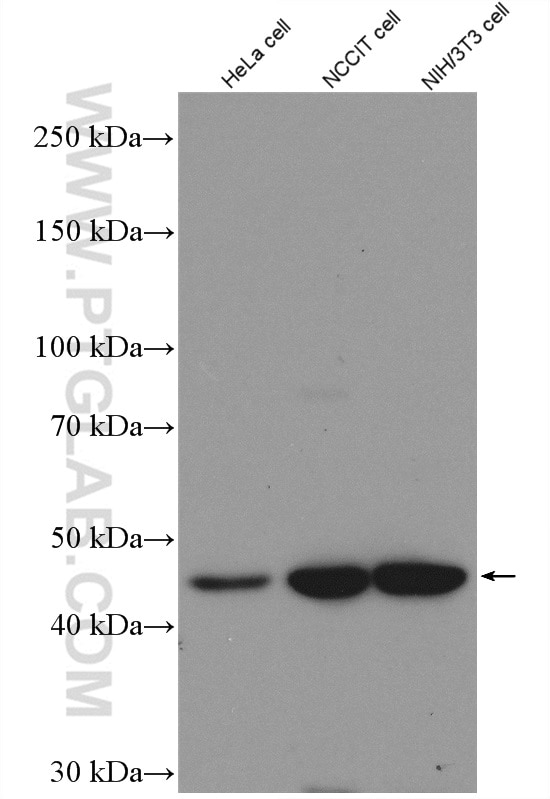
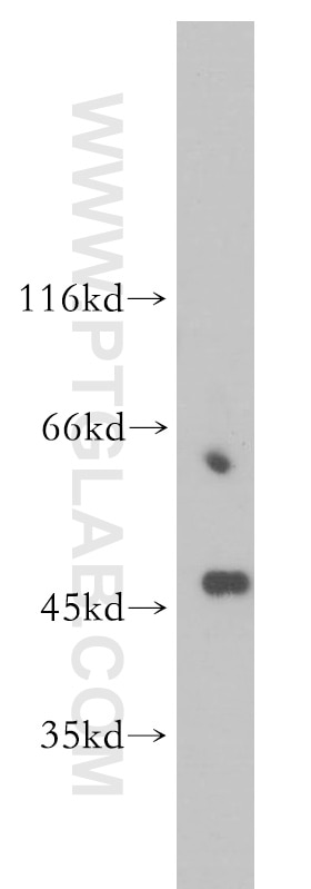
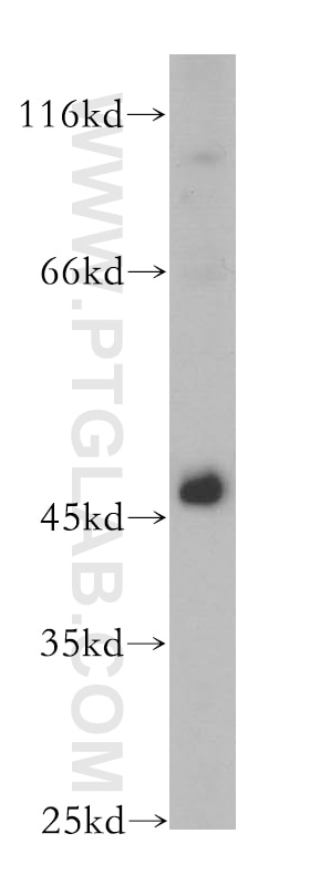
| Positive WB detected in | NIH/3T3 cells, HeLa cells |
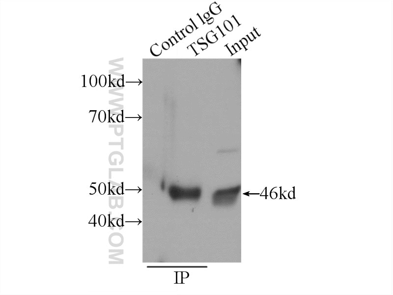
| Positive IP detected in | HeLa cells |
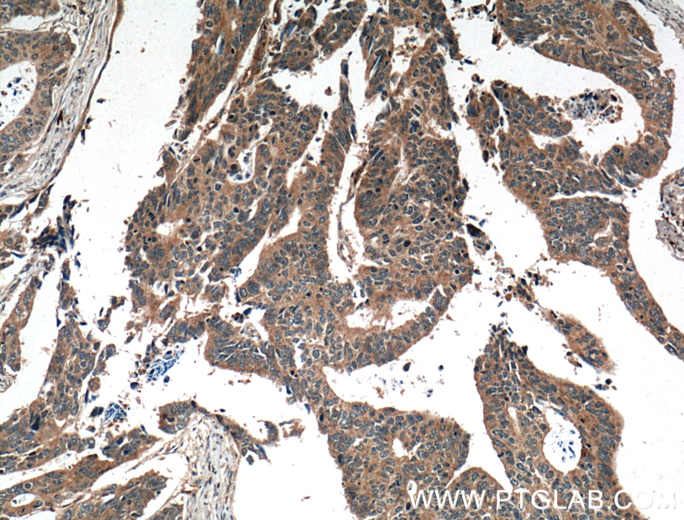
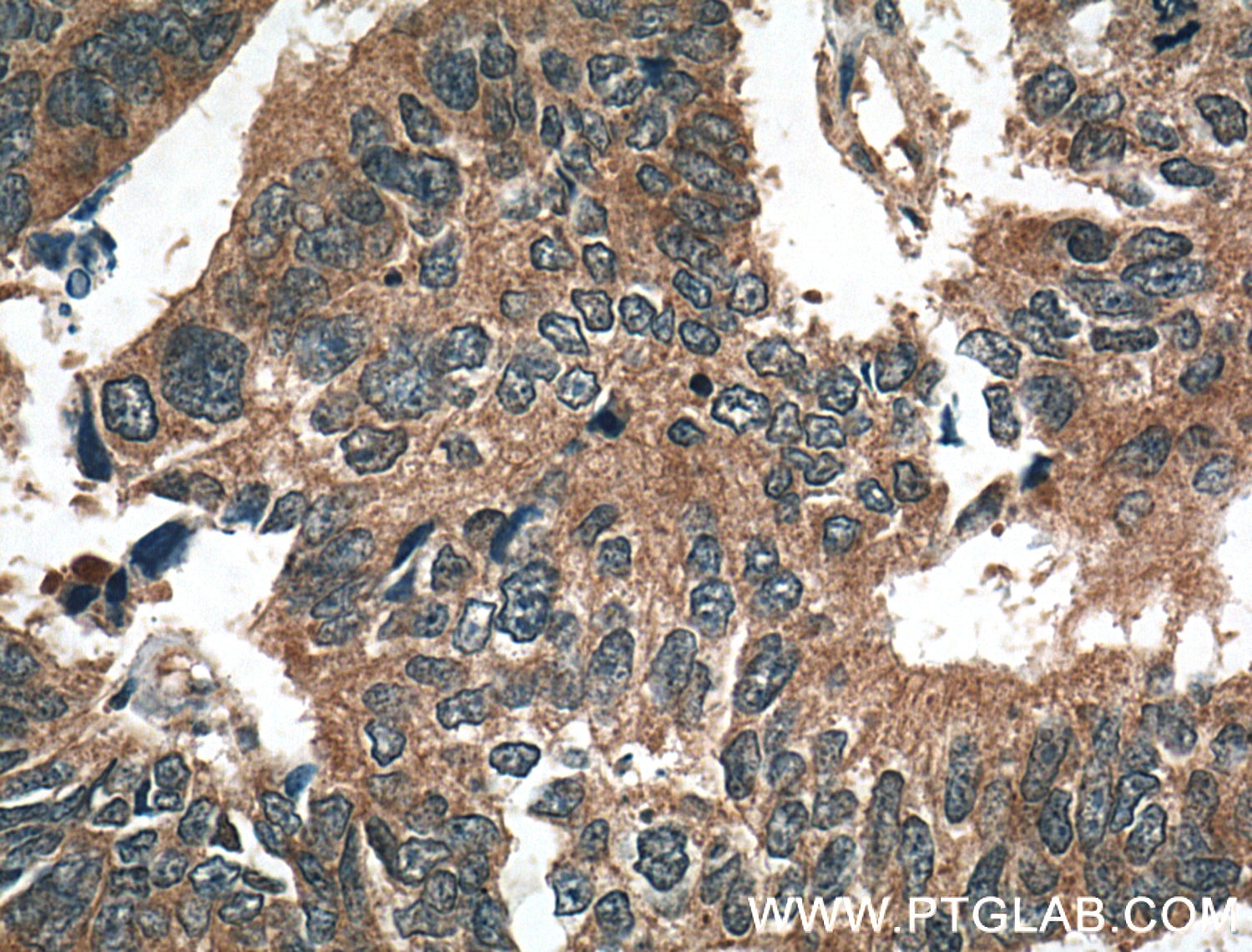
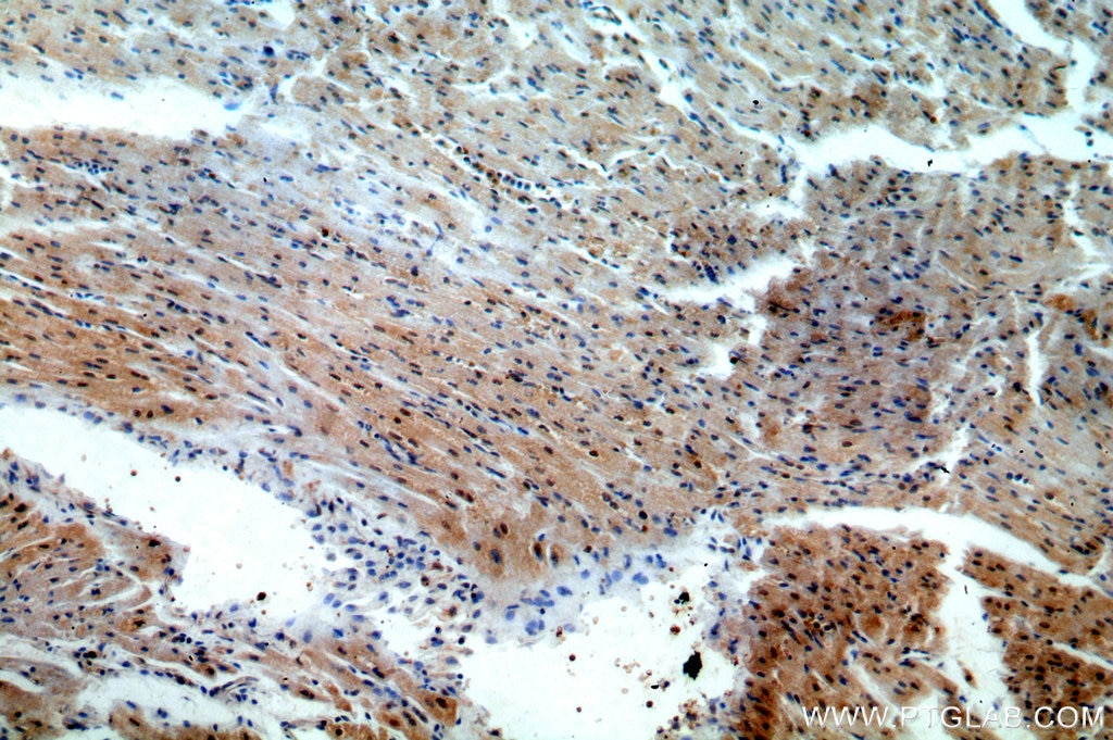
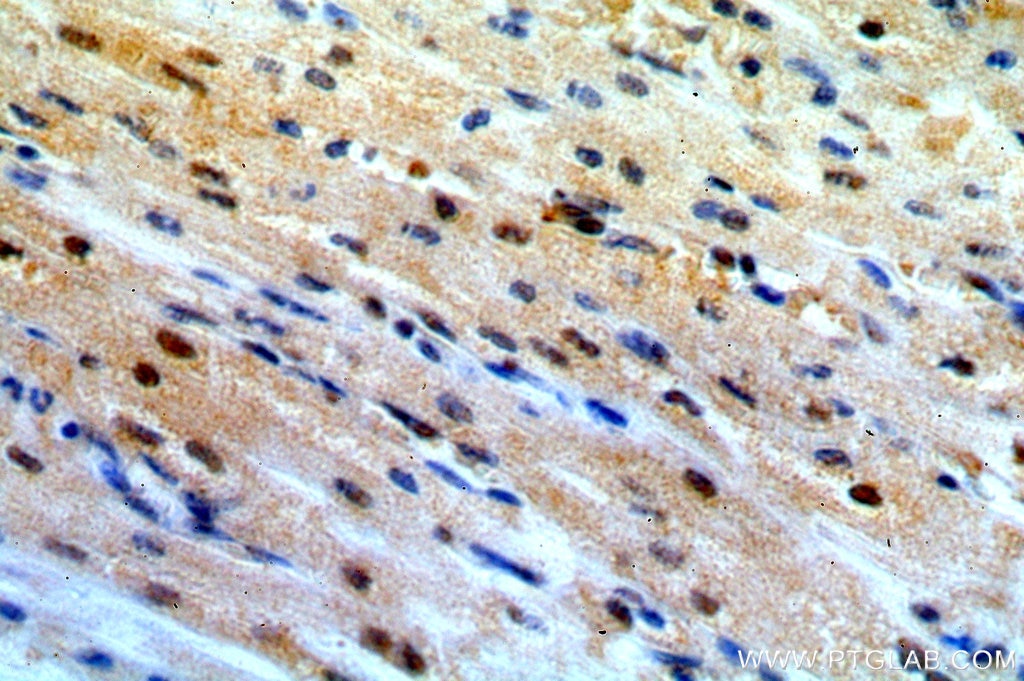
| Positive IHC detected in | human colon cancer tissue, human heart tissue Note: suggested antigen retrieval with TE buffer pH 9.0; (*) Alternatively, antigen retrieval may be performed with citrate buffer pH 6.0 |
| Positive IF/ICC detected in | Hela cells |
Recommended dilution
| Application | Dilution |
|---|---|
| Western Blot (WB) | WB : 1:1000-1:5000 |
| Immunoprecipitation (IP) | IP : 0.5-4.0 ug for 1.0-3.0 mg of total protein lysate |
| Immunohistochemistry (IHC) | IHC : 1:100-1:400 |
| Immunofluorescence (IF)/ICC | IF/ICC : 1:10-1:100 |
| It is recommended that this reagent should be titrated in each testing system to obtain optimal results. | |
| Sample-dependent, Check data in validation data gallery. | |
Product Information
14497-1-AP targets TSG101 in WB, IHC, IF/ICC, IP, RIP, ELISA applications and shows reactivity with human, mouse, rat samples.
| Tested Reactivity | human, mouse, rat |
| Cited Reactivity | human, mouse, rat, pig, rabbit, canine, monkey, bovine |
| Host / Isotype | Rabbit / IgG |
| Class | Polyclonal |
| Type | Antibody |
| Immunogen |
CatNo: Ag5920 Product name: Recombinant human TSG101 protein Source: e coli.-derived, PET28a Tag: 6*His Domain: 144-373 aa of BC002487 Sequence: RPISASYPPYQATGPPNTSYMPGMPGGISPYPSGYPPNPSGYPGCPYPPGGPYPATTSSQYPSQPPVTTVGPSRDGTISEDTIRASLISAVSDKLRWRMKEEMDRAQAELNALKRTEEDLKKGHQKLEEMVTRLDQEVAEVDKNIELLKKKDEELSSALEKMENQSENNDIDEVIIPTAPLYKQILNLYAEENAIEDTILYLGEALRRGVIDLDVFLKHVRLLSRKQFQL Predict reactive species |
| Full Name | tumor susceptibility gene 101 |
| Calculated Molecular Weight | 44 kDa |
| Observed Molecular Weight | 46 kDa |
| GenBank Accession Number | BC002487 |
| Gene Symbol | TSG101 |
| Gene ID (NCBI) | 7251 |
| RRID | AB_2208090 |
| Conjugate | Unconjugated |
| Form | Liquid |
| Purification Method | Antigen affinity purification |
| UNIPROT ID | Q99816 |
| Storage Buffer | PBS with 0.02% sodium azide and 50% glycerol, pH 7.3. |
| Storage Conditions | Store at -20°C. Stable for one year after shipment. Aliquoting is unnecessary for -20oC storage. 20ul sizes contain 0.1% BSA. |
Background Information
TSG101(Tumor susceptibility gene 101 protein) is essential for endosomal sorting, membrane receptor degradation and the final stages of cytokinesis. It plays a crucial role for cell proliferation and cell survival. TSG101 has been identified as a candidate tumor suppressor gene and belongs to the ubiquitin-conjugating enzyme family. TSG101 is a marker for exosome. This protein has 2 isoforms produced by alternative splicing with the molecular mass of 44 and 32 kDa.
Publications
| Species | Application | Title |
|---|---|---|
Nature Microenvironment-induced PTEN loss by exosomal microRNA primes brain metastasis outgrowth. | ||
Cell Exosome transfer from stromal to breast cancer cells regulates therapy resistance pathways. | ||
Cell Exosome RNA Unshielding Couples Stromal Activation to Pattern Recognition Receptor Signaling in Cancer. | ||
Nat Immunol Exosomes mediate the cell-to-cell transmission of IFN-α-induced antiviral activity. | ||
Nat Cell Biol Endosomal membrane tension regulates ESCRT-III-dependent intra-lumenal vesicle formation.
| ||
ACS Nano Mesenchymal Stem Cell-Derived Extracellular Vesicles Attenuate Mitochondrial Damage and Inflammation by Stabilizing Mitochondrial DNA. |
Reviews
The reviews below have been submitted by verified Proteintech customers who received an incentive for providing their feedback.
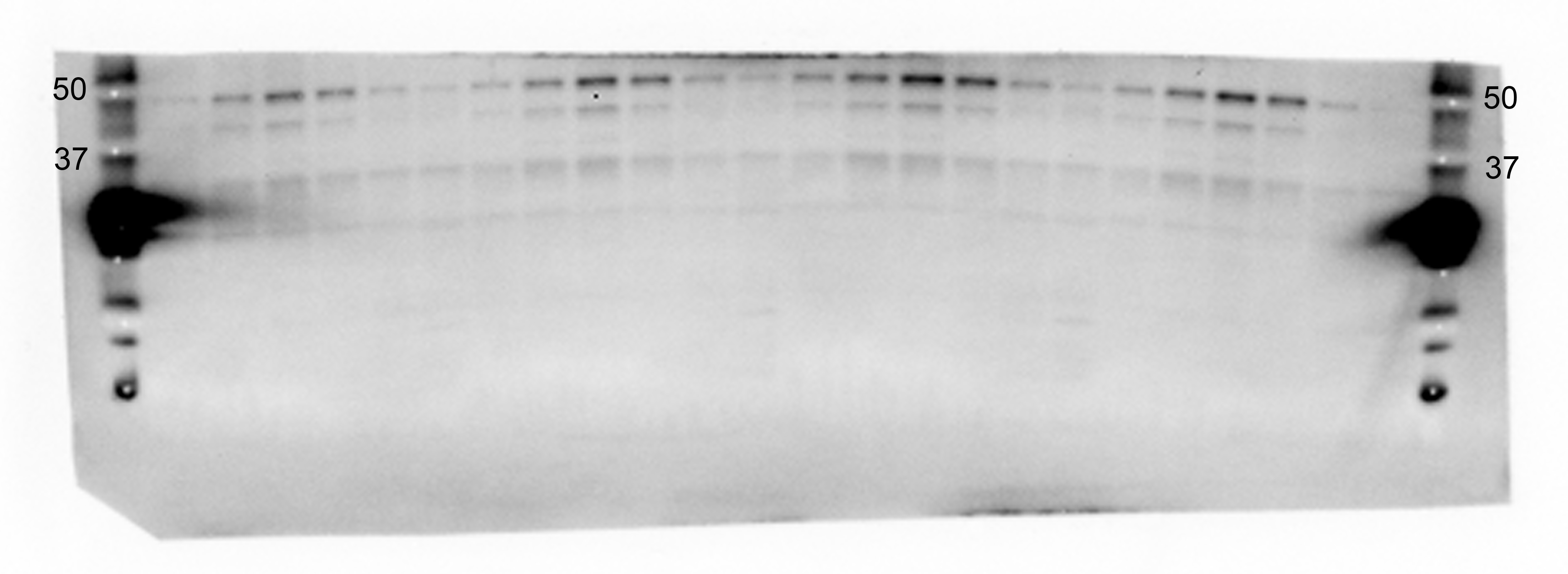
FH Pasquale (Verified Customer) (11-06-2019) | The antibody recognize the exosomal marker TSG101 as claimed. We tested it by Western blotting in our exosome preps isolated from murine brains and it gave the signal at the correct molecular weight in the fractions that are enriched in exosomes, so it seems to be specific. However, we report here some drawbacks that we have experienced using this antibody (see the file attached):1. There is a strong cross-reactivity of the antibody with the molecular weight marker we use (The Bio-Rad Dual Color Standard, cat. number 1610374)2. The signal over noise ratio is a little low (the background coming from the membrane is pretty high)
 |

