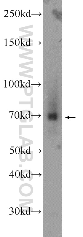
BxPC-3 cells were subjected to SDS PAGE followed by western blot with 18468-1-AP (TRIM41 antibody) at dilution of 1:800 incubated at room temperature for 1.5 hours.
BxPC-3 cells were subjected to SDS PAGE followed by western blot with 18468-1-AP (TRIM41 antibody) at dilution of 1:800 incubated at room temperature for 1.5 hours.
WB analysis of human placenta using 18468-1-AP
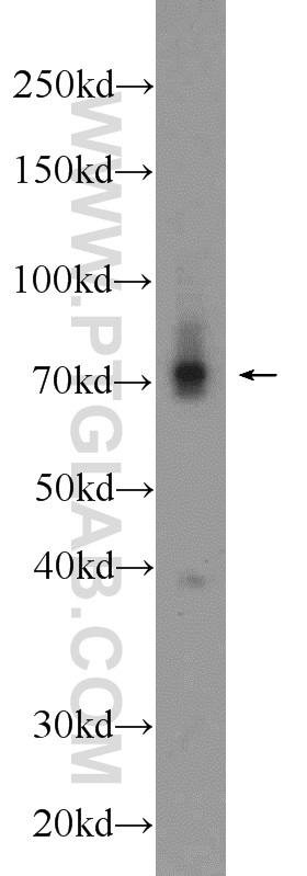
human placenta tissue were subjected to SDS PAGE followed by western blot with 18468-1-AP (TRIM41 antibody) at dilution of 1:1000 incubated at room temperature for 1.5 hours.
human placenta tissue were subjected to SDS PAGE followed by western blot with 18468-1-AP (TRIM41 antibody) at dilution of 1:1000 incubated at room temperature for 1.5 hours.
WB analysis of mouse liver using 18468-1-AP
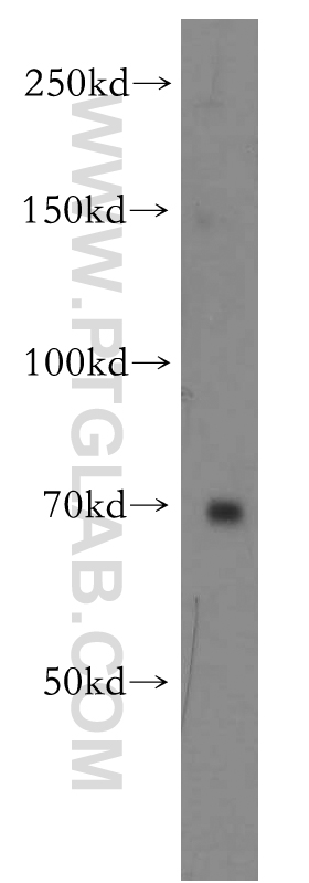
mouse liver tissue were subjected to SDS PAGE followed by western blot with 18468-1-AP (TRIM41 antibody) at dilution of 1:1000 incubated at room temperature for 1.5 hours.
mouse liver tissue were subjected to SDS PAGE followed by western blot with 18468-1-AP (TRIM41 antibody) at dilution of 1:1000 incubated at room temperature for 1.5 hours.
WB analysis of human placenta using 18468-1-AP
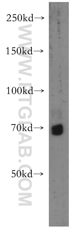
human placenta tissue were subjected to SDS PAGE followed by western blot with 18468-1-AP (TRIM41 antibody) at dilution of 1:500 incubated at room temperature for 1.5 hours.
human placenta tissue were subjected to SDS PAGE followed by western blot with 18468-1-AP (TRIM41 antibody) at dilution of 1:500 incubated at room temperature for 1.5 hours.
WB analysis of mouse liver using 18468-1-AP
mouse liver tissue were subjected to SDS PAGE followed by western blot with 18468-1-AP (TRIM41 antibody) at dilution of 1:500 incubated at room temperature for 1.5 hours.
mouse liver tissue were subjected to SDS PAGE followed by western blot with 18468-1-AP (TRIM41 antibody) at dilution of 1:500 incubated at room temperature for 1.5 hours.
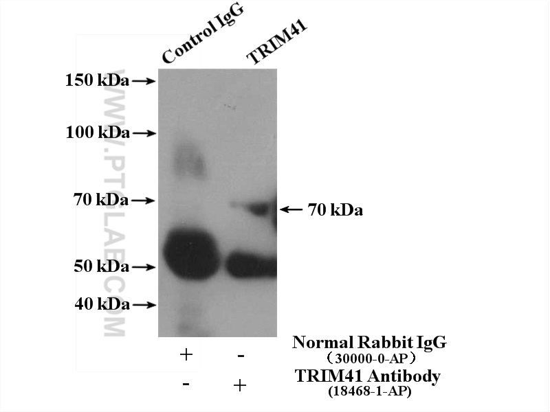
IP experiment of human plasma using 18468-1-AP
IP result of anti-TRIM41 (IP:18468-1-AP, 4ug; Detection:18468-1-AP 1:600) with human plasma lysate 4000ug.
Immunohistochemical analysis of paraffin-embedded human skeletal muscle using 18468-1-AP (TRIM41 antibody) at dilution of 1:50 (under 40x lens).
IHC staining of human breast cancer using 18468-1-AP
Immunohistochemical analysis of paraffin-embedded human breast cancer tissue slide using 18468-1-AP (TRIM41 Antibody) at dilution of 1:200 (under 10x lens). Heat mediated antigen retrieval with Tris-EDTA buffer (pH 9.0).
Immunohistochemical analysis of paraffin-embedded human breast cancer tissue slide using 18468-1-AP (TRIM41 Antibody) at dilution of 1:200 (under 10x lens). Heat mediated antigen retrieval with Tris-EDTA buffer (pH 9.0).
IHC staining of human breast cancer using 18468-1-AP
Immunohistochemical analysis of paraffin-embedded human breast cancer tissue slide using 18468-1-AP (TRIM41 Antibody) at dilution of 1:200 (under 40x lens). Heat mediated antigen retrieval with Tris-EDTA buffer (pH 9.0).
Immunohistochemical analysis of paraffin-embedded human breast cancer tissue slide using 18468-1-AP (TRIM41 Antibody) at dilution of 1:200 (under 40x lens). Heat mediated antigen retrieval with Tris-EDTA buffer (pH 9.0).
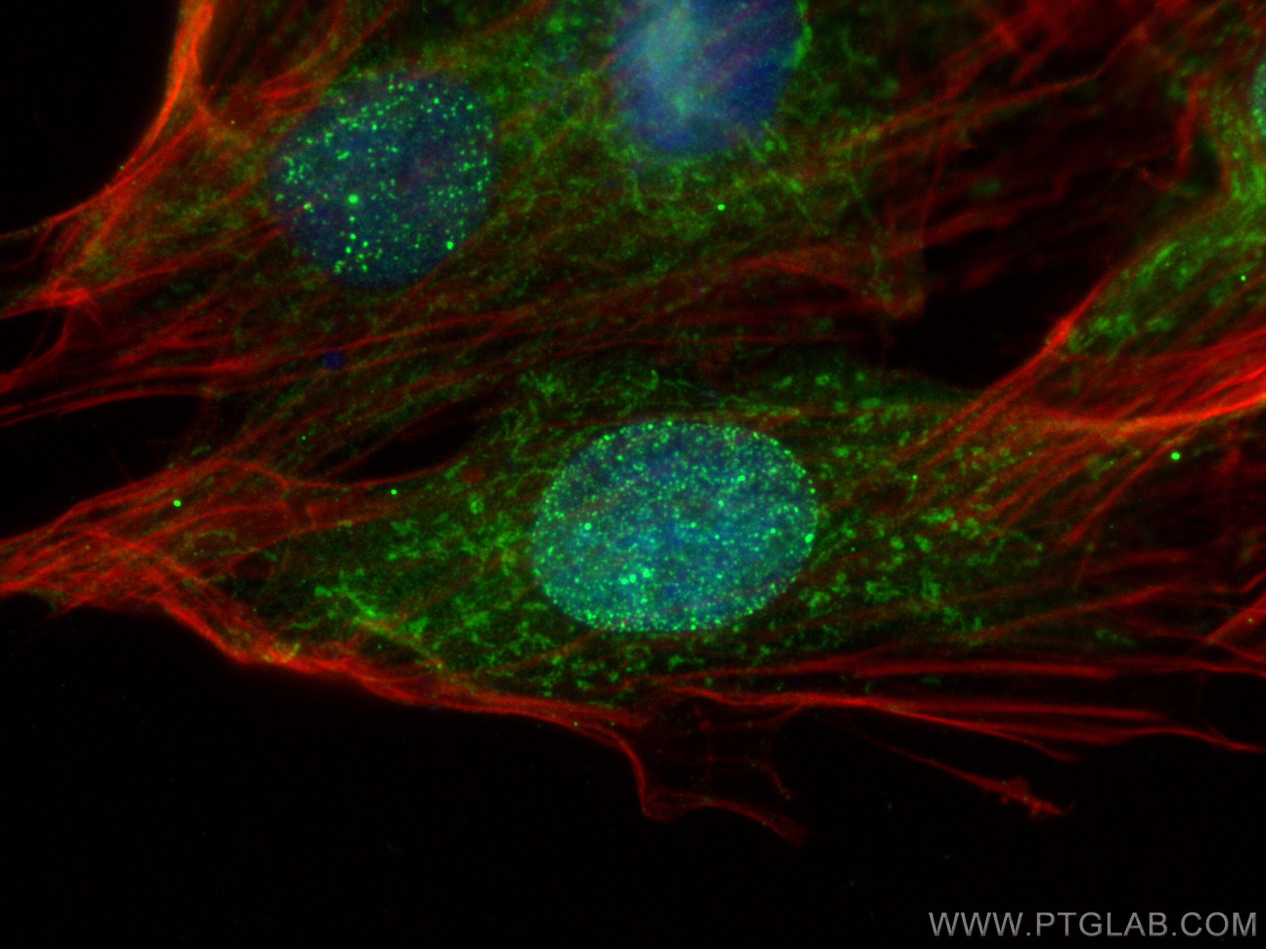
IF Staining of H9C2 using 18468-1-AP
Immunofluorescent analysis of (4% PFA) fixed H9C2 cells using TRIM41 antibody (18468-1-AP) at dilution of 1:400 and Multi-rAb CoraLite ® Plus 488-Goat Anti-Rabbit Recombinant Secondary Antibody (H+L) (RGAR002), CL594-phalloidin (red).
Immunofluorescent analysis of (4% PFA) fixed H9C2 cells using TRIM41 antibody (18468-1-AP) at dilution of 1:400 and Multi-rAb CoraLite ® Plus 488-Goat Anti-Rabbit Recombinant Secondary Antibody (H+L) (RGAR002), CL594-phalloidin (red).
The Proteintech guarantee covers Proteintech antibodies in any species and any application, including those not listed on the datasheet. If the antibody doesn’t perform, you can receive a hassle-free refund or credit note.
BxPC-3 cells, human placenta tissue, mouse liver tissue
Positive IP detected in
human plasma tissue
Positive IHC detected in
human skeletal muscle tissue, human breast cancer tissue Note: suggested antigen retrieval with TE buffer pH 9.0; (*) Alternatively, antigen retrieval may be performed with citrate buffer pH 6.0
Positive IF/ICC detected in
H9C2 cells
Recommended dilution
Application
Dilution
Western Blot (WB)
WB : 1:500-1:2400
Immunoprecipitation (IP)
IP : 0.5-4.0 ug for 1.0-3.0 mg of total protein lysate
Immunohistochemistry (IHC)
IHC : 1:20-1:200
Immunofluorescence (IF)/ICC
IF/ICC : 1:200-1:800
It is recommended that this reagent should be titrated in each testing system to obtain optimal results.
Sample-dependent, Check data in validation data gallery.
BxPC-3 cells were subjected to SDS PAGE followed by western blot with 18468-1-AP (TRIM41 antibody) at dilution of 1:800 incubated at room temperature for 1.5 hours.
WB analysis of human placenta using 18468-1-AP
human placenta tissue were subjected to SDS PAGE followed by western blot with 18468-1-AP (TRIM41 antibody) at dilution of 1:1000 incubated at room temperature for 1.5 hours.
WB analysis of mouse liver using 18468-1-AP
mouse liver tissue were subjected to SDS PAGE followed by western blot with 18468-1-AP (TRIM41 antibody) at dilution of 1:1000 incubated at room temperature for 1.5 hours.
WB analysis of human placenta using 18468-1-AP
human placenta tissue were subjected to SDS PAGE followed by western blot with 18468-1-AP (TRIM41 antibody) at dilution of 1:500 incubated at room temperature for 1.5 hours.
WB analysis of mouse liver using 18468-1-AP
mouse liver tissue were subjected to SDS PAGE followed by western blot with 18468-1-AP (TRIM41 antibody) at dilution of 1:500 incubated at room temperature for 1.5 hours.
IHC Figures
IHC staining of human skeletal muscle using 18468-1-AP
Immunohistochemical analysis of paraffin-embedded human skeletal muscle using 18468-1-AP (TRIM41 antibody) at dilution of 1:50 (under 10x lens).
IHC staining of human skeletal muscle using 18468-1-AP
Immunohistochemical analysis of paraffin-embedded human skeletal muscle using 18468-1-AP (TRIM41 antibody) at dilution of 1:50 (under 40x lens).
IHC staining of human breast cancer using 18468-1-AP
Immunohistochemical analysis of paraffin-embedded human breast cancer tissue slide using 18468-1-AP (TRIM41 Antibody) at dilution of 1:200 (under 10x lens). Heat mediated antigen retrieval with Tris-EDTA buffer (pH 9.0).
IHC staining of human breast cancer using 18468-1-AP
Immunohistochemical analysis of paraffin-embedded human breast cancer tissue slide using 18468-1-AP (TRIM41 Antibody) at dilution of 1:200 (under 40x lens). Heat mediated antigen retrieval with Tris-EDTA buffer (pH 9.0).
IP Figures
IP experiment of human plasma using 18468-1-AP
IP result of anti-TRIM41 (IP:18468-1-AP, 4ug; Detection:18468-1-AP 1:600) with human plasma lysate 4000ug.
IF/ICC Figures
IF Staining of H9C2 using 18468-1-AP
Immunofluorescent analysis of (4% PFA) fixed H9C2 cells using TRIM41 antibody (18468-1-AP) at dilution of 1:400 and Multi-rAb CoraLite ® Plus 488-Goat Anti-Rabbit Recombinant Secondary Antibody (H+L) (RGAR002), CL594-phalloidin (red).
The species listed in Tested Reactivity are in-house verified and applicable species. For unlisted species, please refer to the homology analysis of the immunogen sequence and related species. For rabbit polyclonal antibodies, homology >70% is recommended. For mouse monoclonal antibodies and rabbit recombinant antibodies, homology >90% is recommended. Generally, the higher the homology, the greater the applicability. However, there will be certain differences in protein expression in different species, tissues or cells. Therefore, the homology analysis results are for reference only and do not serve as a guarantee.
At Proteintech, we pride ourselves on our antibody quality, customer service and transparency. As such, we are comparing our antibodies with other vendors, enabling easy identification and comparisons of key data to help you choose the suitable antibody for your needs.
We have selected the top cited antibodies from these vendors for you to compare.
Proteintech
TRIM41 Polyclonal antibody
Catalog Number
18468-1-AP
Citations
1
Dilutions
WB : 1:500-1:2400 IP : 0.5-4.0 ug for IP and 0.5-4.0 ug for 1.0-3.0 mg of total protein lysate for WB IHC : 1:20-1:200 IF/ICC : 1:200-1:800
Applications
WB, IHC, IF/ICC, IP, ELISA
Reactivity
human, mouse, rat
Product Guarantee
Covers any species including not listed on datasheet
Covers any applications including not listed on datasheet
 at dilution of 1:800 incubated at room temperature for 1.5 hours.")
 at dilution of 1:1000 incubated at room temperature for 1.5 hours.")
 at dilution of 1:1000 incubated at room temperature for 1.5 hours.")
 at dilution of 1:500 incubated at room temperature for 1.5 hours.")
 at dilution of 1:500 incubated at room temperature for 1.5 hours.")
 with human plasma lysate 4000ug.")
 at dilution of 1:50 (under 10x lens).")
 at dilution of 1:50 (under 40x lens).")
 at dilution of 1:200 (under 10x lens). Heat mediated antigen retrieval with Tris-EDTA buffer (pH 9.0).")
 at dilution of 1:200 (under 40x lens). Heat mediated antigen retrieval with Tris-EDTA buffer (pH 9.0).")
 fixed H9C2 cells using TRIM41 antibody (18468-1-AP) at dilution of 1:400 and Multi-rAb CoraLite ® Plus 488-Goat Anti-Rabbit Recombinant Secondary Antibody (H+L) (RGAR002), CL594-phalloidin (red).")