Tested Applications
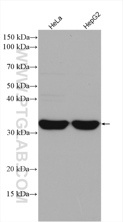
| Positive WB detected in | HeLa cells, HepG2 cells |
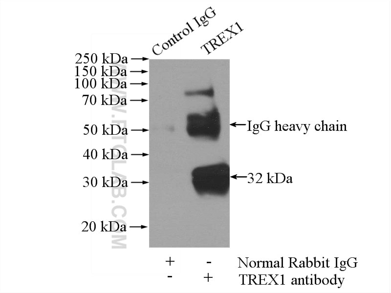
| Positive IP detected in | HeLa cells |
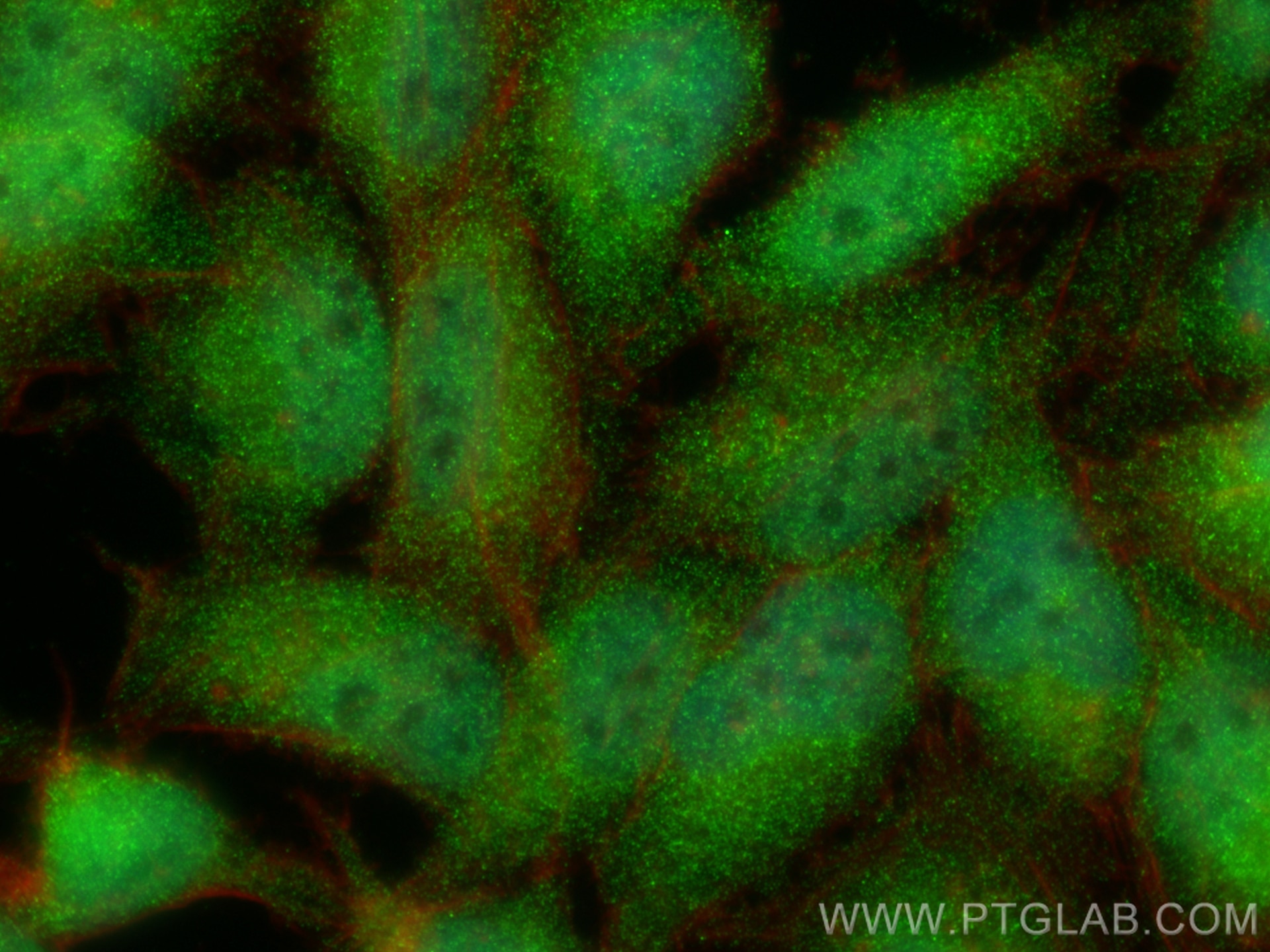
| Positive IF/ICC detected in | HeLa cells |
Recommended dilution
| Application | Dilution |
|---|---|
| Western Blot (WB) | WB : 1:500-1:3000 |
| Immunoprecipitation (IP) | IP : 0.5-4.0 ug for 1.0-3.0 mg of total protein lysate |
| Immunofluorescence (IF)/ICC | IF/ICC : 1:50-1:500 |
| It is recommended that this reagent should be titrated in each testing system to obtain optimal results. | |
| Sample-dependent, Check data in validation data gallery. | |
Published Applications
| KD/KO | See 1 publications below |
| WB | See 4 publications below |
| IF | See 1 publications below |
Product Information
24876-1-AP targets TREX1 in WB, IF/ICC, IP, ELISA applications and shows reactivity with human samples.
| Tested Reactivity | human |
| Cited Reactivity | human, bovine |
| Host / Isotype | Rabbit / IgG |
| Class | Polyclonal |
| Type | Antibody |
| Immunogen | TREX1 fusion protein Ag18325 Predict reactive species |
| Full Name | three prime repair exonuclease 1 |
| Calculated Molecular Weight | 369 aa, 39 kDa |
| Observed Molecular Weight | 32-39 kDa |
| GenBank Accession Number | BC023630 |
| Gene Symbol | TREX1 |
| Gene ID (NCBI) | 11277 |
| RRID | AB_2879772 |
| Conjugate | Unconjugated |
| Form | Liquid |
| Purification Method | Antigen affinity purification |
| UNIPROT ID | Q9NSU2 |
| Storage Buffer | PBS with 0.02% sodium azide and 50% glycerol , pH 7.3 |
| Storage Conditions | Store at -20°C. Stable for one year after shipment. Aliquoting is unnecessary for -20oC storage. 20ul sizes contain 0.1% BSA. |
Background Information
TREX1 (three prime repair exonuclease 1), also known as trophoblast expressed 1, CRV, AGS1, AGS5, DRN3, HERNS or DNase III, is a member of the exonuclease superfamily and belongs to the TREX family. TREX1 may play a role in DNA repair. TREX1 is expressed in thymus, spleen, liver, brain, heart, small intestine and colon. Mutations or defects in the gene encoding TREX1 have been associated with a variety of diseases, including systemic lupus erythematosus, chilblain lupus (CHBL), Aicardi-Goutieres syndrome type 1 (AGS1) and type 5 (AGS5).
Protocols
| Product Specific Protocols | |
|---|---|
| WB protocol for TREX1 antibody 24876-1-AP | Download protocol |
| IF protocol for TREX1 antibody 24876-1-AP | Download protocol |
| IP protocol for TREX1 antibody 24876-1-AP | Download protocol |
| Standard Protocols | |
|---|---|
| Click here to view our Standard Protocols |
Publications
| Species | Application | Title |
|---|---|---|
J Neuropathol Exp Neurol Retinal Vasculopathy With Cerebral Leukodystrophy: Clinicopathologic Features of an Autopsied Patient With a Heterozygous TREX 1 Mutation. | ||
bioRxiv Pathogen-driven CRISPR screens identify TREX1 as a regulator of DNA self-sensing during influenza virus infection
| ||
Cell Host Microbe Pathogen-driven CRISPR screens identify TREX1 as a regulator of DNA self-sensing during influenza virus infection | ||
Cell Rep Pharmacological inhibition of neddylation impairs long interspersed element 1 retrotransposition | ||
Int J Mol Sci Inactivation of Myostatin Delays Senescence via TREX1-SASP in Bovine Skeletal Muscle Cells |
Reviews
The reviews below have been submitted by verified Proteintech customers who received an incentive for providing their feedback.
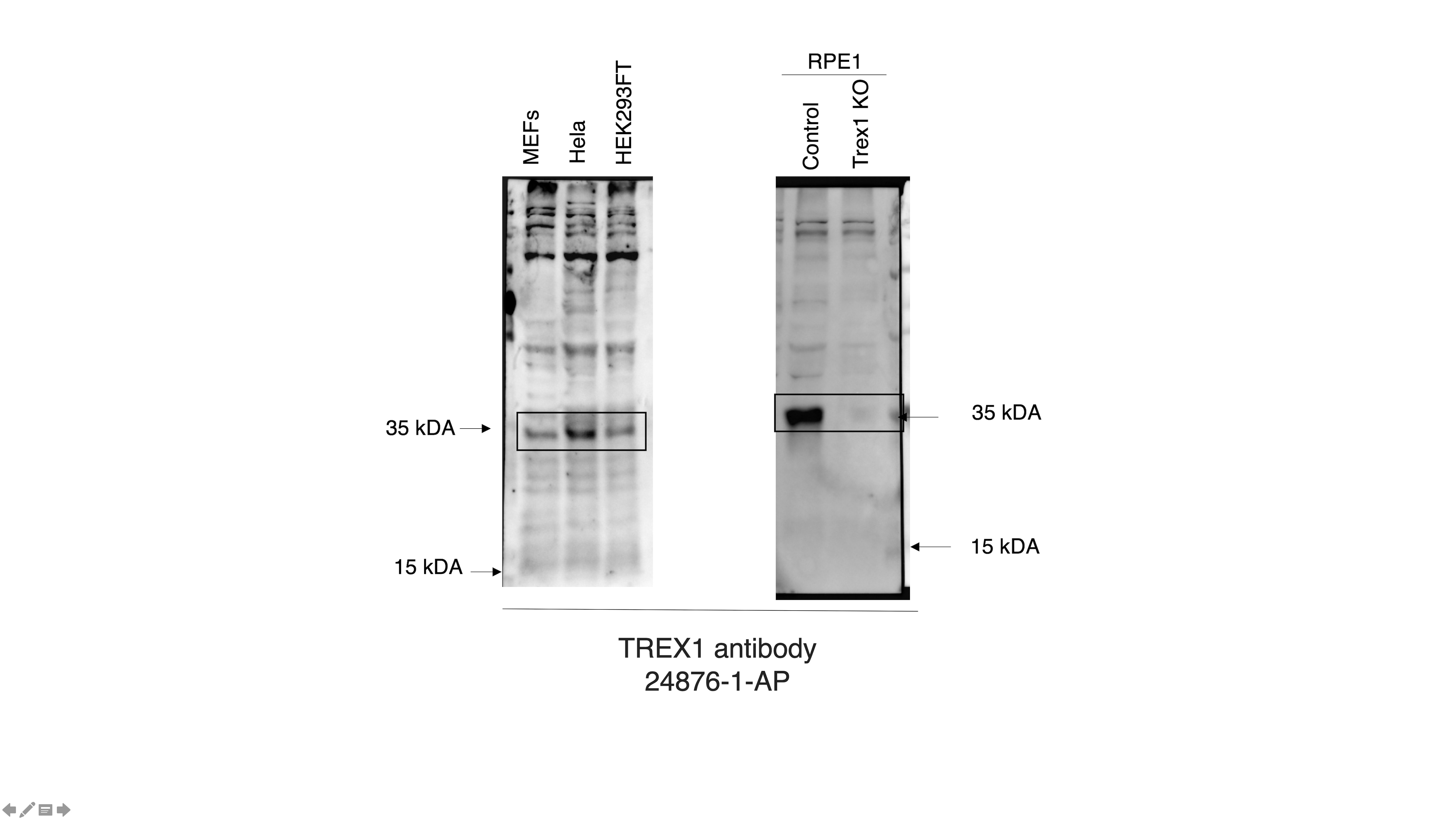
FH Chantal (Verified Customer) (10-04-2022) | reconignises the specific band, validated by KO of TREX1. A little bit of background. the signal is weak at 1:1000 dilution, better at 1:500. Works on human and mouse cell lines.
 |


