Brain tissues were subjected to SDS PAGE followed by western blot with 68048-1-Ig (TPPP antibody) at dilution of 1:10000 incubated at room temperature for 1.5 hours. The membrane was stripped and reblotted with HRP-conjugated GAPDH Monoclonal antibody (HRP-60004) as loading control.
Brain tissues were subjected to SDS PAGE followed by western blot with 68048-1-Ig (TPPP antibody) at dilution of 1:10000 incubated at room temperature for 1.5 hours. The membrane was stripped and reblotted with HRP-conjugated GAPDH Monoclonal antibody (HRP-60004) as loading control.
WB analysis of SH-SY5Y using 68048-1-Ig
WB result of TPPP antibody (68048-1-Ig; 1:10000; incubated at room temperature for 1.5 hours) with sh-Control and sh-TPPP transfected SH-SY5Y cells.
WB result of TPPP antibody (68048-1-Ig; 1:10000; incubated at room temperature for 1.5 hours) with sh-Control and sh-TPPP transfected SH-SY5Y cells.
WB analysis of pig cerebellum using 68048-1-Ig
pig cerebellum tissue were subjected to SDS PAGE followed by western blot with 68048-1-Ig (TPPP antibody) at dilution of 1:10000 incubated at room temperature for 1.5 hours.
pig cerebellum tissue were subjected to SDS PAGE followed by western blot with 68048-1-Ig (TPPP antibody) at dilution of 1:10000 incubated at room temperature for 1.5 hours.
WB analysis of rabbit cerebellum using 68048-1-Ig
rabbit cerebellum tissue were subjected to SDS PAGE followed by western blot with 68048-1-Ig (TPPP antibody) at dilution of 1:10000 incubated at room temperature for 1.5 hours.
rabbit cerebellum tissue were subjected to SDS PAGE followed by western blot with 68048-1-Ig (TPPP antibody) at dilution of 1:10000 incubated at room temperature for 1.5 hours.
WB analysis of rat cerebellum using 68048-1-Ig
rat cerebellum tissue were subjected to SDS PAGE followed by western blot with 68048-1-Ig (TPPP antibody) at dilution of 1:10000 incubated at room temperature for 1.5 hours.
rat cerebellum tissue were subjected to SDS PAGE followed by western blot with 68048-1-Ig (TPPP antibody) at dilution of 1:10000 incubated at room temperature for 1.5 hours.
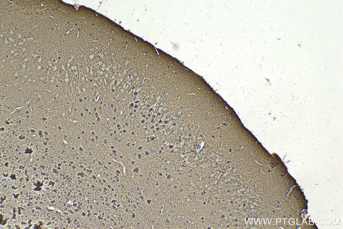
IHC staining of mouse brain using 68048-1-Ig
Immunohistochemical analysis of paraffin-embedded mouse brain tissue slide using 68048-1-Ig (TPPP antibody) at dilution of 1:1000 (under 40x lens). Heat mediated antigen retrieval with Tris-EDTA buffer (pH 9.0).
Immunohistochemical analysis of paraffin-embedded mouse brain tissue slide using 68048-1-Ig (TPPP antibody) at dilution of 1:1000 (under 40x lens). Heat mediated antigen retrieval with Tris-EDTA buffer (pH 9.0).
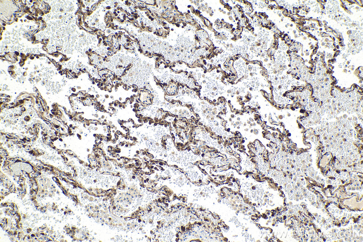
IHC staining of human lung using 68048-1-Ig
Immunohistochemical analysis of paraffin-embedded human lung tissue slide using 68048-1-Ig (TPPP antibody) at dilution of 1:1000 (under 10x lens). Heat mediated antigen retrieval with Tris-EDTA buffer (pH 9.0).
Immunohistochemical analysis of paraffin-embedded human lung tissue slide using 68048-1-Ig (TPPP antibody) at dilution of 1:1000 (under 10x lens). Heat mediated antigen retrieval with Tris-EDTA buffer (pH 9.0).
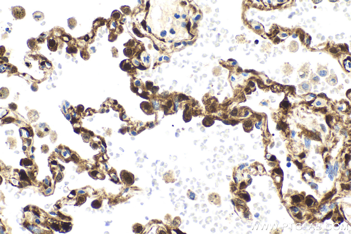
IHC staining of human lung using 68048-1-Ig
Immunohistochemical analysis of paraffin-embedded human lung tissue slide using 68048-1-Ig (TPPP antibody) at dilution of 1:1000 (under 40x lens). Heat mediated antigen retrieval with Tris-EDTA buffer (pH 9.0).
Immunohistochemical analysis of paraffin-embedded human lung tissue slide using 68048-1-Ig (TPPP antibody) at dilution of 1:1000 (under 40x lens). Heat mediated antigen retrieval with Tris-EDTA buffer (pH 9.0).
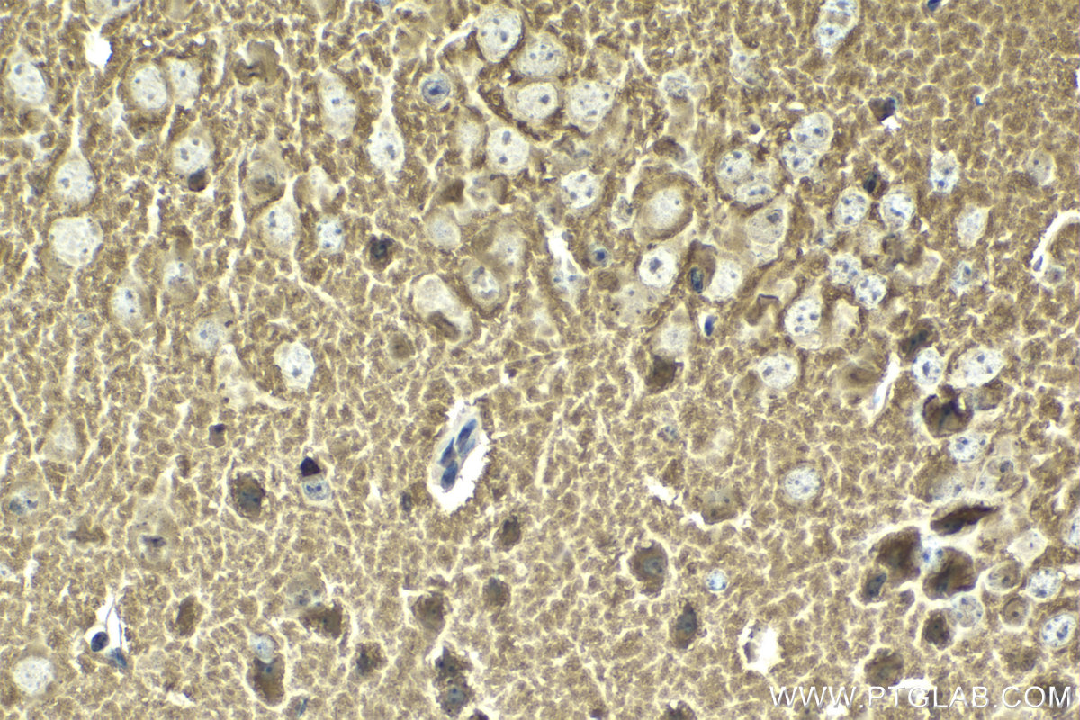
IHC staining of mouse brain using 68048-1-Ig
Immunohistochemical analysis of paraffin-embedded mouse brain tissue slide using 68048-1-Ig (TPPP antibody) at dilution of 1:1000 (under 10x lens). Heat mediated antigen retrieval with Tris-EDTA buffer (pH 9.0).
Immunohistochemical analysis of paraffin-embedded mouse brain tissue slide using 68048-1-Ig (TPPP antibody) at dilution of 1:1000 (under 10x lens). Heat mediated antigen retrieval with Tris-EDTA buffer (pH 9.0).
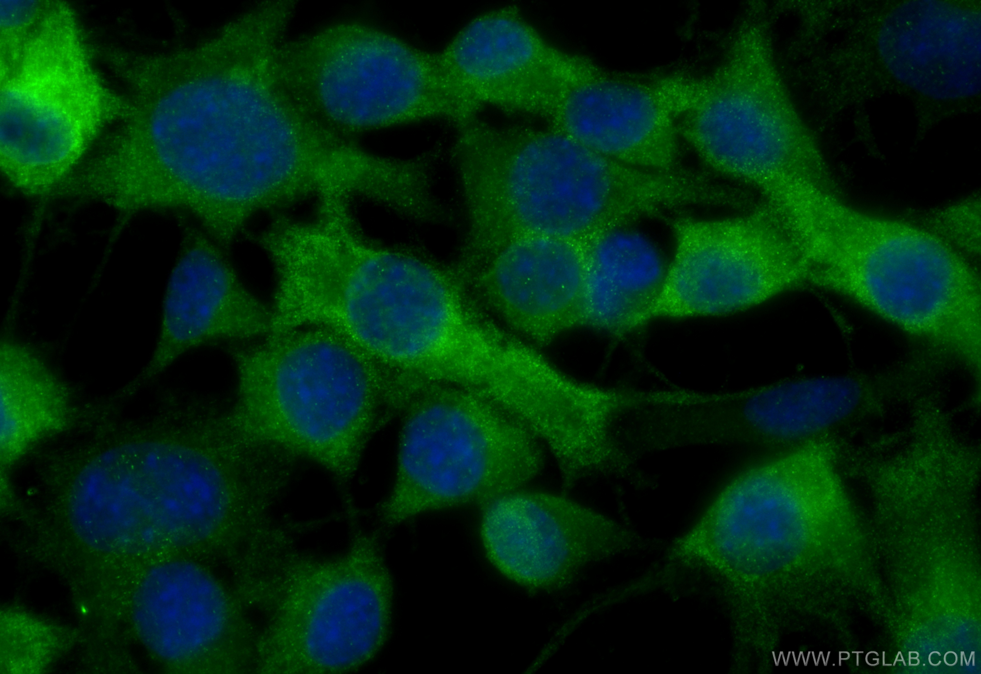
IF Staining of Neuro-2a using 68048-1-Ig
Immunofluorescent analysis of (-20°C Ethanol) fixed Neuro-2a cells using TPPP antibody (68048-1-Ig, Clone: 1E5F5 ) at dilution of 1:800 and CoraLite®488-Conjugated Goat Anti-Mouse IgG(H+L) (SA00013-1).
Immunofluorescent analysis of (-20°C Ethanol) fixed Neuro-2a cells using TPPP antibody (68048-1-Ig, Clone: 1E5F5 ) at dilution of 1:800 and CoraLite®488-Conjugated Goat Anti-Mouse IgG(H+L) (SA00013-1).
The Proteintech guarantee covers Proteintech antibodies in any species and any application, including those not listed on the datasheet. If the antibody doesn’t perform, you can receive a hassle-free refund or credit note.
human lung tissue, mouse brain tissue Note: suggested antigen retrieval with TE buffer pH 9.0; (*) Alternatively, antigen retrieval may be performed with citrate buffer pH 6.0
Positive IF/ICC detected in
Neuro-2a cells
Recommended dilution
Application
Dilution
Western Blot (WB)
WB : 1:5000-1:50000
Immunohistochemistry (IHC)
IHC : 1:500-1:2000
Immunofluorescence (IF)/ICC
IF/ICC : 1:400-1:1600
It is recommended that this reagent should be titrated in each testing system to obtain optimal results.
Sample-dependent, Check data in validation data gallery.
Product Information
68048-1-Ig targets TPPP in WB, IHC, IF/ICC, ELISA applications and shows reactivity with human, mouse, rat, pig, rabbit, chicken samples.
PBS with 0.02% sodium azide and 50% glycerol pH 7.3.
Storage Conditions
Store at -20°C. Stable for one year after shipment. Aliquoting is unnecessary for -20oC storage. 20ul sizes contain 0.1% BSA.
Background Information
TPPP, also named as TPPP1, P24 and P25, belongs to the TPPP family. TPPP promotes in vitro the polymerization of tubulin into double-walled tubules and polymorphic aggregates or bundled stabilized microtubules blocks. When overexpressed, TPPP inhibits mitotic spindle assembly and nuclear envelope breakdown, apparently without affecting other cellular events.
Brain tissues were subjected to SDS PAGE followed by western blot with 68048-1-Ig (TPPP antibody) at dilution of 1:10000 incubated at room temperature for 1.5 hours. The membrane was stripped and reblotted with HRP-conjugated GAPDH Monoclonal antibody (HRP-60004) as loading control.
WB analysis of SH-SY5Y using 68048-1-Ig
WB result of TPPP antibody (68048-1-Ig; 1:10000; incubated at room temperature for 1.5 hours) with sh-Control and sh-TPPP transfected SH-SY5Y cells.
WB analysis of pig cerebellum using 68048-1-Ig
pig cerebellum tissue were subjected to SDS PAGE followed by western blot with 68048-1-Ig (TPPP antibody) at dilution of 1:10000 incubated at room temperature for 1.5 hours.
WB analysis of rabbit cerebellum using 68048-1-Ig
rabbit cerebellum tissue were subjected to SDS PAGE followed by western blot with 68048-1-Ig (TPPP antibody) at dilution of 1:10000 incubated at room temperature for 1.5 hours.
WB analysis of rat cerebellum using 68048-1-Ig
rat cerebellum tissue were subjected to SDS PAGE followed by western blot with 68048-1-Ig (TPPP antibody) at dilution of 1:10000 incubated at room temperature for 1.5 hours.
IHC Figures
IHC staining of mouse brain using 68048-1-Ig
Immunohistochemical analysis of paraffin-embedded mouse brain tissue slide using 68048-1-Ig (TPPP antibody) at dilution of 1:1000 (under 40x lens). Heat mediated antigen retrieval with Tris-EDTA buffer (pH 9.0).
IHC staining of human lung using 68048-1-Ig
Immunohistochemical analysis of paraffin-embedded human lung tissue slide using 68048-1-Ig (TPPP antibody) at dilution of 1:1000 (under 10x lens). Heat mediated antigen retrieval with Tris-EDTA buffer (pH 9.0).
IHC staining of human lung using 68048-1-Ig
Immunohistochemical analysis of paraffin-embedded human lung tissue slide using 68048-1-Ig (TPPP antibody) at dilution of 1:1000 (under 40x lens). Heat mediated antigen retrieval with Tris-EDTA buffer (pH 9.0).
IHC staining of mouse brain using 68048-1-Ig
Immunohistochemical analysis of paraffin-embedded mouse brain tissue slide using 68048-1-Ig (TPPP antibody) at dilution of 1:1000 (under 10x lens). Heat mediated antigen retrieval with Tris-EDTA buffer (pH 9.0).
IF/ICC Figures
IF Staining of Neuro-2a using 68048-1-Ig
Immunofluorescent analysis of (-20°C Ethanol) fixed Neuro-2a cells using TPPP antibody (68048-1-Ig, Clone: 1E5F5 ) at dilution of 1:800 and CoraLite®488-Conjugated Goat Anti-Mouse IgG(H+L) (SA00013-1).
The species listed in Tested Reactivity are in-house verified and applicable species. For unlisted species, please refer to the homology analysis of the immunogen sequence and related species. For rabbit polyclonal antibodies, homology >70% is recommended. For mouse monoclonal antibodies and rabbit recombinant antibodies, homology >90% is recommended. Generally, the higher the homology, the greater the applicability. However, there will be certain differences in protein expression in different species, tissues or cells. Therefore, the homology analysis results are for reference only and do not serve as a guarantee.
At Proteintech, we pride ourselves on our antibody quality, customer service and transparency. As such, we are comparing our antibodies with other vendors, enabling easy identification and comparisons of key data to help you choose the suitable antibody for your needs.
We have selected the top cited antibodies from these vendors for you to compare.
 at dilution of 1:10000 incubated at room temperature for 1.5 hours. The membrane was stripped and reblotted with HRP-conjugated GAPDH Monoclonal antibody (HRP-60004) as loading control.")
 with sh-Control and sh-TPPP transfected SH-SY5Y cells.")
 at dilution of 1:10000 incubated at room temperature for 1.5 hours.")
 at dilution of 1:10000 incubated at room temperature for 1.5 hours.")
 at dilution of 1:10000 incubated at room temperature for 1.5 hours.")
 at dilution of 1:1000 (under 40x lens). Heat mediated antigen retrieval with Tris-EDTA buffer (pH 9.0).")
 at dilution of 1:1000 (under 10x lens). Heat mediated antigen retrieval with Tris-EDTA buffer (pH 9.0).")
 at dilution of 1:1000 (under 40x lens). Heat mediated antigen retrieval with Tris-EDTA buffer (pH 9.0).")
 at dilution of 1:1000 (under 10x lens). Heat mediated antigen retrieval with Tris-EDTA buffer (pH 9.0).")
 fixed Neuro-2a cells using TPPP antibody (68048-1-Ig, Clone: 1E5F5 ) at dilution of 1:800 and CoraLite®488-Conjugated Goat Anti-Mouse IgG(H+L) (SA00013-1).")