Filter:
 at dilution of 1:2000 incubated at room temperature for 1.5 hours.")
 with si-Control and si-TLE3 transfected HEK293 cells.")
 at dilution of 1:1000 incubated at room temperature for 1.5 hours.")
 at dilution of 1:1000 incubated at room temperature for 1.5 hours.")
 at dilution of 1:1000 incubated at room temperature for 1.5 hours.")
 with HepG2 cells lysate 2400ug.")
 at dilution of 1:200 (under 40x lens. Heat mediated antigen retrieval with Tris-EDTA buffer (pH 9.0).")
 at dilution of 1:200 (under 10x lens. Heat mediated antigen retrieval with Tris-EDTA buffer (pH 9.0).")
 at dilution of 1:200 (under 40x lens. Heat mediated antigen retrieval with Tris-EDTA buffer (pH 9.0).")
 at dilution of 1:200 (under 10x lens. Heat mediated antigen retrieval with Tris-EDTA buffer (pH 9.0).")
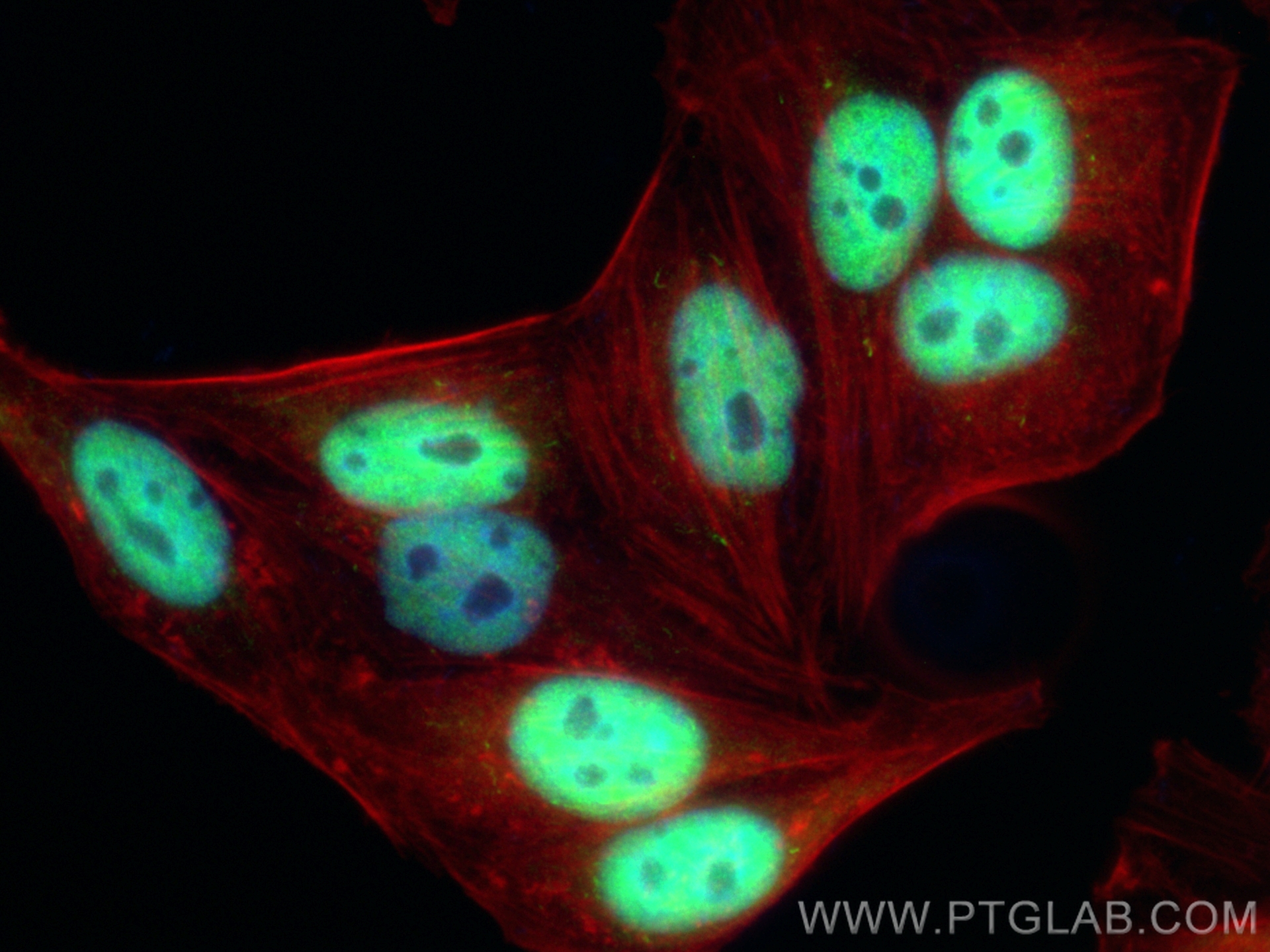
 fixed HepG2 cells using TLE3 antibody (22094-1-AP) at dilution of 1:600 and CoraLite®488-Conjugated AffiniPure Goat Anti-Rabbit IgG(H+L) (SA00013-2), CL594-Phalloidin (red).")
Tested Applications
| Positive WB detected in | HEK-293 cells, A431 cells, HepG2 cells |
| Positive IP detected in | HepG2 cells |
| Positive IHC detected in | human lung cancer tissue, human breast cancer tissue Note: suggested antigen retrieval with TE buffer pH 9.0; (*) Alternatively, antigen retrieval may be performed with citrate buffer pH 6.0 |
| Positive IF/ICC detected in | HepG2 cells |
Recommended dilution
| Application | Dilution |
|---|---|
| Western Blot (WB) | WB : 1:1000-1:4000 |
| Immunoprecipitation (IP) | IP : 0.5-4.0 ug for 1.0-3.0 mg of total protein lysate |
| Immunohistochemistry (IHC) | IHC : 1:50-1:500 |
| Immunofluorescence (IF)/ICC | IF/ICC : 1:300-1:1200 |
| It is recommended that this reagent should be titrated in each testing system to obtain optimal results. | |
| Sample-dependent, Check data in validation data gallery. | |
Published Applications
| KD/KO | See 1 publications below |
| WB | See 1 publications below |
| IF | See 1 publications below |
Product Information
22094-1-AP targets TLE3 in WB, IHC, IF/ICC, IP, ELISA applications and shows reactivity with human, mouse, rat samples.
| Tested Reactivity | human, mouse, rat |
| Cited Reactivity | mouse |
| Host / Isotype | Rabbit / IgG |
| Class | Polyclonal |
| Type | Antibody |
| Immunogen |
CatNo: Ag17300 Product name: Recombinant human TLE3 protein Source: e coli.-derived, PGEX-4T Tag: GST Domain: 158-240 aa of BC015729 Sequence: PVTGSSSGLLALGALGSQAHLTVKDEKNHHELDHRERESSANNSVSPSESLRASEKHRGSADYSMEAKKRKAEEKDSLSRYDS Predict reactive species |
| Full Name | transducin-like enhancer of split 3 (E(sp1) homolog, Drosophila) |
| Calculated Molecular Weight | 772 aa, 83 kDa |
| Observed Molecular Weight | 90 kDa |
| GenBank Accession Number | BC015729 |
| Gene Symbol | TLE3 |
| Gene ID (NCBI) | 7090 |
| RRID | AB_2878993 |
| Conjugate | Unconjugated |
| Form | Liquid |
| Purification Method | Antigen affinity purification |
| UNIPROT ID | Q04726 |
| Storage Buffer | PBS with 0.02% sodium azide and 50% glycerol, pH 7.3. |
| Storage Conditions | Store at -20°C. Stable for one year after shipment. Aliquoting is unnecessary for -20oC storage. 20ul sizes contain 0.1% BSA. |
Background Information
Transducin-like enhancer of split 3(TLE3) is a corepressor that binds to a variant of transciption factor, which mediated by CTNNB1 and TCF family members in Wnt signaling. It's suggested that effect of full-length TLE family members be regulated by association with dominant-negative AES.
Protocols
| Product Specific Protocols | |
|---|---|
| IF protocol for TLE3 antibody 22094-1-AP | Download protocol |
| IHC protocol for TLE3 antibody 22094-1-AP | Download protocol |
| IP protocol for TLE3 antibody 22094-1-AP | Download protocol |
| WB protocol for TLE3 antibody 22094-1-AP | Download protocol |
| Standard Protocols | |
|---|---|
| Click here to view our Standard Protocols |
Publications
| Species | Application | Title |
|---|---|---|
Cells Differential Regulation of TLE3 in Sertoli Cells of the Testes during Postnatal Development.
| ||
J Cell Sci TAZ interactome analysis using nanotrap-based affinity purification-mass spectrometry |