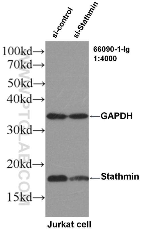
with si-Control and si-Stathmin transfected Jurkat cells.")
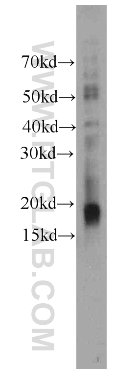
 at dilution of 1:1000 incubated at room temperature for 1.5 hours.")
 at dilution of 1:1000 incubated at room temperature for 1.5 hours.")
 at dilution of 1:1000 incubated at room temperature for 1.5 hours.")

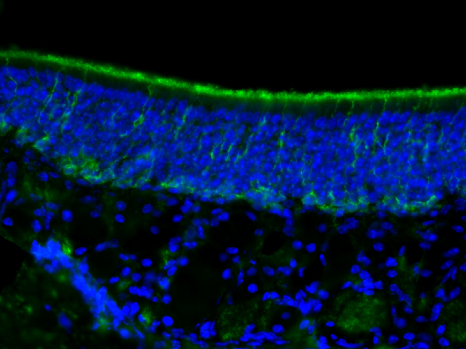
. Basal cell staining and dendritic processes as expected. By Dr. Brian Lin (Schwob Lab).")
Tested Applications
| Positive WB detected in | Jurkat cells, K-562 cells, human brain tissue |
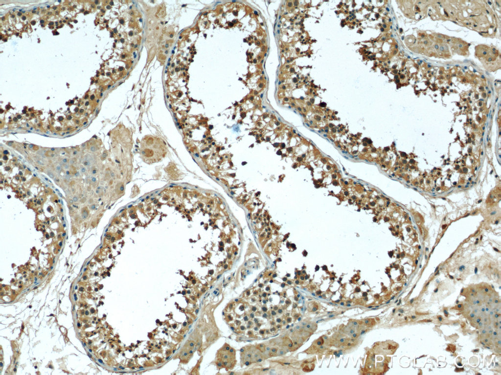
| Positive IHC detected in | human testis tissue Note: suggested antigen retrieval with TE buffer pH 9.0; (*) Alternatively, antigen retrieval may be performed with citrate buffer pH 6.0 |
Recommended dilution
| Application | Dilution |
|---|---|
| Western Blot (WB) | WB : 1:500-1:2000 |
| Immunohistochemistry (IHC) | IHC : 1:20-1:200 |
| It is recommended that this reagent should be titrated in each testing system to obtain optimal results. | |
| Sample-dependent, Check data in validation data gallery. | |
Product Information
66090-1-Ig targets Stathmin 1 in WB, IF, IHC, ELISA applications and shows reactivity with human, mouse samples.
| Tested Reactivity | human, mouse |
| Host / Isotype | Mouse / IgG1 |
| Class | Monoclonal |
| Type | Antibody |
| Immunogen | Stathmin 1 fusion protein Ag19102 Predict reactive species |
| Full Name | stathmin 1/oncoprotein 18 |
| Calculated Molecular Weight | 18 kDa |
| Observed Molecular Weight | 18 kDa |
| GenBank Accession Number | BC014353 |
| Gene Symbol | Stathmin 1 |
| Gene ID (NCBI) | 3925 |
| Conjugate | Unconjugated |
| Form | Liquid |
| Purification Method | Protein G purification |
| UNIPROT ID | P16949 |
| Storage Buffer | PBS with 0.02% sodium azide and 50% glycerol , pH 7.3 |
| Storage Conditions | Store at -20°C. Stable for one year after shipment. Aliquoting is unnecessary for -20oC storage. 20ul sizes contain 0.1% BSA. |
Background Information
Stathmin 1 (STMN1) normally regulates microtubule dynamics either by sequestering free tubulin heterodimers or by promoting microtubule catastrophe. STMN1 is highly expressed in fetal and adult brain, spinal cord, and cerebellum. Many different phosphorylated forms are observed depending on specific combinations among the sites which can be phosphorylated. Phosphorylation of stathmin is involved in response to NGF, neuron polarization and microtubule polymerization inhibition activity. Increased expression of STMN1 has been observed in a variety of human malignancies, such as colorectal primary tumors and metastatic tissues, but its association with melanoma is so far not well known.