HEK-293 cells were subjected to SDS PAGE followed by western blot with 68016-1-Ig (STK11/LKB1 antibody) at dilution of 1:4900 incubated at room temperature for 1.5 hours.
HEK-293 cells were subjected to SDS PAGE followed by western blot with 68016-1-Ig (STK11/LKB1 antibody) at dilution of 1:4900 incubated at room temperature for 1.5 hours.
WB analysis of NIH/3T3 using 68016-1-Ig
NIH/3T3 cells were subjected to SDS PAGE followed by western blot with 68016-1-Ig (STK11/LKB1 antibody) at dilution of 1:5000 incubated at room temperature for 1.5 hours.
NIH/3T3 cells were subjected to SDS PAGE followed by western blot with 68016-1-Ig (STK11/LKB1 antibody) at dilution of 1:5000 incubated at room temperature for 1.5 hours.
WB analysis of PC-3 using 68016-1-Ig
PC-3 cells were subjected to SDS PAGE followed by western blot with 68016-1-Ig (STK11/LKB1 antibody) at dilution of 1:5000 incubated at room temperature for 1.5 hours.
PC-3 cells were subjected to SDS PAGE followed by western blot with 68016-1-Ig (STK11/LKB1 antibody) at dilution of 1:5000 incubated at room temperature for 1.5 hours.
WB analysis of HSC-T6 using 68016-1-Ig
Calyculin A treated HSC-T6 cells were subjected to SDS PAGE followed by western blot with 68016-1-Ig (STK11/LKB1 antibody) at dilution of 1:5000 incubated at room temperature for 1.5 hours.
Calyculin A treated HSC-T6 cells were subjected to SDS PAGE followed by western blot with 68016-1-Ig (STK11/LKB1 antibody) at dilution of 1:5000 incubated at room temperature for 1.5 hours.
IHC staining of mouse testis using 68016-1-Ig
Immunohistochemical analysis of paraffin-embedded mouse testis tissue slide using 68016-1-Ig (STK11/LKB1 antibody) at dilution of 1:500 (under 10x lens). Heat mediated antigen retrieval with Tris-EDTA buffer (pH 9.0).
The Proteintech guarantee covers Proteintech antibodies in any species and any application, including those not listed on the datasheet. If the antibody doesn’t perform, you can receive a hassle-free refund or credit note.
mouse testis tissue Note: suggested antigen retrieval with TE buffer pH 9.0; (*) Alternatively, antigen retrieval may be performed with citrate buffer pH 6.0
Positive IF-P detected in
mouse colon tissue
Positive IF/ICC detected in
SKOV-3 cells
Recommended dilution
Application
Dilution
Western Blot (WB)
WB : 1:1000-1:9800
Immunohistochemistry (IHC)
IHC : 1:250-1:1000
Immunofluorescence (IF)-P
IF-P : 1:200-1:800
Immunofluorescence (IF)/ICC
IF/ICC : 1:250-1:1000
It is recommended that this reagent should be titrated in each testing system to obtain optimal results.
Sample-dependent, Check data in validation data gallery.
Product Information
68016-1-Ig targets STK11/LKB1 in WB, IHC, IF/ICC, IF-P, ELISA applications and shows reactivity with human, mouse, rat samples.
PBS with 0.02% sodium azide and 50% glycerol, pH 7.3.
Storage Conditions
Store at -20°C. Stable for one year after shipment. Aliquoting is unnecessary for -20oC storage. 20ul sizes contain 0.1% BSA.
Background Information
STK11(serine/threonine-protein kinase 11) is also named as LKB1, PJS, and belongs to the protein kinase superfamily. It controls the activity of AMP-activated protein kinase (AMPK) family members, thereby playing a role in various processes such as cell metabolism, cell polarity, apoptosis and DNA damage response. The tumour suppressor protein LKB1 is a serine/threonine kinase that has been causally linked to Peutz-Jeghers syndrome (PJS). Defects in STK11 are a cause of Peutz-Jeghers syndrome (PJS) and defects in STK11 have been associated with testicular germ cell tumor (TGCT) and some sporadic cancers, especially lung cancers. STK11 has 2 isoforms with MW of 49 kDa and 45 kDa, and can be detected as 50-54 kDa after posttranslational modification.
HEK-293 cells were subjected to SDS PAGE followed by western blot with 68016-1-Ig (STK11/LKB1 antibody) at dilution of 1:4900 incubated at room temperature for 1.5 hours.
WB analysis of NIH/3T3 using 68016-1-Ig
NIH/3T3 cells were subjected to SDS PAGE followed by western blot with 68016-1-Ig (STK11/LKB1 antibody) at dilution of 1:5000 incubated at room temperature for 1.5 hours.
WB analysis of PC-3 using 68016-1-Ig
PC-3 cells were subjected to SDS PAGE followed by western blot with 68016-1-Ig (STK11/LKB1 antibody) at dilution of 1:5000 incubated at room temperature for 1.5 hours.
WB analysis of HSC-T6 using 68016-1-Ig
Calyculin A treated HSC-T6 cells were subjected to SDS PAGE followed by western blot with 68016-1-Ig (STK11/LKB1 antibody) at dilution of 1:5000 incubated at room temperature for 1.5 hours.
IHC Figures
IHC staining of mouse testis using 68016-1-Ig
Immunohistochemical analysis of paraffin-embedded mouse testis tissue slide using 68016-1-Ig (STK11/LKB1 antibody) at dilution of 1:500 (under 10x lens). Heat mediated antigen retrieval with Tris-EDTA buffer (pH 9.0).
IHC staining of mouse testis using 68016-1-Ig
Immunohistochemical analysis of paraffin-embedded mouse testis tissue slide using 68016-1-Ig (STK11/LKB1 antibody) at dilution of 1:500 (under 40x lens). Heat mediated antigen retrieval with Tris-EDTA buffer (pH 9.0).
IF-P Figures
IF Staining of mouse colon using 68016-1-Ig
Immunofluorescent analysis of (4% PFA) fixed paraffin-embedded mouse colon tissue using STK11/LKB1 antibody (68016-1-Ig, Clone: 2D11E5 ) at dilution of 1:400 and CoraLite®488-Conjugated AffiniPure Goat Anti-Mouse IgG(H+L) (SA00013-1). Heat mediated antigen retrieval with Tris-EDTA buffer (pH 9.0).
IF/ICC Figures
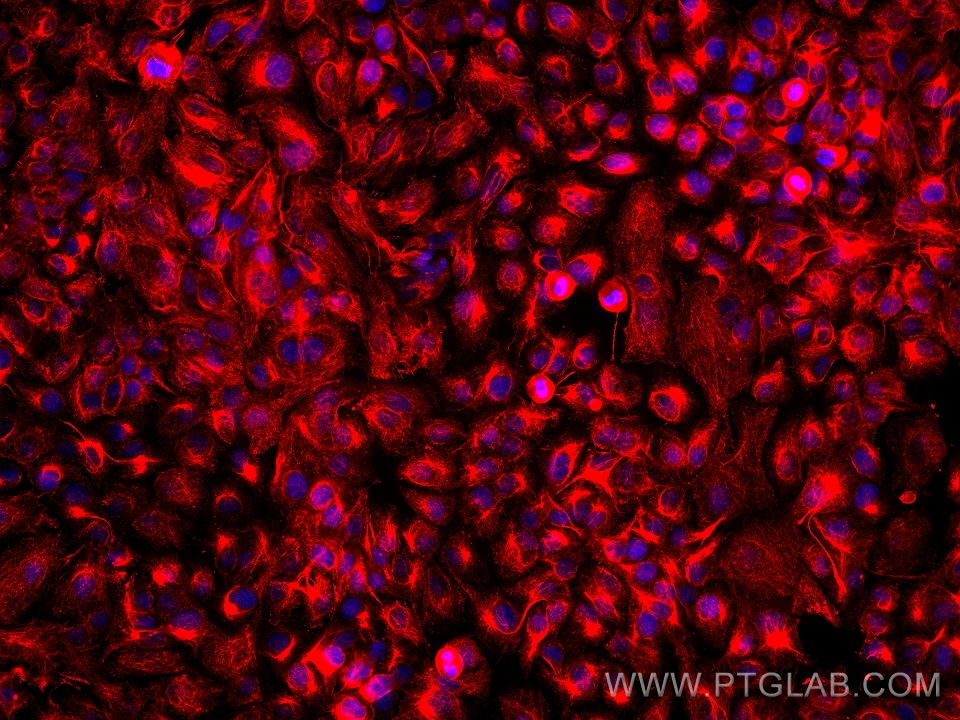
IF Staining of SKOV-3 using 68016-1-Ig
Immunofluorescent analysis of (4% PFA) fixed SKOV-3 cells using STK11/LKB1 antibody (68016-1-Ig, Clone: 2D11E5 ) at dilution of 1:500 and Multi-rAb CoraLite® Plus 594-Goat Anti-Mouse Recombinant Secondary Antibody (H+L) (Cat.NO. RGAM004 ).
The species listed in Tested Reactivity are in-house verified and applicable species. For unlisted species, please refer to the homology analysis of the immunogen sequence and related species. For rabbit polyclonal antibodies, homology >70% is recommended. For mouse monoclonal antibodies and rabbit recombinant antibodies, homology >90% is recommended. Generally, the higher the homology, the greater the applicability. However, there will be certain differences in protein expression in different species, tissues or cells. Therefore, the homology analysis results are for reference only and do not serve as a guarantee.
At Proteintech, we pride ourselves on our antibody quality, customer service and transparency. As such, we are comparing our antibodies with other vendors, enabling easy identification and comparisons of key data to help you choose the suitable antibody for your needs.
We have selected the top cited antibodies from these vendors for you to compare.
 at dilution of 1:4900 incubated at room temperature for 1.5 hours.")
 at dilution of 1:5000 incubated at room temperature for 1.5 hours.")
 at dilution of 1:5000 incubated at room temperature for 1.5 hours.")
 at dilution of 1:5000 incubated at room temperature for 1.5 hours.")
 at dilution of 1:500 (under 10x lens). Heat mediated antigen retrieval with Tris-EDTA buffer (pH 9.0).")
 at dilution of 1:500 (under 40x lens). Heat mediated antigen retrieval with Tris-EDTA buffer (pH 9.0).")
 fixed paraffin-embedded mouse colon tissue using STK11/LKB1 antibody (68016-1-Ig, Clone: 2D11E5 ) at dilution of 1:400 and CoraLite®488-Conjugated AffiniPure Goat Anti-Mouse IgG(H+L) (SA00013-1). Heat mediated antigen retrieval with Tris-EDTA buffer (pH 9.0).")
 fixed SKOV-3 cells using STK11/LKB1 antibody (68016-1-Ig, Clone: 2D11E5 ) at dilution of 1:500 and Multi-rAb CoraLite® Plus 594-Goat Anti-Mouse Recombinant Secondary Antibody (H+L) (Cat.NO. RGAM004 ).")