at dilution of 1:5000 incubated at room temperature for 1.5 hours.")
 at dilution of 1:600 incubated at room temperature for 1.5 hours.")
 at dilution of 1:600 incubated at room temperature for 1.5 hours.")
 at dilution of 1:600 incubated at room temperature for 1.5 hours.")
 at dilution of 1:600 incubated at room temperature for 1.5 hours.")
 with HepG2 cells lysate 4000ug.")
 at dilution of 1:200 (under 10x lens). Heat mediated antigen retrieval with Tris-EDTA buffer (pH 9.0).")
 at dilution of 1:200 (under 40x lens). Heat mediated antigen retrieval with Tris-EDTA buffer (pH 9.0).")
 at dilution of 1:50 (under 10x lens).")
 at dilution of 1:50 (under 40x lens).")
 at dilution of 1:50 (under 10x lens).")
 at dilution of 1:50 (under 40x lens).")
 at dilution of 1:200 (under 10x lens). Heat mediated antigen retrieval with Tris-EDTA buffer (pH 9.0).")
 at dilution of 1:200 (under 40x lens). Heat mediated antigen retrieval with Tris-EDTA buffer (pH 9.0).")
 fixed HepG2 cells using SNX6 antibody (10114-1-AP) at dilution of 1:200 and CoraLite®488-Conjugated AffiniPure Goat Anti-Rabbit IgG(H+L), CL594-Phalloidin (red).")
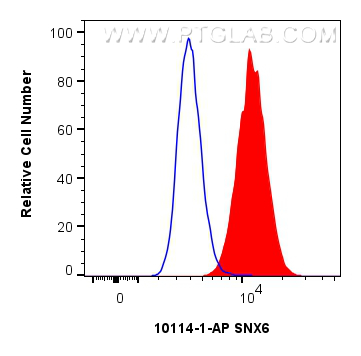
 and CoraLite®488-Conjugated Goat Anti-Rabbit IgG(H+L) (SA00013-2)(red), or 0.25 ug rabbit IgG isotype control (blue). Cells were fixed with 4% PFA and permeabilized with Flow Cytometry Perm Buffer.")
Tested Applications
| Positive WB detected in | HeLa cells, HepG2 cells, human placenta tissue, mouse brain tissue, RAW 264.7 cells |
| Positive IP detected in | HepG2 cells |
| Positive IHC detected in | mouse brain tissue, human prostate cancer tissue, human heart tissue Note: suggested antigen retrieval with TE buffer pH 9.0; (*) Alternatively, antigen retrieval may be performed with citrate buffer pH 6.0 |
| Positive IF/ICC detected in | HepG2 cells |
| Positive FC (Intra) detected in | RAW 264.7 cells |
Recommended dilution
| Application | Dilution |
|---|---|
| Western Blot (WB) | WB : 1:2000-1:10000 |
| Immunoprecipitation (IP) | IP : 0.5-4.0 ug for 1.0-3.0 mg of total protein lysate |
| Immunohistochemistry (IHC) | IHC : 1:50-1:500 |
| Immunofluorescence (IF)/ICC | IF/ICC : 1:50-1:500 |
| Flow Cytometry (FC) (INTRA) | FC (INTRA) : 0.25 ug per 10^6 cells in a 100 µl suspension |
| It is recommended that this reagent should be titrated in each testing system to obtain optimal results. | |
| Sample-dependent, Check data in validation data gallery. | |
Published Applications
| KD/KO | See 1 publications below |
| WB | See 3 publications below |
| IHC | See 1 publications below |
Product Information
10114-1-AP targets SNX6 in WB, IHC, IF/ICC, FC (Intra), IP, ELISA applications and shows reactivity with human, mouse, rat samples.
| Tested Reactivity | human, mouse, rat |
| Cited Reactivity | human |
| Host / Isotype | Rabbit / IgG |
| Class | Polyclonal |
| Type | Antibody |
| Immunogen |
CatNo: Ag0168 Product name: Recombinant human SNX6 protein Source: e coli.-derived, PGEX-4T Tag: GST Domain: 19-290 aa of BC001798 Sequence: AIFKKTVAMHEVFLCRVAAHPILRRDLNFHVFLEYNQDLSVRGKNKKEKLEDFFKNMVKSADGVIVSGVKDVDDFFEHERTFLLEYHNRVKDASAKSDRMTRSHKSAADDYNRIGSSLYALGTQDSTDICKFFLKVSELFDKTRKIEARVSADEDLKLSDLLKYYLRESQAAKDLLYRRSRSLVDYENANKALDKARAKNKDVLQAETSQQLCCQKFEKISESAKQELIDFKTRRVAAFRKNLVELAELELKHAKGNLQLLQNCLAVLNGDT Predict reactive species |
| Full Name | sorting nexin 6 |
| Calculated Molecular Weight | 34 kDa |
| Observed Molecular Weight | 47 kDa |
| GenBank Accession Number | BC001798 |
| Gene Symbol | SNX6 |
| Gene ID (NCBI) | 58533 |
| RRID | AB_2192720 |
| Conjugate | Unconjugated |
| Form | Liquid |
| Purification Method | Antigen affinity purification |
| UNIPROT ID | Q9UNH7 |
| Storage Buffer | PBS with 0.02% sodium azide and 50% glycerol, pH 7.3. |
| Storage Conditions | Store at -20°C. Stable for one year after shipment. Aliquoting is unnecessary for -20oC storage. 20ul sizes contain 0.1% BSA. |
Background Information
Sorting nexins (SNX) have previously been shown to regulate the cell-surface expression of the human epidermal growth factor receptor. On the basis of the predicted protein sequences, several members of this family, including SNX6, have been identified. SNX6, containing coiled-coil regions within its large C-terminal domain and is found distributed in both membrane and cytosolic fractions, typical of hydrophilic peripheral membrane proteins. The functions of SNX6 are likely to be unique to endosomes, mediated in part by interactions with SNX6-specific C-terminal sequences and membrane-associated determinants
Protocols
| Product Specific Protocols | |
|---|---|
| IHC protocol for SNX6 antibody 10114-1-AP | Download protocol |
| IP protocol for SNX6 antibody 10114-1-AP | Download protocol |
| WB protocol for SNX6 antibody 10114-1-AP | Download protocol |
| FC protocol for SNX6 antibody 10114-1-AP | Download protocol |
| IF protocol for SNX6 antibody 10114-1-AP | Download protocol |
| Standard Protocols | |
|---|---|
| Click here to view our Standard Protocols |
Publications
| Species | Application | Title |
|---|---|---|
Nat Cell Biol Recycling of autophagosomal components from autolysosomes by the recycler complex. | ||
Acta Biochim Biophys Sin (Shanghai) SNX6 predicts poor prognosis and contributes to the metastasis of pancreatic cancer cells via activating epithelial-mesenchymal transition.
| ||
Front Immunol Zika virus modulates mitochondrial dynamics, mitophagy, and mitochondria-derived vesicles to facilitate viral replication in trophoblast cells |