Various lysates were subjected to SDS PAGE followed by western blot with 25402-1-AP (SLC25A22 antibody) at dilution of 1:3000 incubated at room temperature for 1.5 hours.
Various lysates were subjected to SDS PAGE followed by western blot with 25402-1-AP (SLC25A22 antibody) at dilution of 1:3000 incubated at room temperature for 1.5 hours.
WB analysis using 25402-1-AP
Various lysates were subjected to SDS PAGE followed by western blot with 25402-1-AP (SLC25A22 antibody) at dilution of 1:5000 incubated at room temperature for 1.5 hours.
Various lysates were subjected to SDS PAGE followed by western blot with 25402-1-AP (SLC25A22 antibody) at dilution of 1:5000 incubated at room temperature for 1.5 hours.
WB analysis using 25402-1-AP
Various lysates were subjected to SDS PAGE followed by western blot with 25402-1-AP (SLC25A22 antibody) at dilution of 1:3000 incubated at room temperature for 1.5 hours.
Various lysates were subjected to SDS PAGE followed by western blot with 25402-1-AP (SLC25A22 antibody) at dilution of 1:3000 incubated at room temperature for 1.5 hours.
IHC staining of mouse brain using 25402-1-AP
Immunohistochemical analysis of paraffin-embedded mouse brain tissue slide using 25402-1-AP (SLC25A22 antibody) at dilution of 1:200 (under 10x lens). Heat mediated antigen retrieval with Tris-EDTA buffer (pH 9.0).
Immunohistochemical analysis of paraffin-embedded mouse brain tissue slide using 25402-1-AP (SLC25A22 antibody) at dilution of 1:200 (under 40x lens). Heat mediated antigen retrieval with Tris-EDTA buffer (pH 9.0).
IHC staining of human liver cancer using 25402-1-AP
Immunohistochemical analysis of paraffin-embedded human liver cancer tissue slide using 25402-1-AP (SLC25A22 antibody) at dilution of 1:200 (under 40x lens). Heat mediated antigen retrieval with Tris-EDTA buffer (pH 9.0).
Immunohistochemical analysis of paraffin-embedded human liver cancer tissue slide using 25402-1-AP (SLC25A22 antibody) at dilution of 1:200 (under 40x lens). Heat mediated antigen retrieval with Tris-EDTA buffer (pH 9.0).
IHC staining of human colon cancer using 25402-1-AP
Immunohistochemical analysis of paraffin-embedded human colon cancer tissue slide using 25402-1-AP (SLC25A22 antibody) at dilution of 1:200 (under 10x lens). Heat mediated antigen retrieval with Tris-EDTA buffer (pH 9.0).
Immunohistochemical analysis of paraffin-embedded human colon cancer tissue slide using 25402-1-AP (SLC25A22 antibody) at dilution of 1:200 (under 10x lens). Heat mediated antigen retrieval with Tris-EDTA buffer (pH 9.0).
IHC staining of human colon cancer using 25402-1-AP
Immunohistochemical analysis of paraffin-embedded human colon cancer tissue slide using 25402-1-AP (SLC25A22 antibody) at dilution of 1:200 (under 40x lens). Heat mediated antigen retrieval with Tris-EDTA buffer (pH 9.0).
Immunohistochemical analysis of paraffin-embedded human colon cancer tissue slide using 25402-1-AP (SLC25A22 antibody) at dilution of 1:200 (under 40x lens). Heat mediated antigen retrieval with Tris-EDTA buffer (pH 9.0).
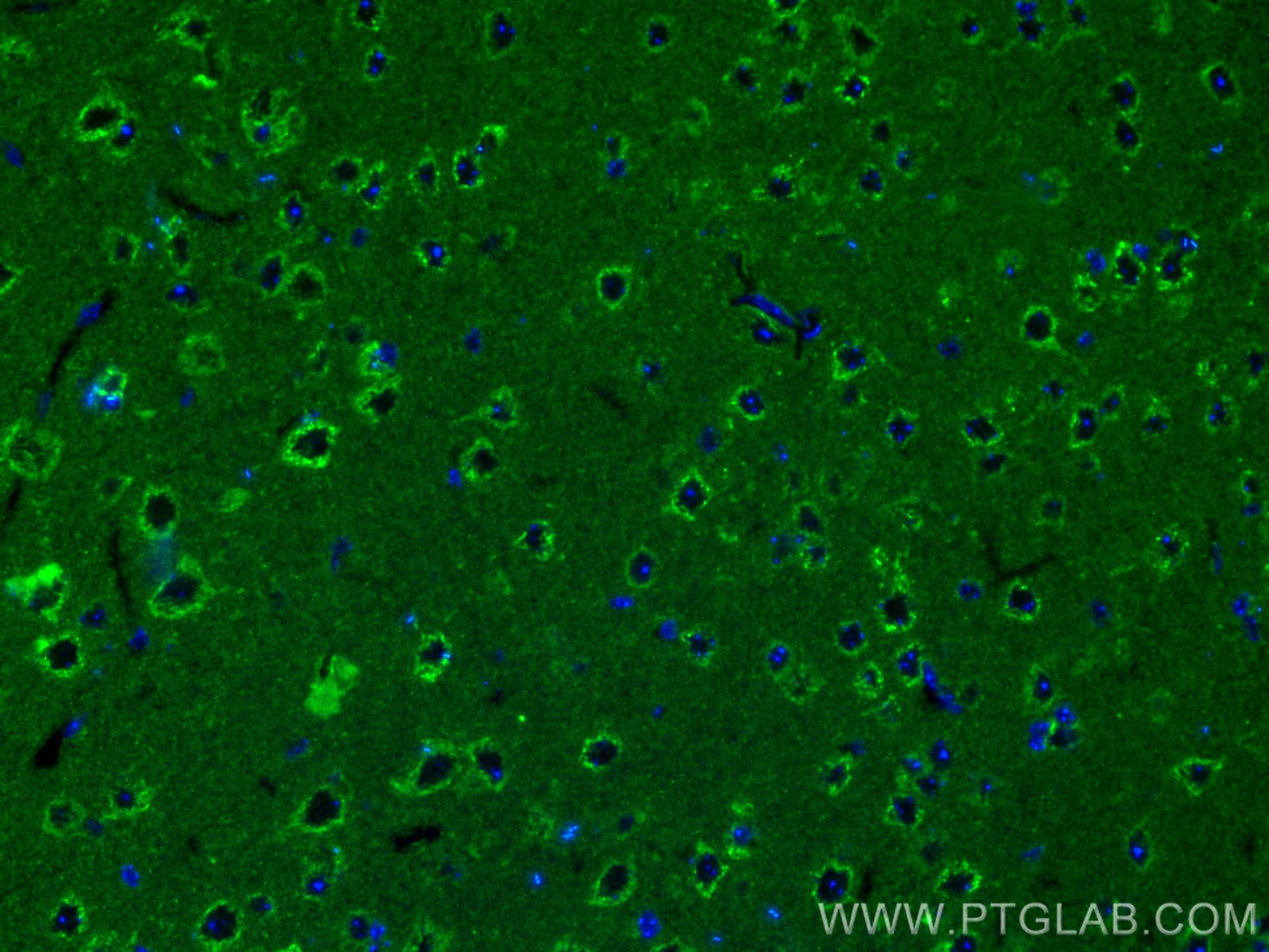
IF Staining of mouse brain using 25402-1-AP
Immunofluorescent analysis of (4% PFA) fixed mouse brain tissue using SLC25A22 antibody (25402-1-AP) at dilution of 1:200 and CoraLite®488-Conjugated Goat Anti-Rabbit IgG(H+L).
Immunofluorescent analysis of (4% PFA) fixed mouse brain tissue using SLC25A22 antibody (25402-1-AP) at dilution of 1:200 and CoraLite®488-Conjugated Goat Anti-Rabbit IgG(H+L).
IF Staining of mouse brain using 25402-1-AP
Immunofluorescent analysis of (4% PFA) fixed mouse brain tissue using 25402-1-AP (SLC25A22 antibody) at dilution of 1:50 and CoraLite488-Conjugated Goat Anti-Rabbit IgG(H+L).
Immunofluorescent analysis of (4% PFA) fixed mouse brain tissue using 25402-1-AP (SLC25A22 antibody) at dilution of 1:50 and CoraLite488-Conjugated Goat Anti-Rabbit IgG(H+L).
IF Staining of COS-7 using 25402-1-AP
Immunofluorescent analysis of (4% PFA) fixed COS-7 cells using 25402-1-AP (SLC25A22 antibody), at dilution of 1:200 and CoraLite®488-Conjugated Goat Anti-Rabbit IgG(H+L).
Immunofluorescent analysis of (4% PFA) fixed COS-7 cells using 25402-1-AP (SLC25A22 antibody), at dilution of 1:200 and CoraLite®488-Conjugated Goat Anti-Rabbit IgG(H+L).
The Proteintech guarantee covers Proteintech antibodies in any species and any application, including those not listed on the datasheet. If the antibody doesn’t perform, you can receive a hassle-free refund or credit note.
mouse brain tissue, human colon cancer tissue, human liver cancer tissue Note: suggested antigen retrieval with TE buffer pH 9.0; (*) Alternatively, antigen retrieval may be performed with citrate buffer pH 6.0
Positive IF-P detected in
mouse brain tissue
Positive IF/ICC detected in
COS-7 cells
Recommended dilution
Application
Dilution
Western Blot (WB)
WB : 1:1000-1:6000
Immunohistochemistry (IHC)
IHC : 1:50-1:500
Immunofluorescence (IF)-P
IF-P : 1:50-1:500
Immunofluorescence (IF)/ICC
IF/ICC : 1:50-1:500
It is recommended that this reagent should be titrated in each testing system to obtain optimal results.
Sample-dependent, Check data in validation data gallery.
PBS with 0.02% sodium azide and 50% glycerol , pH 7.3
Storage Conditions
Store at -20°C. Stable for one year after shipment. Aliquoting is unnecessary for -20oC storage. 20ul sizes contain 0.1% BSA.
Background Information
SLC25A22 encodes a mitochondrial glutamate/H+ symporter with highly expression in brain, especially in astrocytes, where it controls glutamate uptake. The mutations of SLC25A22 are associated with the early/infantile epileptic encephalopathies. It has been shown that SLC25A22 plays a key role in promoting proliferation and migration in cancer, such as human colorectal cancer (CRC) and Gallbladder cancer (GBC). And there is a result that the expression of SLC25A22 in GBC was significantly higher than that in adjacent tissues. 25402-1-AP antibody is specific to SLC25A22.(PMID: 30814911, 25033742, 24596948)
Various lysates were subjected to SDS PAGE followed by western blot with 25402-1-AP (SLC25A22 antibody) at dilution of 1:3000 incubated at room temperature for 1.5 hours.
WB analysis using 25402-1-AP
Various lysates were subjected to SDS PAGE followed by western blot with 25402-1-AP (SLC25A22 antibody) at dilution of 1:5000 incubated at room temperature for 1.5 hours.
WB analysis using 25402-1-AP
Various lysates were subjected to SDS PAGE followed by western blot with 25402-1-AP (SLC25A22 antibody) at dilution of 1:3000 incubated at room temperature for 1.5 hours.
IHC Figures
IHC staining of mouse brain using 25402-1-AP
Immunohistochemical analysis of paraffin-embedded mouse brain tissue slide using 25402-1-AP (SLC25A22 antibody) at dilution of 1:200 (under 10x lens). Heat mediated antigen retrieval with Tris-EDTA buffer (pH 9.0).
IHC staining of mouse brain using 25402-1-AP
Immunohistochemical analysis of paraffin-embedded mouse brain tissue slide using 25402-1-AP (SLC25A22 antibody) at dilution of 1:200 (under 40x lens). Heat mediated antigen retrieval with Tris-EDTA buffer (pH 9.0).
IHC staining of human liver cancer using 25402-1-AP
Immunohistochemical analysis of paraffin-embedded human liver cancer tissue slide using 25402-1-AP (SLC25A22 antibody) at dilution of 1:200 (under 40x lens). Heat mediated antigen retrieval with Tris-EDTA buffer (pH 9.0).
IHC staining of human liver cancer using 25402-1-AP
Immunohistochemical analysis of paraffin-embedded human liver cancer tissue slide using 25402-1-AP (SLC25A22 antibody) at dilution of 1:200 (under 40x lens). Heat mediated antigen retrieval with Tris-EDTA buffer (pH 9.0).
IHC staining of human colon cancer using 25402-1-AP
Immunohistochemical analysis of paraffin-embedded human colon cancer tissue slide using 25402-1-AP (SLC25A22 antibody) at dilution of 1:200 (under 10x lens). Heat mediated antigen retrieval with Tris-EDTA buffer (pH 9.0).
IHC staining of human colon cancer using 25402-1-AP
Immunohistochemical analysis of paraffin-embedded human colon cancer tissue slide using 25402-1-AP (SLC25A22 antibody) at dilution of 1:200 (under 40x lens). Heat mediated antigen retrieval with Tris-EDTA buffer (pH 9.0).
IF-P Figures
IF Staining of mouse brain using 25402-1-AP
Immunofluorescent analysis of (4% PFA) fixed mouse brain tissue using SLC25A22 antibody (25402-1-AP) at dilution of 1:200 and CoraLite®488-Conjugated Goat Anti-Rabbit IgG(H+L).
IF Staining of mouse brain using 25402-1-AP
Immunofluorescent analysis of (4% PFA) fixed mouse brain tissue using 25402-1-AP (SLC25A22 antibody) at dilution of 1:50 and CoraLite488-Conjugated Goat Anti-Rabbit IgG(H+L).
IF/ICC Figures
IF Staining of COS-7 using 25402-1-AP
Immunofluorescent analysis of (4% PFA) fixed COS-7 cells using 25402-1-AP (SLC25A22 antibody), at dilution of 1:200 and CoraLite®488-Conjugated Goat Anti-Rabbit IgG(H+L).
The species listed in Tested Reactivity are in-house verified and applicable species. For unlisted species, please refer to the homology analysis of the immunogen sequence and related species. For rabbit polyclonal antibodies, homology >70% is recommended. For mouse monoclonal antibodies and rabbit recombinant antibodies, homology >90% is recommended. Generally, the higher the homology, the greater the applicability. However, there will be certain differences in protein expression in different species, tissues or cells. Therefore, the homology analysis results are for reference only and do not serve as a guarantee.
At Proteintech, we pride ourselves on our antibody quality, customer service and transparency. As such, we are comparing our antibodies with other vendors, enabling easy identification and comparisons of key data to help you choose the suitable antibody for your needs.
We have selected the top cited antibodies from these vendors for you to compare.
 at dilution of 1:3000 incubated at room temperature for 1.5 hours.")
 at dilution of 1:5000 incubated at room temperature for 1.5 hours.")
 at dilution of 1:3000 incubated at room temperature for 1.5 hours.")
 at dilution of 1:200 (under 10x lens). Heat mediated antigen retrieval with Tris-EDTA buffer (pH 9.0).")
 at dilution of 1:200 (under 40x lens). Heat mediated antigen retrieval with Tris-EDTA buffer (pH 9.0).")
 at dilution of 1:200 (under 40x lens). Heat mediated antigen retrieval with Tris-EDTA buffer (pH 9.0).")
 at dilution of 1:200 (under 40x lens). Heat mediated antigen retrieval with Tris-EDTA buffer (pH 9.0).")
 at dilution of 1:200 (under 10x lens). Heat mediated antigen retrieval with Tris-EDTA buffer (pH 9.0).")
 at dilution of 1:200 (under 40x lens). Heat mediated antigen retrieval with Tris-EDTA buffer (pH 9.0).")
 fixed mouse brain tissue using SLC25A22 antibody (25402-1-AP) at dilution of 1:200 and CoraLite®488-Conjugated Goat Anti-Rabbit IgG(H+L).")
 fixed mouse brain tissue using 25402-1-AP (SLC25A22 antibody) at dilution of 1:50 and CoraLite488-Conjugated Goat Anti-Rabbit IgG(H+L).")
 fixed COS-7 cells using 25402-1-AP (SLC25A22 antibody), at dilution of 1:200 and CoraLite®488-Conjugated Goat Anti-Rabbit IgG(H+L).")