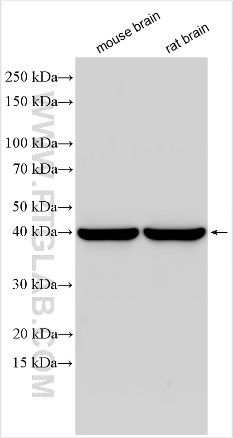
Various lysates were subjected to SDS PAGE followed by western blot with 30146-1-AP (SEPT3 antibody) at dilution of 1:3000 incubated at room temperature for 1.5 hours.
Various lysates were subjected to SDS PAGE followed by western blot with 30146-1-AP (SEPT3 antibody) at dilution of 1:3000 incubated at room temperature for 1.5 hours.
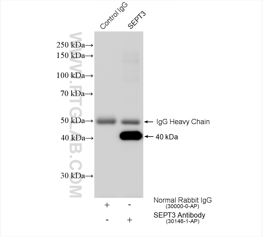
IP experiment of mouse brain using 30146-1-AP
IP result of anti-SEPT3 (IP:30146-1-AP, 4ug; Detection:30146-1-AP 1:2000) with mouse brain tissue lysate 1280 ug.
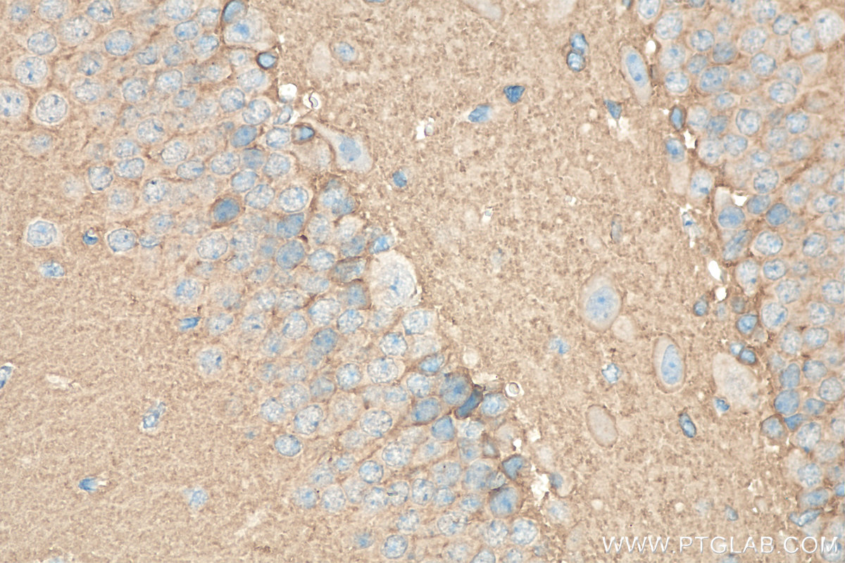
Immunohistochemical analysis of paraffin-embedded mouse brain tissue slide using 30146-1-AP (SEPT3 antibody) at dilution of 1:200 (under 40x lens). Heat mediated antigen retrieval with Tris-EDTA buffer (pH 9.0).
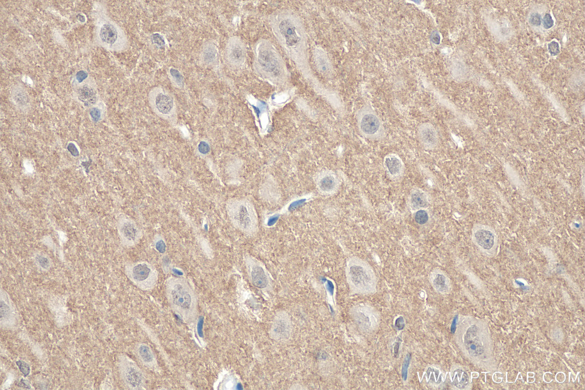
IHC staining of rat brain using 30146-1-AP
Immunohistochemical analysis of paraffin-embedded rat brain tissue slide using 30146-1-AP (SEPT3 antibody) at dilution of 1:200 (under 40x lens). Heat mediated antigen retrieval with Tris-EDTA buffer (pH 9.0).
Immunohistochemical analysis of paraffin-embedded rat brain tissue slide using 30146-1-AP (SEPT3 antibody) at dilution of 1:200 (under 40x lens). Heat mediated antigen retrieval with Tris-EDTA buffer (pH 9.0).
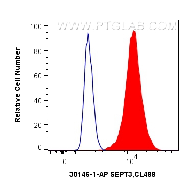
FC experiment of HEK-293 using 30146-1-AP
1X10^6 HEK-293 cells were intracellularly stained with 0.4 ug Anti-Human SEPT3 (30146-1-AP) and CoraLite®488-Conjugated Goat Anti-Rabbit IgG(H+L) at dilution 1:1000 (red), or 0.4 ug Control Antibody. Cells were fixed with 4% PFA and permeabilized with Flow Cytometry Perm Buffer (PF00011-C).
1X10^6 HEK-293 cells were intracellularly stained with 0.4 ug Anti-Human SEPT3 (30146-1-AP) and CoraLite®488-Conjugated Goat Anti-Rabbit IgG(H+L) at dilution 1:1000 (red), or 0.4 ug Control Antibody. Cells were fixed with 4% PFA and permeabilized with Flow Cytometry Perm Buffer (PF00011-C).
The Proteintech guarantee covers Proteintech antibodies in any species and any application, including those not listed on the datasheet. If the antibody doesn’t perform, you can receive a hassle-free refund or credit note.
mouse brain tissue, rat brain tissue Note: suggested antigen retrieval with TE buffer pH 9.0; (*) Alternatively, antigen retrieval may be performed with citrate buffer pH 6.0
Positive FC (Intra) detected in
HEK-293 cells
Recommended dilution
Application
Dilution
Western Blot (WB)
WB : 1:1000-1:6000
Immunoprecipitation (IP)
IP : 0.5-4.0 ug for 1.0-3.0 mg of total protein lysate
Immunohistochemistry (IHC)
IHC : 1:50-1:500
Flow Cytometry (FC) (INTRA)
FC (INTRA) : 0.40 ug per 10^6 cells in a 100 µl suspension
It is recommended that this reagent should be titrated in each testing system to obtain optimal results.
Sample-dependent, Check data in validation data gallery.
Product Information
30146-1-AP targets SEPT3 in WB, IHC, FC (Intra), IP, ELISA applications and shows reactivity with human, mouse, rat samples.
PBS with 0.02% sodium azide and 50% glycerol , pH 7.3
Storage Conditions
Store at -20°C. Stable for one year after shipment. Aliquoting is unnecessary for -20oC storage. 20ul sizes contain 0.1% BSA.
Background Information
SEPT3 is a developmentally regulated phosphoprotein. SEPT3 is a member of the septin GTPase family, which can form polymers and contribute to the cytoskeleton. SEPT3 is involved in neuronal autophagy. In the process of autophagy regulation, the level of SEPT3 will change according to the autophagy protein. SEPT3 is specifically expressed in the brain (PMID: 15485489; 35932293).
Various lysates were subjected to SDS PAGE followed by western blot with 30146-1-AP (SEPT3 antibody) at dilution of 1:3000 incubated at room temperature for 1.5 hours.
IHC Figures
IHC staining of mouse brain using 30146-1-AP
Immunohistochemical analysis of paraffin-embedded mouse brain tissue slide using 30146-1-AP (SEPT3 antibody) at dilution of 1:200 (under 40x lens). Heat mediated antigen retrieval with Tris-EDTA buffer (pH 9.0).
IHC staining of rat brain using 30146-1-AP
Immunohistochemical analysis of paraffin-embedded rat brain tissue slide using 30146-1-AP (SEPT3 antibody) at dilution of 1:200 (under 40x lens). Heat mediated antigen retrieval with Tris-EDTA buffer (pH 9.0).
IP Figures
IP experiment of mouse brain using 30146-1-AP
IP result of anti-SEPT3 (IP:30146-1-AP, 4ug; Detection:30146-1-AP 1:2000) with mouse brain tissue lysate 1280 ug.
FC (INTRA) Figures
FC experiment of HEK-293 using 30146-1-AP
1X10^6 HEK-293 cells were intracellularly stained with 0.4 ug Anti-Human SEPT3 (30146-1-AP) and CoraLite®488-Conjugated Goat Anti-Rabbit IgG(H+L) at dilution 1:1000 (red), or 0.4 ug Control Antibody. Cells were fixed with 4% PFA and permeabilized with Flow Cytometry Perm Buffer (PF00011-C).
The species listed in Tested Reactivity are in-house verified and applicable species. For unlisted species, please refer to the homology analysis of the immunogen sequence and related species. For rabbit polyclonal antibodies, homology >70% is recommended. For mouse monoclonal antibodies and rabbit recombinant antibodies, homology >90% is recommended. Generally, the higher the homology, the greater the applicability. However, there will be certain differences in protein expression in different species, tissues or cells. Therefore, the homology analysis results are for reference only and do not serve as a guarantee.
At Proteintech, we pride ourselves on our antibody quality, customer service and transparency. As such, we are comparing our antibodies with other vendors, enabling easy identification and comparisons of key data to help you choose the suitable antibody for your needs.
We have selected the top cited antibodies from these vendors for you to compare.
Proteintech
SEPT3 Polyclonal antibody
Catalog Number
30146-1-AP
Citations
-
Dilutions
WB : 1:1000-1:6000 IP : 0.5-4.0 ug for IP and 0.5-4.0 ug for 1.0-3.0 mg of total protein lysate for WB IHC : 1:50-1:500 FC (INTRA) : 0.40 ug per 10^6 cells in a 100 µl suspension
Applications
WB, IHC, FC (Intra), IP, ELISA
Reactivity
human, mouse, rat
Product Guarantee
Covers any species including not listed on datasheet
Covers any applications including not listed on datasheet
 at dilution of 1:3000 incubated at room temperature for 1.5 hours.")
 with mouse brain tissue lysate 1280 ug.")
 at dilution of 1:200 (under 40x lens). Heat mediated antigen retrieval with Tris-EDTA buffer (pH 9.0).")
 at dilution of 1:200 (under 40x lens). Heat mediated antigen retrieval with Tris-EDTA buffer (pH 9.0).")
 and CoraLite®488-Conjugated Goat Anti-Rabbit IgG(H+L) at dilution 1:1000 (red), or 0.4 ug Control Antibody. Cells were fixed with 4% PFA and permeabilized with Flow Cytometry Perm Buffer (PF00011-C).")