Filter:
 at dilution of 1:300 incubated at room temperature for 1.5 hours.")
 at dilution of 1:5000 incubated at room temperature for 1.5 hours.")
 with HeLa cells lysate 840 ug.")
 at dilution of 1:1000 (under 40x lens). Heat mediated antigen retrieval with Tris-EDTA buffer (pH 9.0).")
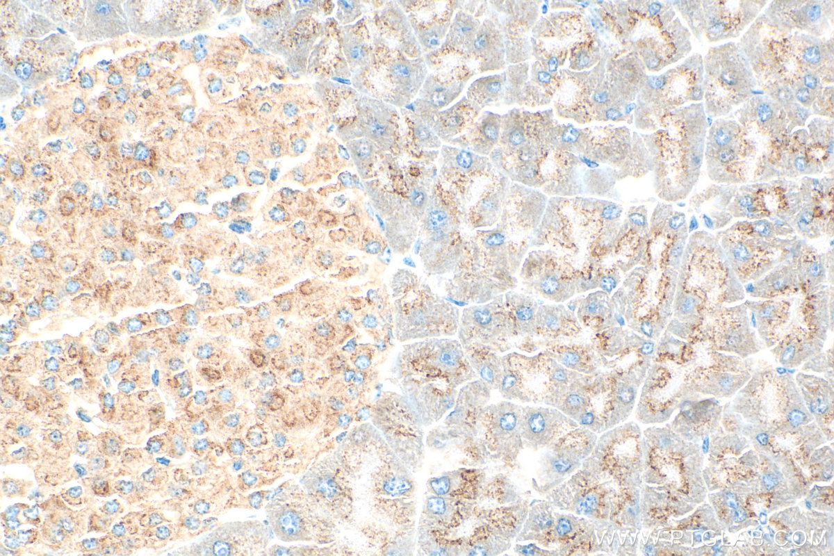
 at dilution of 1:200 (under 10x lens). Heat mediated antigen retrieval with Tris-EDTA buffer (pH 9.0).")
 at dilution of 1:200 (under 40x lens). Heat mediated antigen retrieval with Tris-EDTA buffer (pH 9.0).")
 fixed A431 cells using SEC16A antibody (20025-1-AP) at dilution of 1:200 and CoraLite®488-Conjugated AffiniPure Goat Anti-Rabbit IgG(H+L).")
 and CoraLite®488-Conjugated AffiniPure Goat Anti-Rabbit IgG(H+L) at dilution 1:1000 (red), or 0.4 ug Control Antibody. Cells were fixed with 4% PFA and permeabilized with Flow Cytometry Perm Buffer (PF00011-C).")
Tested Applications
| Positive WB detected in | HEK-293 cells, HeLa cells |
| Positive IP detected in | HeLa cells |
| Positive IHC detected in | mouse pancreas tissue Note: suggested antigen retrieval with TE buffer pH 9.0; (*) Alternatively, antigen retrieval may be performed with citrate buffer pH 6.0 |
| Positive IF/ICC detected in | A431 cells |
| Positive FC (Intra) detected in | HEK-293 cells |
Recommended dilution
| Application | Dilution |
|---|---|
| Western Blot (WB) | WB : 1:2000-1:10000 |
| Immunoprecipitation (IP) | IP : 0.5-4.0 ug for 1.0-3.0 mg of total protein lysate |
| Immunohistochemistry (IHC) | IHC : 1:200-1:1000 |
| Immunofluorescence (IF)/ICC | IF/ICC : 1:50-1:500 |
| Flow Cytometry (FC) (INTRA) | FC (INTRA) : 0.40 ug per 10^6 cells in a 100 µl suspension |
| It is recommended that this reagent should be titrated in each testing system to obtain optimal results. | |
| Sample-dependent, Check data in validation data gallery. | |
Published Applications
| KD/KO | See 1 publications below |
| WB | See 2 publications below |
| IF | See 2 publications below |
Product Information
20025-1-AP targets SEC16A in WB, IHC, IF/ICC, FC (Intra), IP, ELISA applications and shows reactivity with human, mouse, rat samples.
| Tested Reactivity | human, mouse, rat |
| Cited Reactivity | human, mouse, rat |
| Host / Isotype | Rabbit / IgG |
| Class | Polyclonal |
| Type | Antibody |
| Immunogen | Peptide Predict reactive species |
| Full Name | SEC16 homolog A (S. cerevisiae) |
| Calculated Molecular Weight | 234 kDa |
| Observed Molecular Weight | 250-300 kDa |
| GenBank Accession Number | NM_014866 |
| Gene Symbol | SEC16A |
| Gene ID (NCBI) | 9919 |
| RRID | AB_2878635 |
| Conjugate | Unconjugated |
| Form | Liquid |
| Purification Method | Antigen affinity purification |
| UNIPROT ID | O15027 |
| Storage Buffer | PBS with 0.02% sodium azide and 50% glycerol , pH 7.3 |
| Storage Conditions | Store at -20°C. Stable for one year after shipment. Aliquoting is unnecessary for -20oC storage. 20ul sizes contain 0.1% BSA. |
Background Information
SEC16A, also named as KIAA0310, SEC16 and SEC16L, is required for secretory cargo trafic from the endoplasmic reticulum to the Golgi apparatus. SAR1A-GTP-dependent assembly of SEC16A on the ER membrane forms an organized scaffold defining an ERES. SEC16A is required for normal transitional endoplasmic reticulum (tER) organization.
Protocols
| Product Specific Protocols | |
|---|---|
| WB protocol for SEC16A antibody 20025-1-AP | Download protocol |
| IHC protocol for SEC16A antibody 20025-1-AP | Download protocol |
| IF protocol for SEC16A antibody 20025-1-AP | Download protocol |
| IP protocol for SEC16A antibody 20025-1-AP | Download protocol |
| Standard Protocols | |
|---|---|
| Click here to view our Standard Protocols |
Publications
| Species | Application | Title |
|---|---|---|
Nat Chem Biol Functional dissection of the retrograde Shiga toxin trafficking inhibitor Retro-2.
| ||
Cell Death Differ C9orf72 controls hepatic lipid metabolism by regulating SREBP1 transport | ||
Mol Neurodegener Pathological characteristics of axons and alterations of proteomic and lipidomic profiles in midbrain dopaminergic neurodegeneration induced by WDR45-deficiency |