rat retina tissue were subjected to SDS PAGE followed by western blot with 24430-1-AP (RS1 antibody) at dilution of 1:5000 incubated at room temperature for 1.5 hours.
rat retina tissue were subjected to SDS PAGE followed by western blot with 24430-1-AP (RS1 antibody) at dilution of 1:5000 incubated at room temperature for 1.5 hours.
WB analysis of mouse eye using 24430-1-AP
mouse eye tissue were subjected to SDS PAGE followed by western blot with 24430-1-AP (RS1 Antibody) at dilution of 1:600 incubated at room temperature for 1.5 hours.
mouse eye tissue were subjected to SDS PAGE followed by western blot with 24430-1-AP (RS1 Antibody) at dilution of 1:600 incubated at room temperature for 1.5 hours.
IHC staining of mouse eye using 24430-1-AP
Immunohistochemical analysis of paraffin-embedded mouse eye tissue slide using 24430-1-AP (RS1 antibody) at dilution of 1:200 (under 40x lens). Heat mediated antigen retrieval with Tris-EDTA buffer (pH 9.0).
Immunohistochemical analysis of paraffin-embedded mouse eye tissue slide using 24430-1-AP (RS1 antibody) at dilution of 1:200 (under 4x lens). Heat mediated antigen retrieval with Tris-EDTA buffer (pH 9.0).
IF Staining of mouse eye using 24430-1-AP
Immunofluorescent analysis of (4% PFA) fixed mouse eye tissue using RS1 antibody (24430-1-AP) at dilution of 1:200 and CoraLite®488-Conjugated AffiniPure Goat Anti-Rabbit IgG(H+L).
Immunofluorescent analysis of (4% PFA) fixed mouse eye tissue using RS1 antibody (24430-1-AP) at dilution of 1:200 and CoraLite®488-Conjugated AffiniPure Goat Anti-Rabbit IgG(H+L).
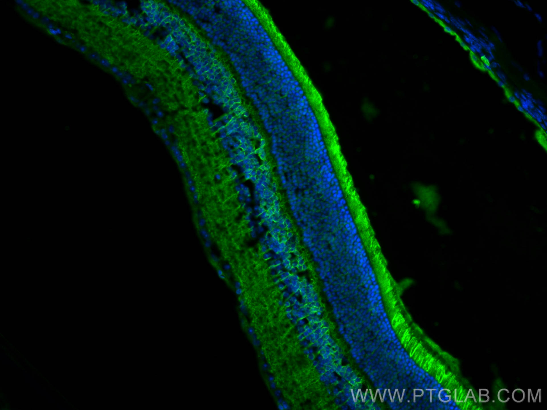
IF Staining of mouse eye using 24430-1-AP
Immunofluorescent analysis of (4% PFA) fixed frozen OCT-embedded mouse eye tissue using 24430-1-AP (RS1 antibody) at dilution of 1:200 and CoraLite488-Conjugated AffiniPure Goat Anti-Rabbit IgG(H+L).
Immunofluorescent analysis of (4% PFA) fixed frozen OCT-embedded mouse eye tissue using 24430-1-AP (RS1 antibody) at dilution of 1:200 and CoraLite488-Conjugated AffiniPure Goat Anti-Rabbit IgG(H+L).
The Proteintech guarantee covers Proteintech antibodies in any species and any application, including those not listed on the datasheet. If the antibody doesn’t perform, you can receive a hassle-free refund or credit note.
mouse eye tissue Note: suggested antigen retrieval with TE buffer pH 9.0; (*) Alternatively, antigen retrieval may be performed with citrate buffer pH 6.0
Positive IF-P detected in
mouse eye tissue
Positive IF-Fro detected in
mouse eye tissue
Recommended dilution
Application
Dilution
Western Blot (WB)
WB : 1:2000-1:10000
Immunohistochemistry (IHC)
IHC : 1:50-1:500
Immunofluorescence (IF)-P
IF-P : 1:50-1:500
Immunofluorescence (IF)-FRO
IF-FRO : 1:50-1:500
It is recommended that this reagent should be titrated in each testing system to obtain optimal results.
Sample-dependent, Check data in validation data gallery.
PBS with 0.02% sodium azide and 50% glycerol , pH 7.3
Storage Conditions
Store at -20°C. Stable for one year after shipment. Aliquoting is unnecessary for -20oC storage. 20ul sizes contain 0.1% BSA.
Background Information
RS1 is also named as XLRS1. RS1 can bind negatively charged membrane lipids, such as phosphatidylserine and phosphoinositides. RS1 is required for normal structure and function of the retina (PMID:19093009). It may play a role in cell-cell adhesion processes in the retina (PMID:27114531). RS1 is high expression in the retina (at protein level) (PMID:10915776, PMID:9326935). It is detected in the inner segment of the photoreceptors, the inner nuclear layer, the inner plexiform layer and the ganglion cell layer. At the macula, it is expressed in both the outer and inner nuclear layers and in the inner plexiform layer (PMID:10915776, PMID:10915776). And RS1 is undetectable in the inner plexiform layers and the inner nuclear layer (PMID:10915776)
rat retina tissue were subjected to SDS PAGE followed by western blot with 24430-1-AP (RS1 antibody) at dilution of 1:5000 incubated at room temperature for 1.5 hours.
WB analysis of mouse eye using 24430-1-AP
mouse eye tissue were subjected to SDS PAGE followed by western blot with 24430-1-AP (RS1 Antibody) at dilution of 1:600 incubated at room temperature for 1.5 hours.
IHC Figures
IHC staining of mouse eye using 24430-1-AP
Immunohistochemical analysis of paraffin-embedded mouse eye tissue slide using 24430-1-AP (RS1 antibody) at dilution of 1:200 (under 40x lens). Heat mediated antigen retrieval with Tris-EDTA buffer (pH 9.0).
IHC staining of mouse eye using 24430-1-AP
Immunohistochemical analysis of paraffin-embedded mouse eye tissue slide using 24430-1-AP (RS1 antibody) at dilution of 1:200 (under 10x lens). Heat mediated antigen retrieval with Tris-EDTA buffer (pH 9.0).
IHC staining of mouse eye using 24430-1-AP
Immunohistochemical analysis of paraffin-embedded mouse eye tissue slide using 24430-1-AP (RS1 antibody) at dilution of 1:200 (under 4x lens). Heat mediated antigen retrieval with Tris-EDTA buffer (pH 9.0).
IF-P Figures
IF Staining of mouse eye using 24430-1-AP
Immunofluorescent analysis of (4% PFA) fixed mouse eye tissue using RS1 antibody (24430-1-AP) at dilution of 1:200 and CoraLite®488-Conjugated AffiniPure Goat Anti-Rabbit IgG(H+L).
IF Staining of mouse eye using 24430-1-AP
Immunofluorescent analysis of (4% PFA) fixed mouse eye tissue using RS1 antibody (24430-1-AP) at dilution of 1:200 and CoraLite®488-Conjugated AffiniPure Goat Anti-Rabbit IgG(H+L).
IF-FRO Figures
IF Staining of mouse eye using 24430-1-AP
Immunofluorescent analysis of (4% PFA) fixed frozen OCT-embedded mouse eye tissue using 24430-1-AP (RS1 antibody) at dilution of 1:200 and CoraLite488-Conjugated AffiniPure Goat Anti-Rabbit IgG(H+L).
The species listed in Tested Reactivity are in-house verified and applicable species. For unlisted species, please refer to the homology analysis of the immunogen sequence and related species. For rabbit polyclonal antibodies, homology >70% is recommended. For mouse monoclonal antibodies and rabbit recombinant antibodies, homology >90% is recommended. Generally, the higher the homology, the greater the applicability. However, there will be certain differences in protein expression in different species, tissues or cells. Therefore, the homology analysis results are for reference only and do not serve as a guarantee.
At Proteintech, we pride ourselves on our antibody quality, customer service and transparency. As such, we are comparing our antibodies with other vendors, enabling easy identification and comparisons of key data to help you choose the suitable antibody for your needs.
We have selected the top cited antibodies from these vendors for you to compare.
 at dilution of 1:5000 incubated at room temperature for 1.5 hours.")
 at dilution of 1:600 incubated at room temperature for 1.5 hours.")
 at dilution of 1:200 (under 40x lens). Heat mediated antigen retrieval with Tris-EDTA buffer (pH 9.0).")
 at dilution of 1:200 (under 10x lens). Heat mediated antigen retrieval with Tris-EDTA buffer (pH 9.0).")
 at dilution of 1:200 (under 4x lens). Heat mediated antigen retrieval with Tris-EDTA buffer (pH 9.0).")
 fixed mouse eye tissue using RS1 antibody (24430-1-AP) at dilution of 1:200 and CoraLite®488-Conjugated AffiniPure Goat Anti-Rabbit IgG(H+L).")
 fixed mouse eye tissue using RS1 antibody (24430-1-AP) at dilution of 1:200 and CoraLite®488-Conjugated AffiniPure Goat Anti-Rabbit IgG(H+L).")
 fixed frozen OCT-embedded mouse eye tissue using 24430-1-AP (RS1 antibody) at dilution of 1:200 and CoraLite488-Conjugated AffiniPure Goat Anti-Rabbit IgG(H+L).")