Filter:
 at dilution of 1:3000 incubated at room temperature for 1.5 hours.")
 at dilution of 1:400 incubated at room temperature for 1.5 hours.")
 with NIH/3T3 cells lysate 4000ug.")
 at dilution of 1:200 (under 40x lens). Heat mediated antigen retrieval with Tris-EDTA buffer (pH 9.0).")
 at dilution of 1:200 (under 10x lens). Heat mediated antigen retrieval with Tris-EDTA buffer (pH 9.0).")
 at dilution of 1:200 (under 10x lens. Heat mediated antigen retrieval with Tris-EDTA buffer (pH 9.0).")
 at dilution of 1:200 (under 40x lens. Heat mediated antigen retrieval with Tris-EDTA buffer (pH 9.0).")
 at dilution of 1:50 (under 10x lens).")
 at dilution of 1:50 (under 40x lens).")
 at dilution of 1:50 (under 10x lens).")
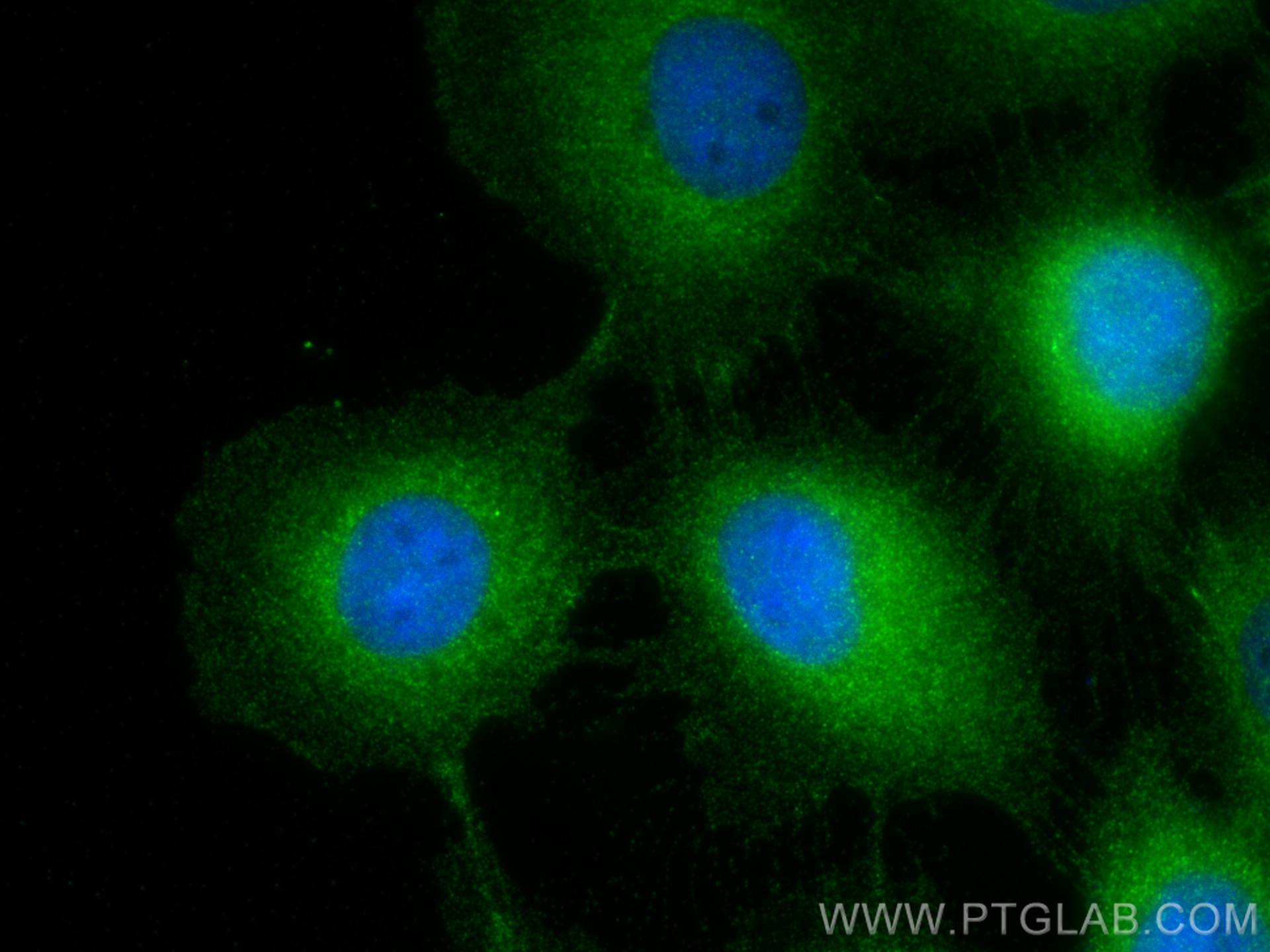
 fixed A431 cells using RRAS2 antibody (12530-1-AP) at dilution of 1:400 and CoraLite®488-Conjugated Goat Anti-Rabbit IgG(H+L).")
 and CoraLite®488-Conjugated Goat Anti-Rabbit IgG(H+L) at dilution 1:1000 (red), or 0.4 ug Control Antibody. Cells were fixed with 4% PFA and permeabilized with Flow Cytometry Perm Buffer (PF00011-C).")
Tested Applications
| Positive WB detected in | NIH/3T3 cells, mouse pancreas tissue |
| Positive IP detected in | NIH/3T3 cells |
| Positive IHC detected in | mouse heart tissue, human heart tissue, human lung cancer tissue Note: suggested antigen retrieval with TE buffer pH 9.0; (*) Alternatively, antigen retrieval may be performed with citrate buffer pH 6.0 |
| Positive IF/ICC detected in | A431 cells |
| Positive FC (Intra) detected in | A431 cells |
Recommended dilution
| Application | Dilution |
|---|---|
| Western Blot (WB) | WB : 1:1000-1:6000 |
| Immunoprecipitation (IP) | IP : 0.5-4.0 ug for 1.0-3.0 mg of total protein lysate |
| Immunohistochemistry (IHC) | IHC : 1:50-1:500 |
| Immunofluorescence (IF)/ICC | IF/ICC : 1:200-1:800 |
| Flow Cytometry (FC) (INTRA) | FC (INTRA) : 0.40 ug per 10^6 cells in a 100 µl suspension |
| It is recommended that this reagent should be titrated in each testing system to obtain optimal results. | |
| Sample-dependent, Check data in validation data gallery. | |
Published Applications
| WB | See 4 publications below |
Product Information
12530-1-AP targets RRAS2 in WB, IHC, IF/ICC, FC (Intra), IP, ELISA applications and shows reactivity with human, mouse samples.
| Tested Reactivity | human, mouse |
| Cited Reactivity | human, monkey |
| Host / Isotype | Rabbit / IgG |
| Class | Polyclonal |
| Type | Antibody |
| Immunogen | RRAS2 fusion protein Ag3226 Predict reactive species |
| Full Name | related RAS viral (r-ras) oncogene homolog 2 |
| Calculated Molecular Weight | 204 aa, 23 kDa |
| Observed Molecular Weight | 23 kDa |
| GenBank Accession Number | BC013106 |
| Gene Symbol | RRAS2 |
| Gene ID (NCBI) | 22800 |
| RRID | AB_2180206 |
| Conjugate | Unconjugated |
| Form | Liquid |
| Purification Method | Antigen affinity purification |
| UNIPROT ID | P62070 |
| Storage Buffer | PBS with 0.02% sodium azide and 50% glycerol pH 7.3. |
| Storage Conditions | Store at -20°C. Stable for one year after shipment. Aliquoting is unnecessary for -20oC storage. 20ul sizes contain 0.1% BSA. |
Protocols
| Product Specific Protocols | |
|---|---|
| WB protocol for RRAS2 antibody 12530-1-AP | Download protocol |
| IHC protocol for RRAS2 antibody 12530-1-AP | Download protocol |
| IF protocol for RRAS2 antibody 12530-1-AP | Download protocol |
| IP protocol for RRAS2 antibody 12530-1-AP | Download protocol |
| FC protocol for RRAS2 antibody 12530-1-AP | Download protocol |
| Standard Protocols | |
|---|---|
| Click here to view our Standard Protocols |
Publications
| Species | Application | Title |
|---|---|---|
Cell Death Differ Global identification of phospho-dependent SCF substrates reveals a FBXO22 phosphodegron and an ERK-FBXO22-BAG3 axis in tumorigenesis. | ||
Fertil Steril Long-term effects of repeated superovulation on ovarian structure and function in rhesus monkeys. | ||
J Biol Chem Competition for cysteine acylation by C16:0 and C18:0 derived lipids is a global phenomenon in the proteome | ||
Cells Co-Inhibition of tGLI1 and GP130 Using FDA-Approved Ketoconazole and Bazedoxifene Is Synergistic Against the Growth and Metastasis of HER2-Enriched and Triple-Negative Breast Cancers |