


Tested Applications
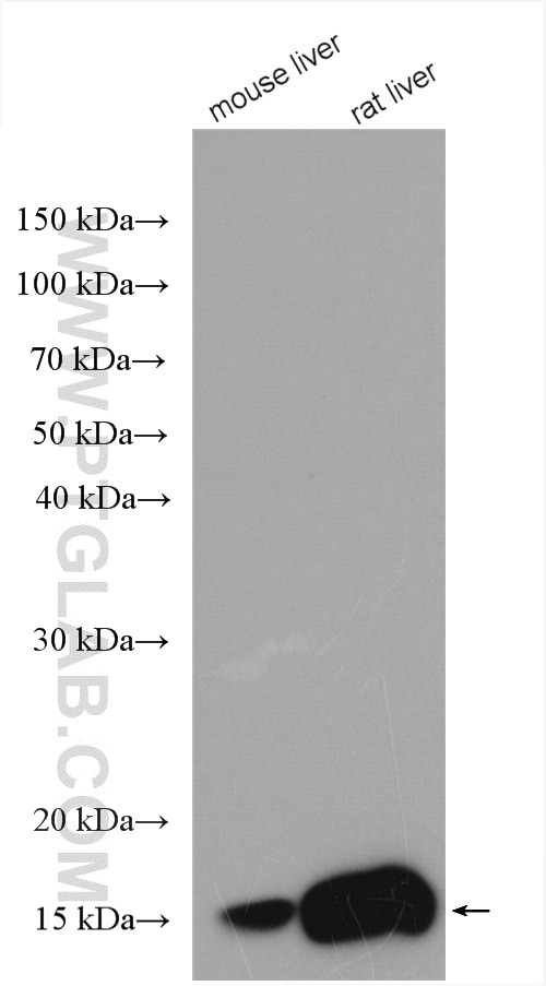
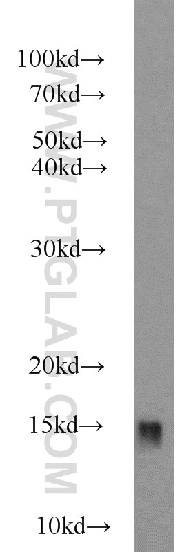
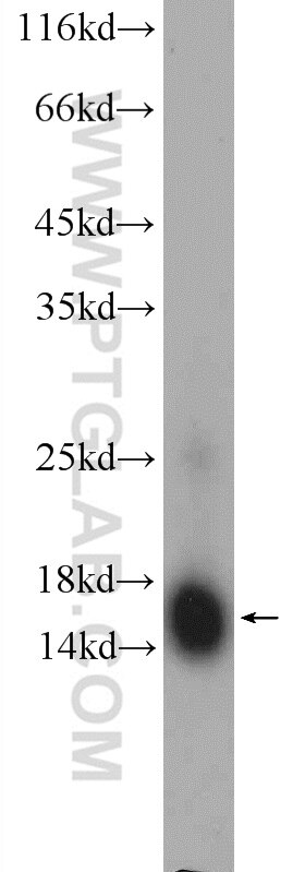
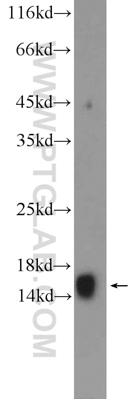
| Positive WB detected in | mouse liver tissue, HEK-293 cells, human brain tissue, human liver tissue, rat liver tissue |
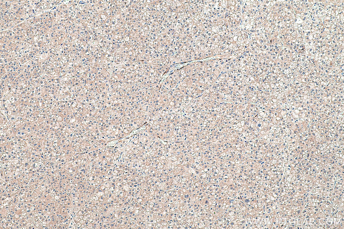
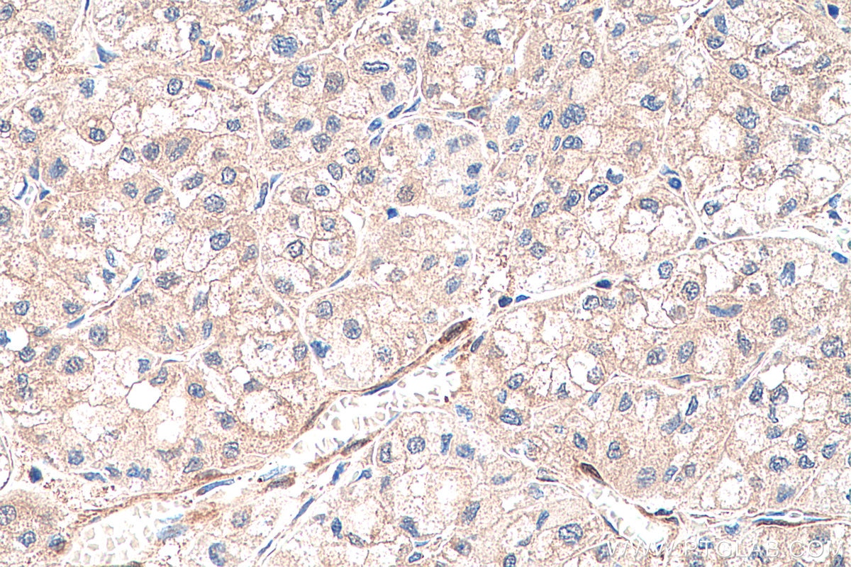
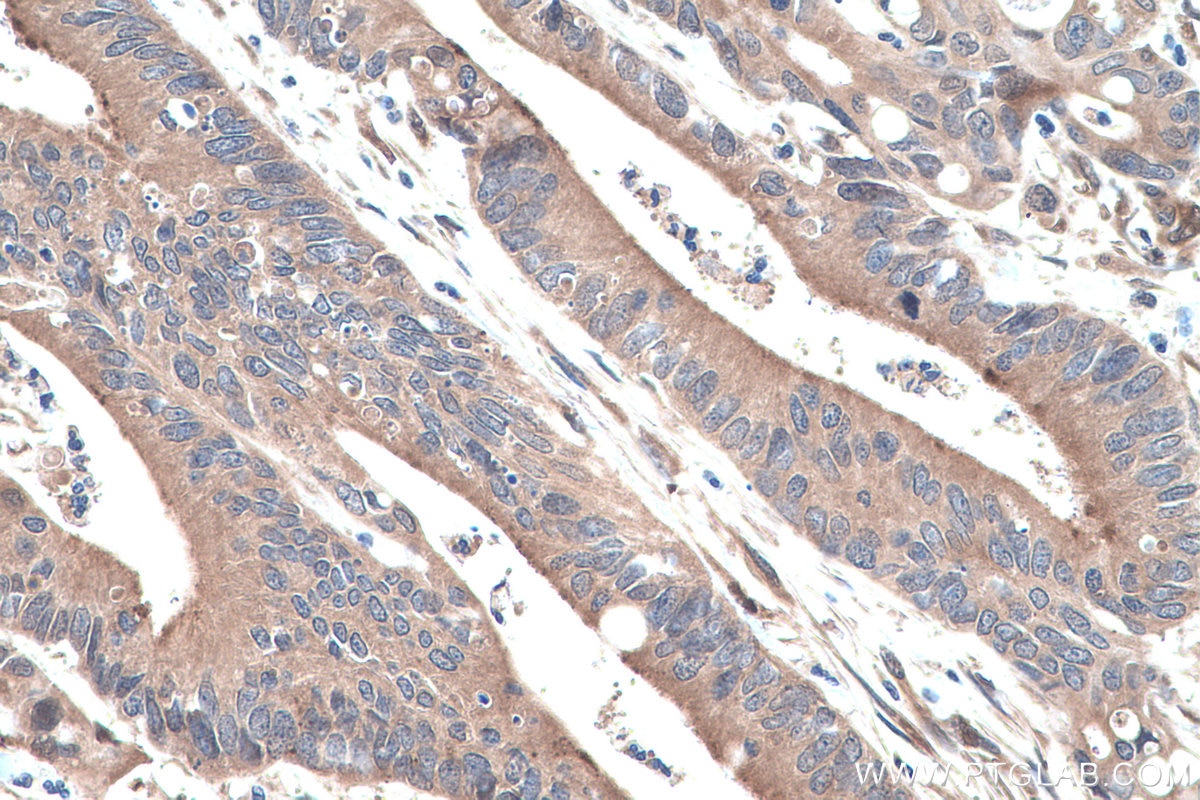
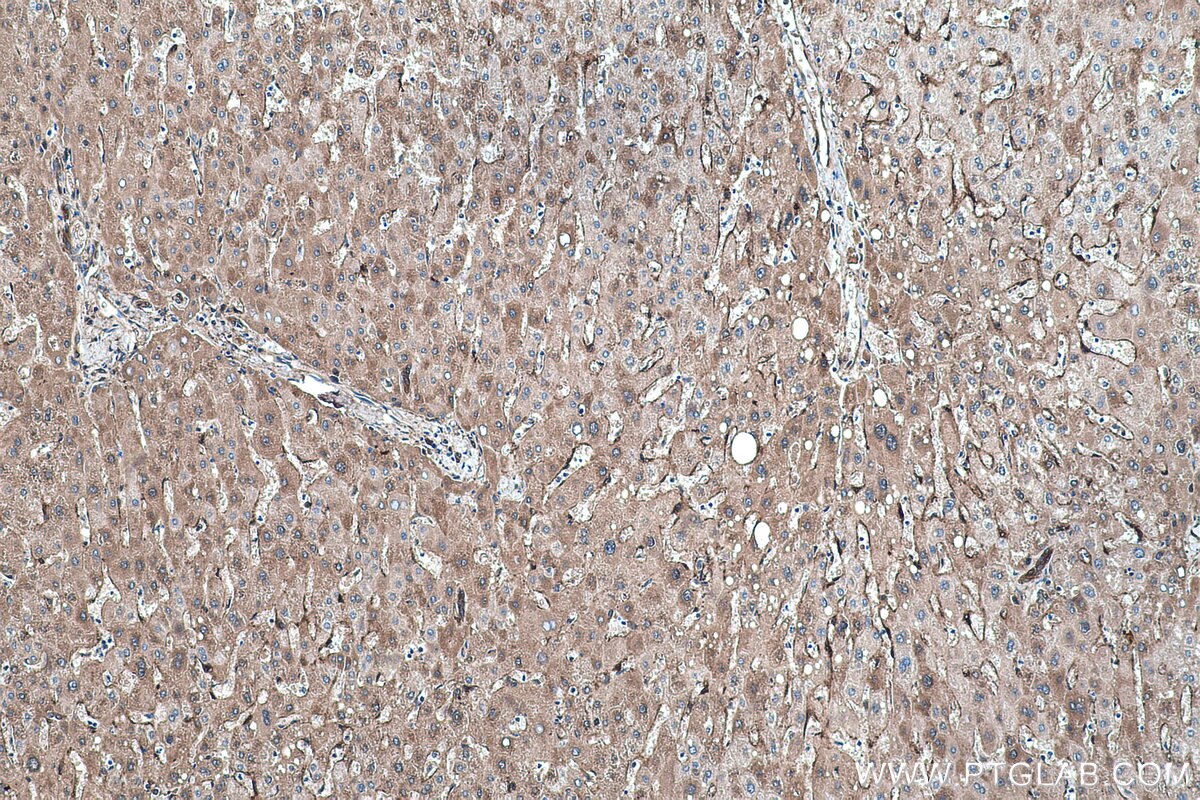
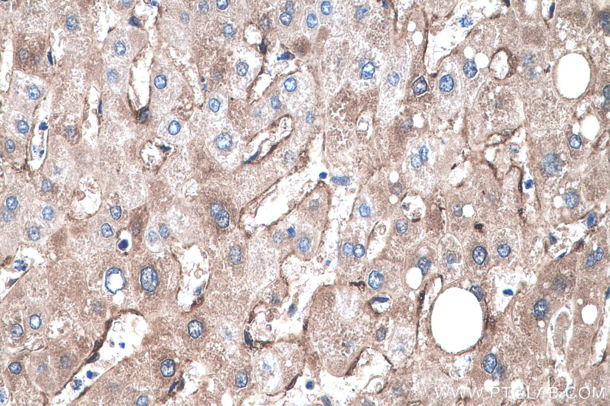
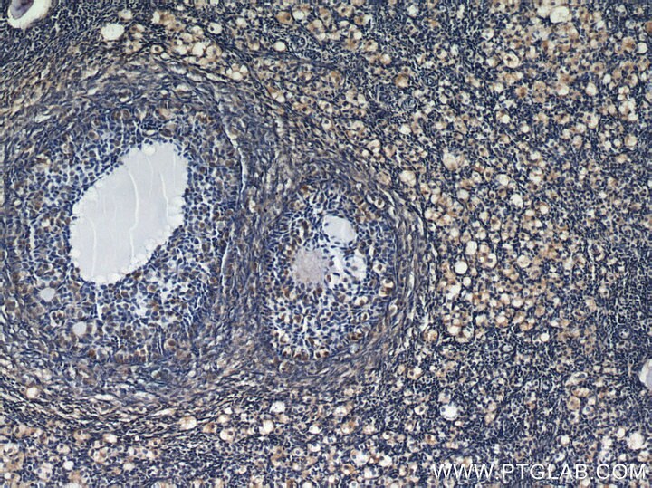
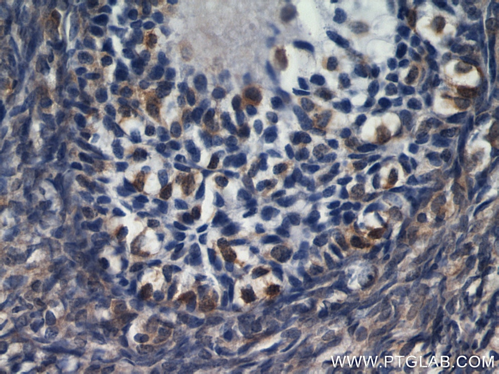
| Positive IHC detected in | human liver cancer tissue, human colon cancer tissue, human liver tissue, human ovary tissue Note: suggested antigen retrieval with TE buffer pH 9.0; (*) Alternatively, antigen retrieval may be performed with citrate buffer pH 6.0 |
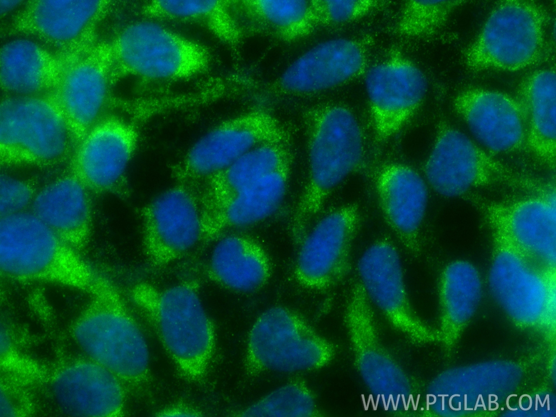
| Positive IF/ICC detected in | HEK-293 cells |
Recommended dilution
| Application | Dilution |
|---|---|
| Western Blot (WB) | WB : 1:500-1:2000 |
| Immunohistochemistry (IHC) | IHC : 1:1000-1:4000 |
| Immunofluorescence (IF)/ICC | IF/ICC : 1:50-1:500 |
| It is recommended that this reagent should be titrated in each testing system to obtain optimal results. | |
| Sample-dependent, Check data in validation data gallery. | |
Product Information
22683-1-AP targets RBP1 in WB, IHC, IF/ICC, ELISA applications and shows reactivity with human, mouse, rat samples.
| Tested Reactivity | human, mouse, rat |
| Cited Reactivity | human, mouse, rat |
| Host / Isotype | Rabbit / IgG |
| Class | Polyclonal |
| Type | Antibody |
| Immunogen |
CatNo: Ag18572 Product name: Recombinant human RBP1 protein Source: e coli.-derived, PGEX-4T Tag: GST Domain: 1-135 aa of BC121052 Sequence: MPVDFTGYWKMLVNENFEEYLRALDVNVALRKIANLLKPDKEIVQDGDHMIIRTLSTFRNYIMDFQVGKEFEEDLTGIDDRKCMTTVSWDGDKLQCVQKGEKEGRGWTQWIEGDELHLEMRVEGVVCKQVFKKVQ Predict reactive species |
| Full Name | retinol binding protein 1, cellular |
| Calculated Molecular Weight | 135 aa, 16 kDa |
| Observed Molecular Weight | 14-21 kDa |
| GenBank Accession Number | BC121052 |
| Gene Symbol | RBP1 |
| Gene ID (NCBI) | 5947 |
| RRID | AB_11182381 |
| Conjugate | Unconjugated |
| Form | Liquid |
| Purification Method | Antigen affinity purification |
| UNIPROT ID | P09455 |
| Storage Buffer | PBS with 0.02% sodium azide and 50% glycerol, pH 7.3. |
| Storage Conditions | Store at -20°C. Stable for one year after shipment. Aliquoting is unnecessary for -20oC storage. 20ul sizes contain 0.1% BSA. |
Protocols
| Product Specific Protocols | |
|---|---|
| IF protocol for RBP1 antibody 22683-1-AP | Download protocol |
| IHC protocol for RBP1 antibody 22683-1-AP | Download protocol |
| WB protocol for RBP1 antibody 22683-1-AP | Download protocol |
| Standard Protocols | |
|---|---|
| Click here to view our Standard Protocols |
Publications
What published studies show
The anti-RBP1 antibody (Cat# 22683-1-AP) has 14 citations published in journals including Nature Communications, Metabolism, Cell Death & Disease, Cell Communication and Signaling, Journal of Biological Chemistry, FASEB Journal, Journal of Molecular and Cellular Cardiology, Translational Oncology, Neural Regeneration Research, Molecular Neurobiology, Journal of Translational Medicine, Brain Sciences, Journal of Food Biochemistry, and European Journal of Histochemistry. Research fields span cancer biology, retinal degeneration, cardiac hypertrophy, diabetes, developmental biology, and neurology.
| Species | Application | Title |
|---|---|---|
Nat Commun TGFβ-mediated dural progenitor cell migration into the coronal suture is crucial for preventing craniosynostosis. | ||
Metabolism Disruption of retinoid homeostasis induces RBP4 overproduction in diabetes: O-GlcNAcylation involved. | ||
Cell Death Dis The RBP1-CKAP4 axis activates oncogenic autophagy and promotes cancer progression in oral squamous cell carcinoma.
| ||
Cell Commun Signal In vivo CRISPR screening identifies POU3F3 as a novel regulator of ferroptosis resistance in hepatocellular carcinoma via retinoic acid signaling | ||
J Transl Med Notch3 enhances the synergistic effect of all-trans retinoic acid and calcipotriol in pancreatic stellate cell activation | ||
Neural Regen Res AAV2-PDE6B restores retinal structure and function in the retinal degeneration 10 mouse model of retinitis pigmentosa by promoting phototransduction and inhibiting apoptosis |
Reviews
What customers say
The product has one verified review (5/5 stars) from a researcher who used it for Western Blot and Immunofluorescence on endothelial cells at a 1:1 dilution. The reviewer reported that the antibody "worked out" as expected, indicating satisfactory performance in both applications. Overall, the limited feedback reflects positive results, though the sample size is small.
The reviews below have been submitted by verified Proteintech customers who received an incentive for providing their feedback.
Melissa (Verified Customer) (06-13-2022) | We have used the antibody and it worked out
|


