Tested Applications
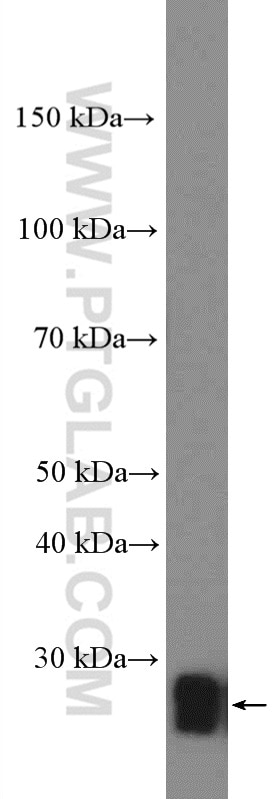
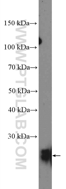
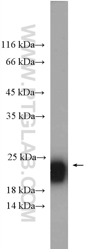
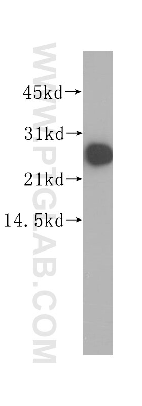
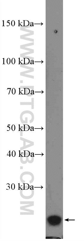
| Positive WB detected in | fetal human brain tissue, mouse brain tissue, Y79 cells, human brain tissue |
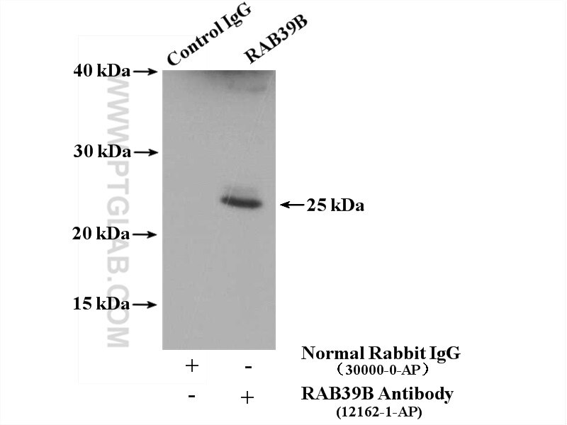
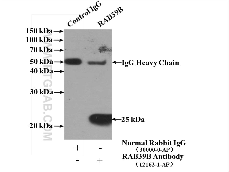
| Positive IP detected in | SH-SY5Y cells, mouse brain tissue |
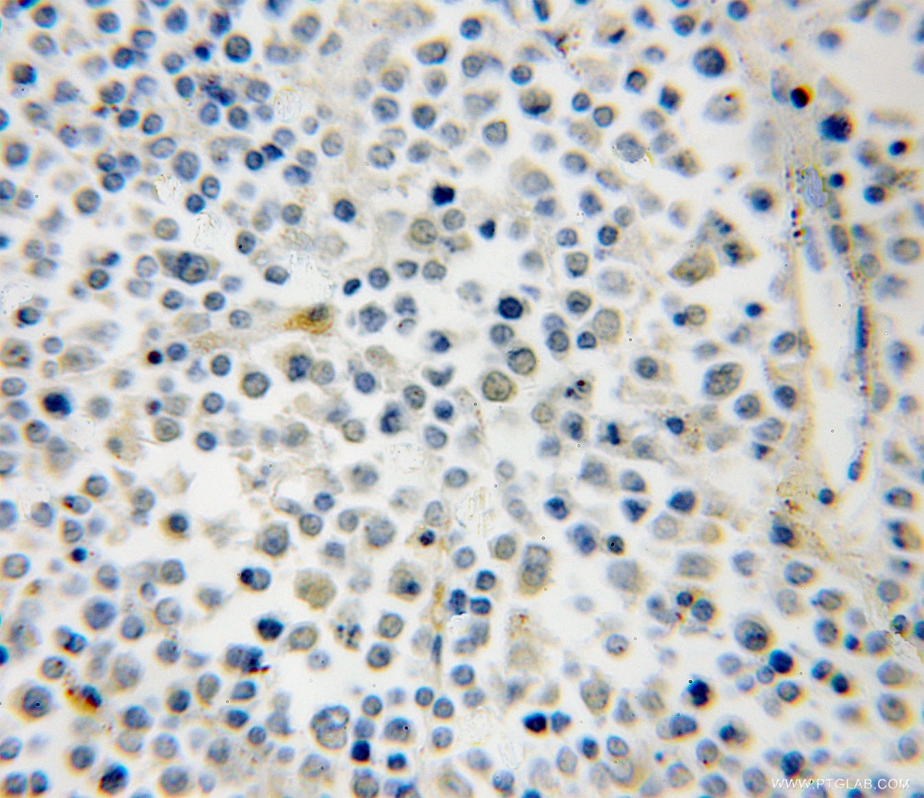
| Positive IHC detected in | human lymphoma tissue Note: suggested antigen retrieval with TE buffer pH 9.0; (*) Alternatively, antigen retrieval may be performed with citrate buffer pH 6.0 |
Recommended dilution
| Application | Dilution |
|---|---|
| Western Blot (WB) | WB : 1:500-1:2000 |
| Immunoprecipitation (IP) | IP : 0.5-4.0 ug for 1.0-3.0 mg of total protein lysate |
| Immunohistochemistry (IHC) | IHC : 1:20-1:200 |
| It is recommended that this reagent should be titrated in each testing system to obtain optimal results. | |
| Sample-dependent, Check data in validation data gallery. | |
Published Applications
| KD/KO | See 2 publications below |
| WB | See 9 publications below |
| IF | See 4 publications below |
| IP | See 1 publications below |
Product Information
12162-1-AP targets RAB39B in WB, IHC, IF, IP, ELISA applications and shows reactivity with human, mouse samples.
| Tested Reactivity | human, mouse |
| Cited Reactivity | human, mouse |
| Host / Isotype | Rabbit / IgG |
| Class | Polyclonal |
| Type | Antibody |
| Immunogen | RAB39B fusion protein Ag2803 Predict reactive species |
| Full Name | RAB39B, member RAS oncogene family |
| Calculated Molecular Weight | 213 aa, 25 kDa |
| Observed Molecular Weight | 25 kDa |
| GenBank Accession Number | BC009714 |
| Gene Symbol | RAB39B |
| Gene ID (NCBI) | 116442 |
| RRID | AB_2177360 |
| Conjugate | Unconjugated |
| Form | Liquid |
| Purification Method | Antigen affinity purification |
| UNIPROT ID | Q96DA2 |
| Storage Buffer | PBS with 0.02% sodium azide and 50% glycerol pH 7.3. |
| Storage Conditions | Store at -20°C. Stable for one year after shipment. Aliquoting is unnecessary for -20oC storage. 20ul sizes contain 0.1% BSA. |
Protocols
| Product Specific Protocols | |
|---|---|
| WB protocol for RAB39B antibody 12162-1-AP | Download protocol |
| IHC protocol for RAB39B antibody 12162-1-AP | Download protocol |
| IP protocol for RAB39B antibody 12162-1-AP | Download protocol |
| Standard Protocols | |
|---|---|
| Click here to view our Standard Protocols |
Publications
| Species | Application | Title |
|---|---|---|
Nat Commun C9orf72-catalyzed GTP loading of Rab39A enables HOPS-mediated membrane tethering and fusion in mammalian autophagy
| ||
Am J Hum Genet Mutations in RAB39B Cause X-Linked Intellectual Disability and Early-Onset Parkinson Disease with α-Synuclein Pathology. | ||
EMBO J Loss of C9ORF72 impairs autophagy and synergizes with polyQ Ataxin-2 to induce motor neuron dysfunction and cell death. | ||
Acta Neuropathol Commun Synaptic localization of C9orf72 regulates post-synaptic glutamate receptor 1 levels. | ||
Brain Pathol RAB39B is redistributed in dementia with Lewy bodies and is sequestered within aβ plaques and Lewy bodies. |
Reviews
The reviews below have been submitted by verified Proteintech customers who received an incentive for providing their feedback.
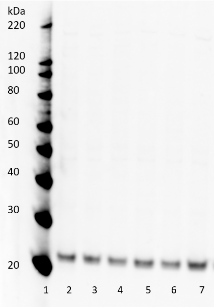
FH David (Verified Customer) (09-25-2019) | The antibodiy works well in membrane enriched fractionates generated from human brain homogenate. Note that several higher molecular weight bands are seen when using crude lysates generated from the same samples.Image details: lane 1 = molecular weight ladder, lanes 2-7 = membrane enriched fractionates (2µg/lane).Membrane blocked in 5% milk powder containing TBST overnight at 4oC. Primary antibody: RAB39B (1:1000) in 5% BSA containing TBST, 1hr incubation at RT. Secondar antibody: y Goat anti Rabbit-HRP (1:5000, 1hr RT) in 5% milk powder containing TBST.
 |
