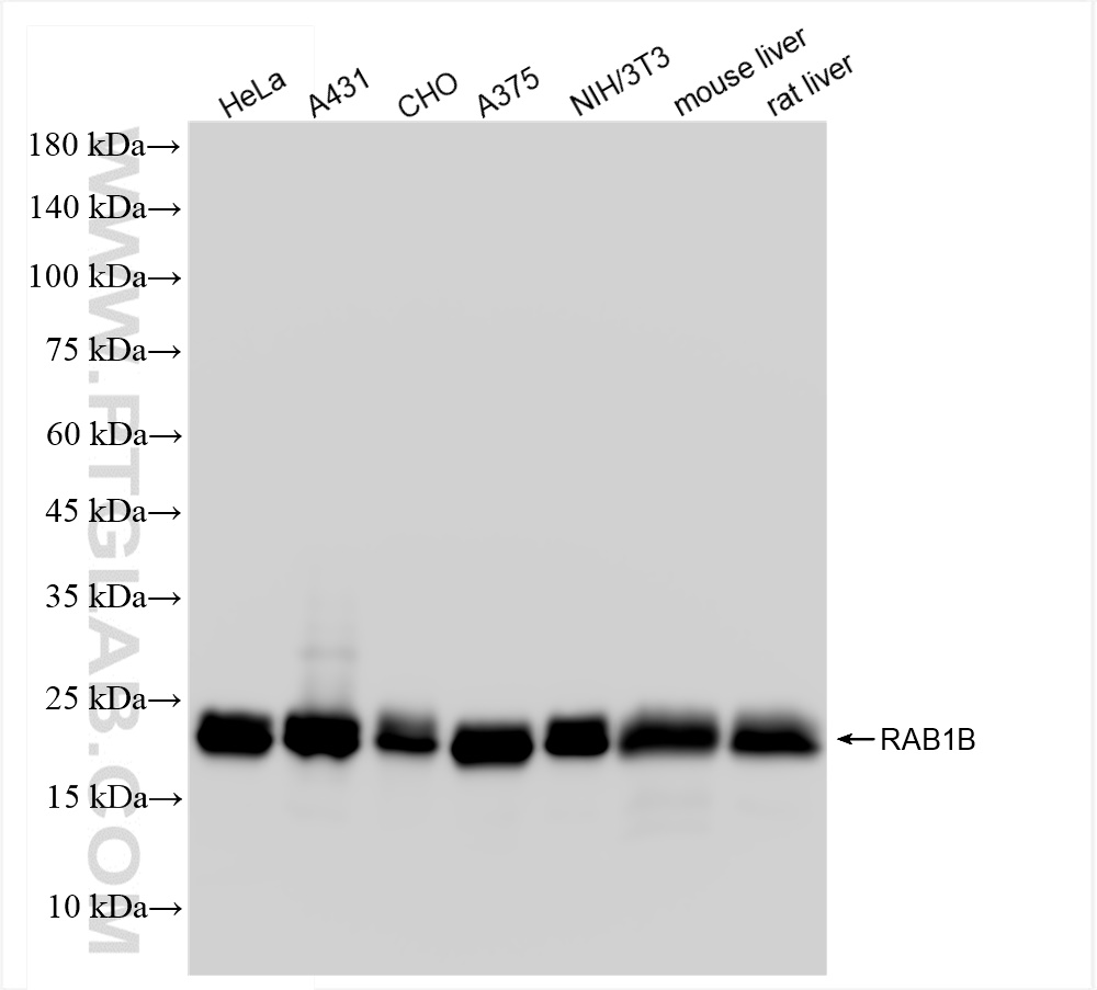
Various lysates were subjected to SDS PAGE followed by western blot with 83687-4-RR (RAB1B antibody) at dilution of 1:10000 incubated at room temperature for 1.5 hours.
Various lysates were subjected to SDS PAGE followed by western blot with 83687-4-RR (RAB1B antibody) at dilution of 1:10000 incubated at room temperature for 1.5 hours.
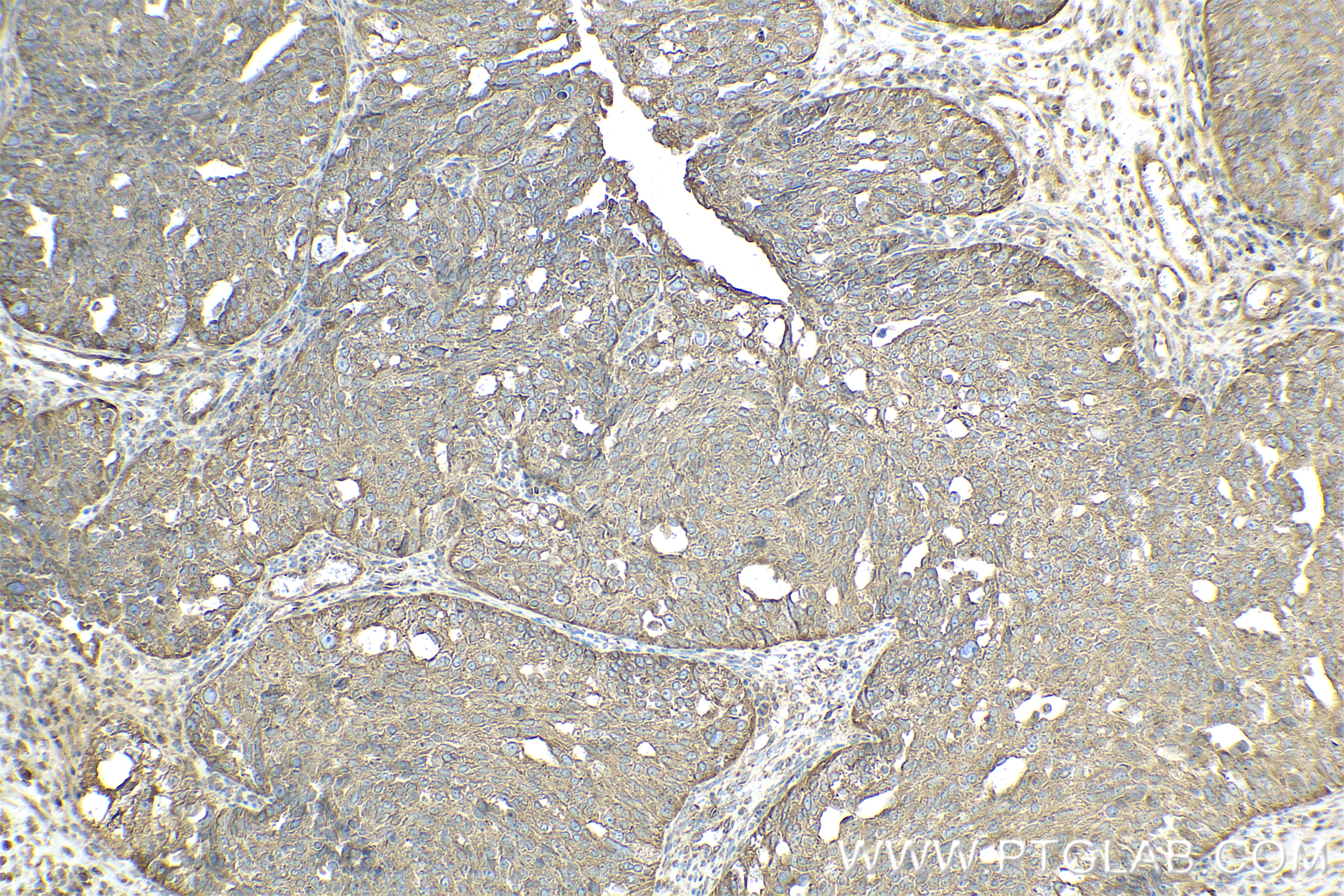
IHC staining of human ovary cancer using 83687-4-RR
Immunohistochemical analysis of paraffin-embedded human ovary cancer tissue slide using 83687-4-RR (RAB1B antibody) at dilution of 1:400 (under 10x lens). Heat mediated antigen retrieval with Tris-EDTA buffer (pH 9.0).
Immunohistochemical analysis of paraffin-embedded human ovary cancer tissue slide using 83687-4-RR (RAB1B antibody) at dilution of 1:400 (under 10x lens). Heat mediated antigen retrieval with Tris-EDTA buffer (pH 9.0).
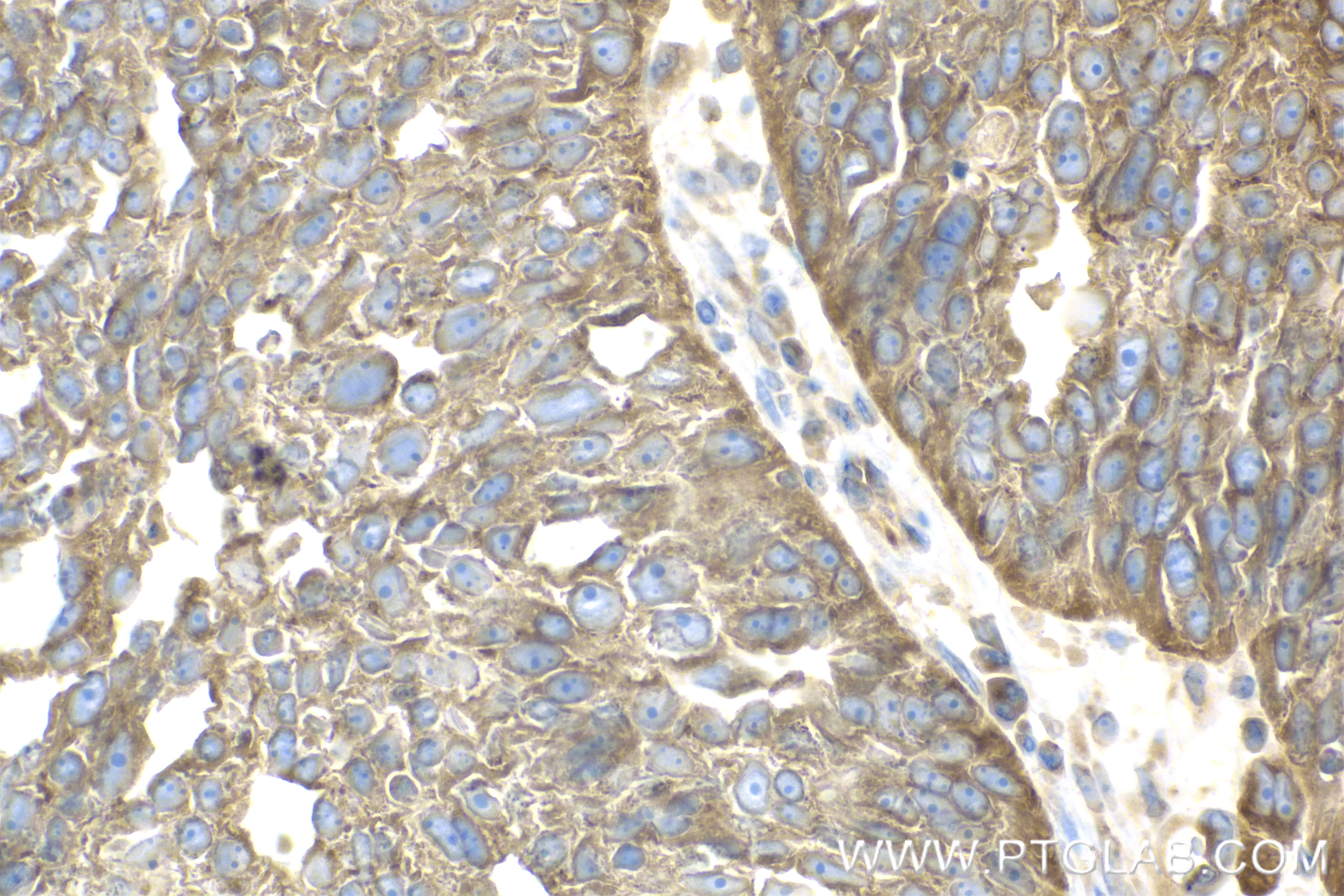
IHC staining of human ovary cancer using 83687-4-RR
Immunohistochemical analysis of paraffin-embedded human ovary cancer tissue slide using 83687-4-RR (RAB1B antibody) at dilution of 1:400 (under 40x lens). Heat mediated antigen retrieval with Tris-EDTA buffer (pH 9.0).
Immunohistochemical analysis of paraffin-embedded human ovary cancer tissue slide using 83687-4-RR (RAB1B antibody) at dilution of 1:400 (under 40x lens). Heat mediated antigen retrieval with Tris-EDTA buffer (pH 9.0).
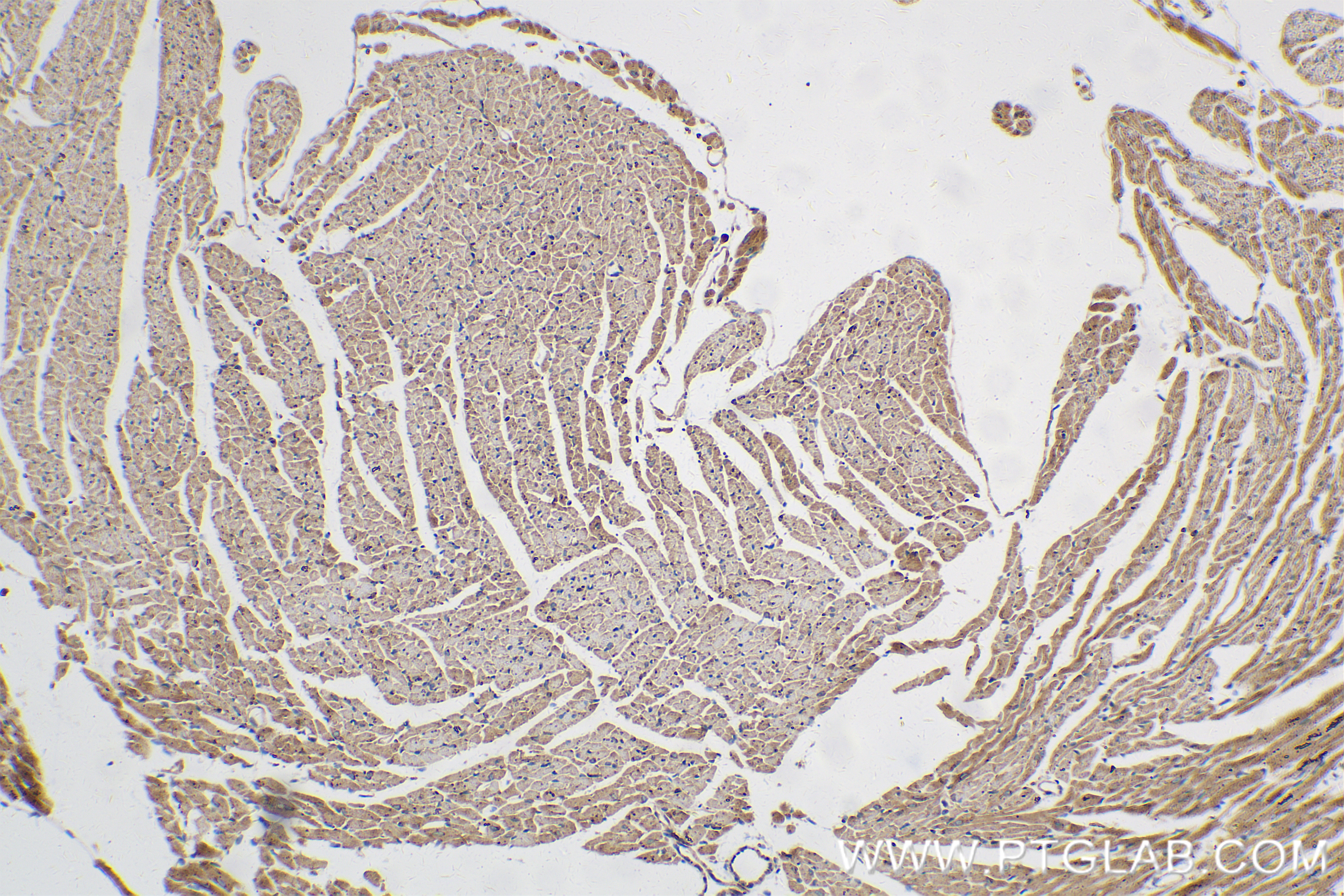
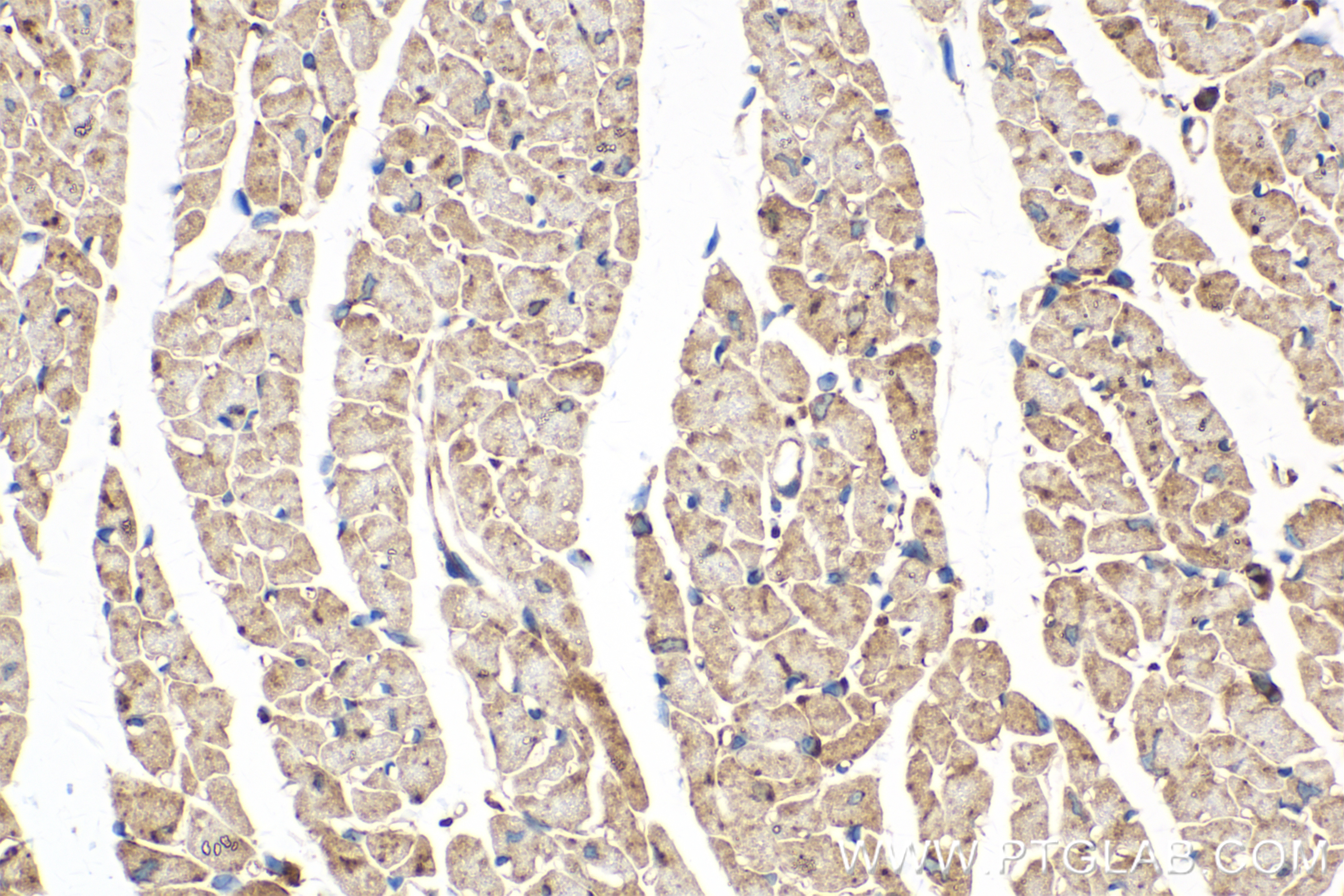
IHC staining of mouse heart using 83687-4-RR
Immunohistochemical analysis of paraffin-embedded mouse heart tissue slide using 83687-4-RR (RAB1B antibody) at dilution of 1:400 (under 10x lens). Heat mediated antigen retrieval with Tris-EDTA buffer (pH 9.0).
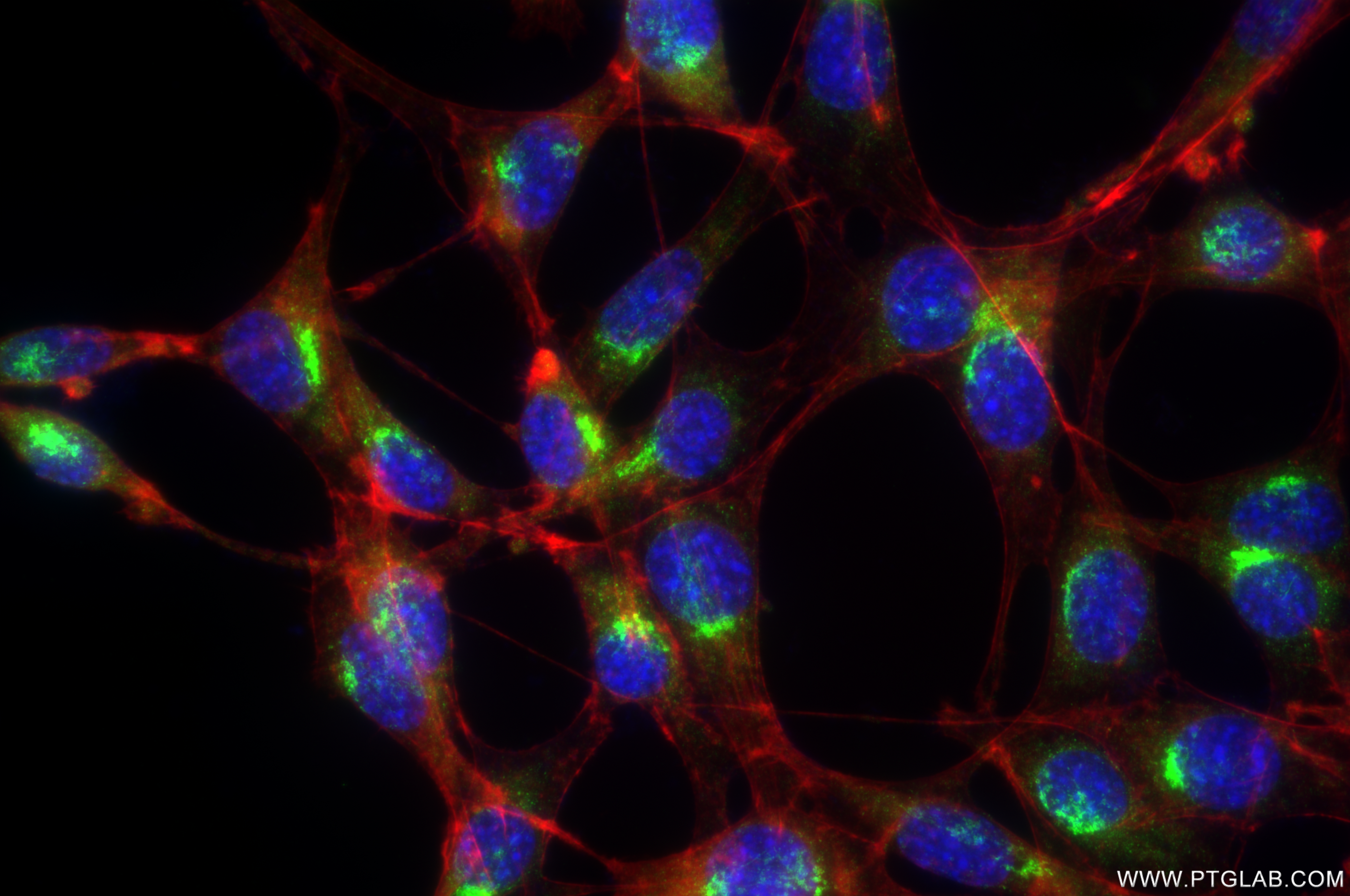
Immunofluorescent analysis of (4% PFA) fixed NIH/3T3 cells using RAB1B antibody (83687-4-RR, Clone: 240635D9 ) at dilution of 1:400 and CoraLite®488-Conjugated Goat Anti-Rabbit IgG(H+L) (SA00013-2), CL594-Phalloidin (red).
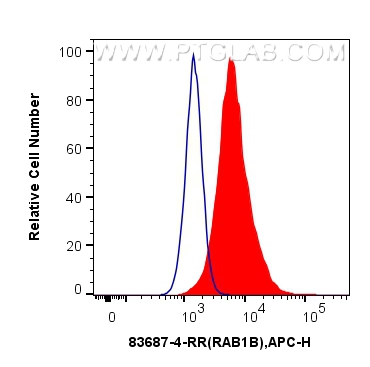
FC experiment of U-251 using 83687-4-RR
1x10^6 U-251 cells were intracellularly stained with 0.25 ug RAB1B Recombinant antibody (83687-4-RR, Clone:240635D9) and APC-Conjugated AffiniPure Goat Anti-Rabbit IgG(H+L)(red), or 0.25 ug Isotype Control (blue). Cells were fixed with 4% PFA and permeabilized with Flow Cytometry Perm Buffer (PF00011-C).
1x10^6 U-251 cells were intracellularly stained with 0.25 ug RAB1B Recombinant antibody (83687-4-RR, Clone:240635D9) and APC-Conjugated AffiniPure Goat Anti-Rabbit IgG(H+L)(red), or 0.25 ug Isotype Control (blue). Cells were fixed with 4% PFA and permeabilized with Flow Cytometry Perm Buffer (PF00011-C).
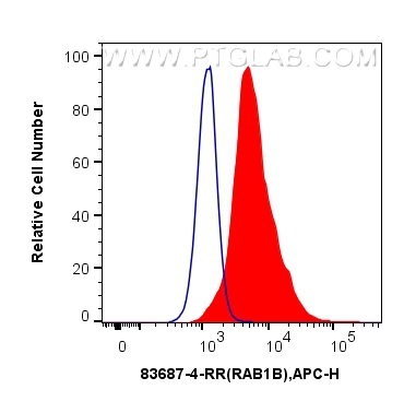
FC experiment of HeLa using 83687-4-RR
1x10^6 HeLa cells were intracellularly stained with 0.25 ug RAB1B Recombinant antibody (83687-4-RR, Clone:240635D9) and APC-Conjugated AffiniPure Goat Anti-Rabbit IgG(H+L)(red), or 0.25 ug Isotype Control (blue). Cells were fixed with 4% PFA and permeabilized with Flow Cytometry Perm Buffer (PF00011-C).
1x10^6 HeLa cells were intracellularly stained with 0.25 ug RAB1B Recombinant antibody (83687-4-RR, Clone:240635D9) and APC-Conjugated AffiniPure Goat Anti-Rabbit IgG(H+L)(red), or 0.25 ug Isotype Control (blue). Cells were fixed with 4% PFA and permeabilized with Flow Cytometry Perm Buffer (PF00011-C).
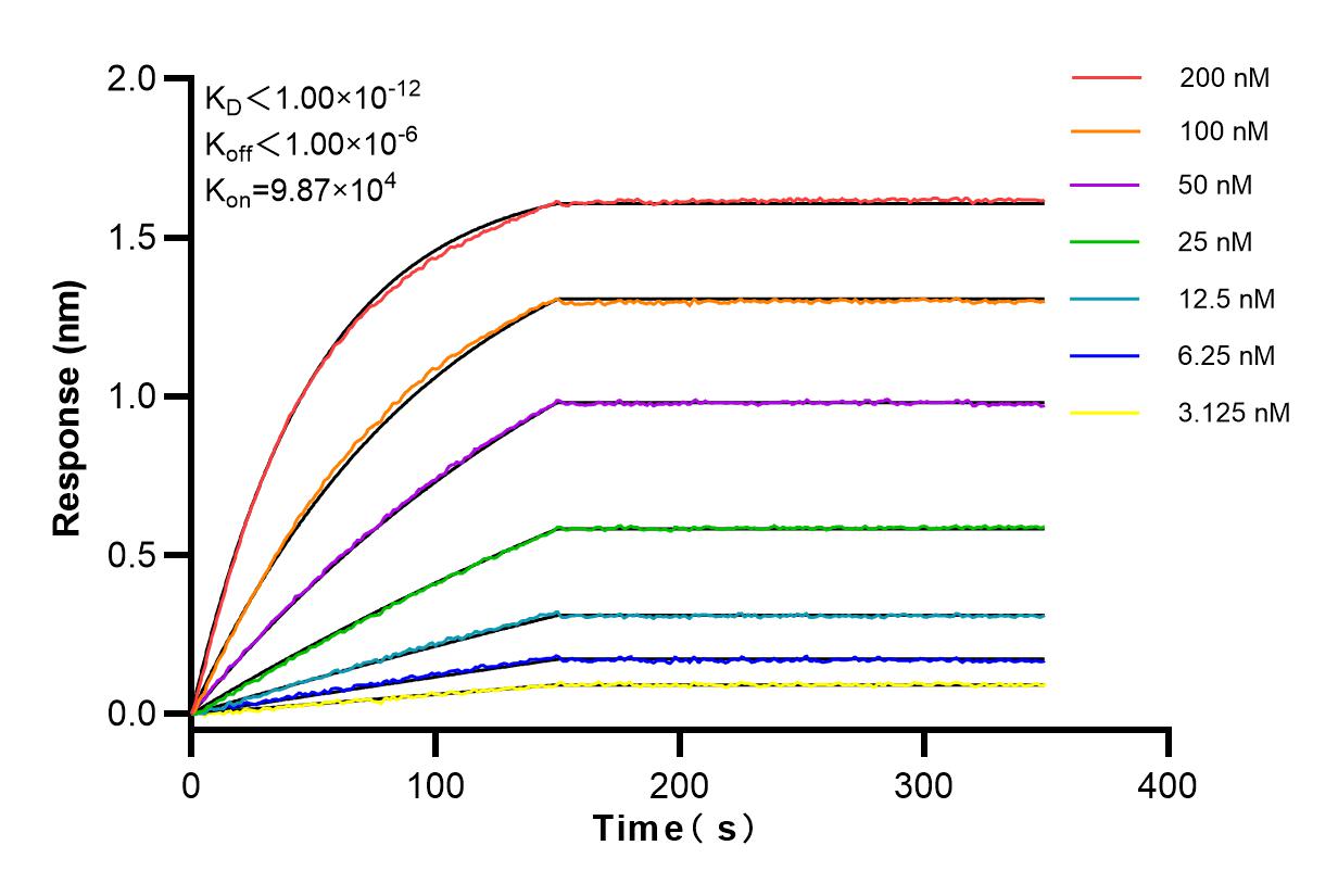
Affinity and Kinetic Characterization of 83687-4-RR
Biolayer interferometry (BLl) kinetic assays of 83687-4-RR
against Human RAB1B were performed. The affinity constant is below 1 pM.
The Proteintech guarantee covers Proteintech antibodies in any species and any application, including those not listed on the datasheet. If the antibody doesn’t perform, you can receive a hassle-free refund or credit note.
HeLa cells, A431 cells, CHO cells, A375 cells, NIH/3T3 cells, mouse liver tissue, rat liver tissue
Positive IHC detected in
human ovary cancer tissue, mouse heart tissue Note: suggested antigen retrieval with TE buffer pH 9.0; (*) Alternatively, antigen retrieval may be performed with citrate buffer pH 6.0
Positive IF/ICC detected in
NIH/3T3 cells
Positive FC (Intra) detected in
U-251 cells, HeLa cells
Recommended dilution
Application
Dilution
Western Blot (WB)
WB : 1:5000-1:50000
Immunohistochemistry (IHC)
IHC : 1:200-1:800
Immunofluorescence (IF)/ICC
IF/ICC : 1:200-1:800
Flow Cytometry (FC) (INTRA)
FC (INTRA) : 0.25 ug per 10^6 cells in a 100 µl suspension
It is recommended that this reagent should be titrated in each testing system to obtain optimal results.
Sample-dependent, Check data in validation data gallery.
Product Information
83687-4-RR targets RAB1B in WB, IHC, IF/ICC, FC (Intra), ELISA applications and shows reactivity with human, mouse, rat samples.
PBS with 0.02% sodium azide and 50% glycerol, pH 7.3.
Storage Conditions
Store at -20°C. Stable for one year after shipment. Aliquoting is unnecessary for -20oC storage. 20ul sizes contain 0.1% BSA.
Background Information
Ras-related protein Rab-1B (RAB1B) belongs to the small GTPase superfamily and Rab family. Rab1b, a GTPase regulating secretory transport, was recently associated with targeting proteins to LDs in a Drosophila RNAi screen. Rab1b recruits lipid-synthesizing enzymes, facilitating Lipid droplets (LDs) growth. The small GTPase Rab1b interacts with diacylglycerol acyltransferase 2 to facilitate its targeting to the surface of lipid droplets. (PMID: 38809969). Rab1b, an extensively studied and established master regulator of ER-to-Golgi transport. The mechanism of Rab1b's function is to promote ER to LD surface targeting of those triglyceride-synthesizing enzymes that can associate with the unique lipid monolayer comprising the LD surface (PMID: 38809969). Rab1b is a regulatory protein involved in both COPI and COPII transport (PMID: 17429068).
Various lysates were subjected to SDS PAGE followed by western blot with 83687-4-RR (RAB1B antibody) at dilution of 1:10000 incubated at room temperature for 1.5 hours.
IHC Figures
IHC staining of human ovary cancer using 83687-4-RR
Immunohistochemical analysis of paraffin-embedded human ovary cancer tissue slide using 83687-4-RR (RAB1B antibody) at dilution of 1:400 (under 10x lens). Heat mediated antigen retrieval with Tris-EDTA buffer (pH 9.0).
IHC staining of human ovary cancer using 83687-4-RR
Immunohistochemical analysis of paraffin-embedded human ovary cancer tissue slide using 83687-4-RR (RAB1B antibody) at dilution of 1:400 (under 40x lens). Heat mediated antigen retrieval with Tris-EDTA buffer (pH 9.0).
IHC staining of mouse heart using 83687-4-RR
Immunohistochemical analysis of paraffin-embedded mouse heart tissue slide using 83687-4-RR (RAB1B antibody) at dilution of 1:400 (under 10x lens). Heat mediated antigen retrieval with Tris-EDTA buffer (pH 9.0).
IHC staining of mouse heart using 83687-4-RR
Immunohistochemical analysis of paraffin-embedded mouse heart tissue slide using 83687-4-RR (RAB1B antibody) at dilution of 1:400 (under 40x lens). Heat mediated antigen retrieval with Tris-EDTA buffer (pH 9.0).
IF/ICC Figures
IF Staining of NIH/3T3 using 83687-4-RR
Immunofluorescent analysis of (4% PFA) fixed NIH/3T3 cells using RAB1B antibody (83687-4-RR, Clone: 240635D9 ) at dilution of 1:400 and CoraLite®488-Conjugated Goat Anti-Rabbit IgG(H+L) (SA00013-2), CL594-Phalloidin (red).
FC (INTRA) Figures
FC experiment of U-251 using 83687-4-RR
1x10^6 U-251 cells were intracellularly stained with 0.25 ug RAB1B Recombinant antibody (83687-4-RR, Clone:240635D9) and APC-Conjugated AffiniPure Goat Anti-Rabbit IgG(H+L)(red), or 0.25 ug Isotype Control (blue). Cells were fixed with 4% PFA and permeabilized with Flow Cytometry Perm Buffer (PF00011-C).
FC experiment of HeLa using 83687-4-RR
1x10^6 HeLa cells were intracellularly stained with 0.25 ug RAB1B Recombinant antibody (83687-4-RR, Clone:240635D9) and APC-Conjugated AffiniPure Goat Anti-Rabbit IgG(H+L)(red), or 0.25 ug Isotype Control (blue). Cells were fixed with 4% PFA and permeabilized with Flow Cytometry Perm Buffer (PF00011-C).
AFFINITY Figures
Affinity and Kinetic Characterization of 83687-4-RR
Biolayer interferometry (BLl) kinetic assays of 83687-4-RR
against Human RAB1B were performed. The affinity constant is below 1 pM.
The species listed in Tested Reactivity are in-house verified and applicable species. For unlisted species, please refer to the homology analysis of the immunogen sequence and related species. For rabbit polyclonal antibodies, homology >70% is recommended. For mouse monoclonal antibodies and rabbit recombinant antibodies, homology >90% is recommended. Generally, the higher the homology, the greater the applicability. However, there will be certain differences in protein expression in different species, tissues or cells. Therefore, the homology analysis results are for reference only and do not serve as a guarantee.
At Proteintech, we pride ourselves on our antibody quality, customer service and transparency. As such, we are comparing our antibodies with other vendors, enabling easy identification and comparisons of key data to help you choose the suitable antibody for your needs.
We have selected the top cited antibodies from these vendors for you to compare.
Proteintech
RAB1B Recombinant antibody
Catalog Number
83687-4-RR
Citations
-
Dilutions
WB : 1:5000-1:50000 IHC : 1:200-1:800 IF/ICC : 1:200-1:800 FC (INTRA) : 0.25 ug per 10^6 cells in a 100 µl suspension
Applications
WB, IHC, IF/ICC, FC (Intra), ELISA
Reactivity
human, mouse, rat
Product Guarantee
Covers any species including not listed on datasheet
Covers any applications including not listed on datasheet
 at dilution of 1:10000 incubated at room temperature for 1.5 hours.")
 at dilution of 1:400 (under 10x lens). Heat mediated antigen retrieval with Tris-EDTA buffer (pH 9.0).")
 at dilution of 1:400 (under 40x lens). Heat mediated antigen retrieval with Tris-EDTA buffer (pH 9.0).")
 at dilution of 1:400 (under 10x lens). Heat mediated antigen retrieval with Tris-EDTA buffer (pH 9.0).")
 at dilution of 1:400 (under 40x lens). Heat mediated antigen retrieval with Tris-EDTA buffer (pH 9.0).")
 fixed NIH/3T3 cells using RAB1B antibody (83687-4-RR, Clone: 240635D9 ) at dilution of 1:400 and CoraLite®488-Conjugated Goat Anti-Rabbit IgG(H+L) (SA00013-2), CL594-Phalloidin (red).")
 and APC-Conjugated AffiniPure Goat Anti-Rabbit IgG(H+L)(red), or 0.25 ug Isotype Control (blue). Cells were fixed with 4% PFA and permeabilized with Flow Cytometry Perm Buffer (PF00011-C).")
 and APC-Conjugated AffiniPure Goat Anti-Rabbit IgG(H+L)(red), or 0.25 ug Isotype Control (blue). Cells were fixed with 4% PFA and permeabilized with Flow Cytometry Perm Buffer (PF00011-C).")
 kinetic assays of 83687-4-RR
against Human RAB1B were performed. The affinity constant is below 1 pM.")