Filter:
 at dilution of 1:1500 incubated at room temperature for 1.5 hours.")
 with HEK-293 cells lysate 3200ug.")
 at dilution of 1:50 (under 10x lens).")
 at dilution of 1:50 (under 40x lens).")
 at dilution of 1:1000 (under 10x lens). Heat mediated antigen retrieval with Tris-EDTA buffer (pH 9.0).")
 at dilution of 1:1000 (under 40x lens). Heat mediated antigen retrieval with Tris-EDTA buffer (pH 9.0).")
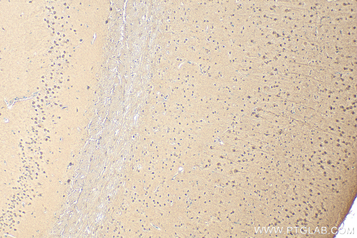
 at dilution of 1:1000 (under 10x lens). Heat mediated antigen retrieval with Tris-EDTA buffer (pH 9.0).")
 at dilution of 1:1000 (under 10x lens). Heat mediated antigen retrieval with Tris-EDTA buffer (pH 9.0).")
 at dilution of 1:1000 (under 40x lens). Heat mediated antigen retrieval with Tris-EDTA buffer (pH 9.0).")
 at dilution of 1:1000 (under 40x lens). Heat mediated antigen retrieval with Tris-EDTA buffer (pH 9.0).")
 at dilution of 1:1000 (under 40x lens). Heat mediated antigen retrieval with Tris-EDTA buffer (pH 9.0).")
 at dilution of 1:50 (under 10x lens).")
 at dilution of 1:50 (under 40x lens).")
 at dilution of 1:50 and Alexa Fluor 488-conjugated AffiniPure Goat Anti-Rabbit IgG(H+L).")
 at dilution of 1:25 and Alexa Fluor 488-conjugated AffiniPure Goat Anti-Rabbit IgG(H+L).")
 at dilution of 1:25 and Alexa Fluor 488-conjugated AffiniPure Goat Anti-Rabbit IgG(H+L).")
 and CoraLite®488-Conjugated AffiniPure Goat Anti-Rabbit IgG(H+L) at dilution 1:1000 (red), or 0.2 ug Control Antibody. Cells were fixed with 4% PFA and permeabilized with Flow Cytometry Perm Buffer (PF00011-C).")
Tested Applications
| Positive WB detected in | A549 cells, HEK-293 cells, HepG2 cells, Jurkat cells, MCF-7 cells, Neuro-2a cells |
| Positive IP detected in | HEK-293 cells |
| Positive IHC detected in | human lung cancer tissue, human testis tissue, human thyroid cancer tissue, mouse brain tissue, mouse colon tissue, mouse testis tissue, rat small intestine tissue, rat testis tissue Note: suggested antigen retrieval with TE buffer pH 9.0; (*) Alternatively, antigen retrieval may be performed with citrate buffer pH 6.0 |
| Positive IF/ICC detected in | HeLa cells, MCF-7 cells, HepG2 cells |
| Positive FC (Intra) detected in | HepG2 cells |
Recommended dilution
| Application | Dilution |
|---|---|
| Western Blot (WB) | WB : 1:500-1:3000 |
| Immunoprecipitation (IP) | IP : 0.5-4.0 ug for 1.0-3.0 mg of total protein lysate |
| Immunohistochemistry (IHC) | IHC : 1:50-1:500 |
| Immunofluorescence (IF)/ICC | IF/ICC : 1:20-1:200 |
| Flow Cytometry (FC) (INTRA) | FC (INTRA) : 0.20 ug per 10^6 cells in a 100 µl suspension |
| It is recommended that this reagent should be titrated in each testing system to obtain optimal results. | |
| Sample-dependent, Check data in validation data gallery. | |
Published Applications
| WB | See 3 publications below |
| IF | See 2 publications below |
Product Information
11573-1-AP targets PSMA6 in WB, IHC, IF/ICC, FC (Intra), IP, ELISA applications and shows reactivity with human, mouse, rat samples.
| Tested Reactivity | human, mouse, rat |
| Cited Reactivity | human, oyster |
| Host / Isotype | Rabbit / IgG |
| Class | Polyclonal |
| Type | Antibody |
| Immunogen | PSMA6 fusion protein Ag2154 Predict reactive species |
| Full Name | proteasome (prosome, macropain) subunit, alpha type, 6 |
| Calculated Molecular Weight | 246 aa, 27 kDa |
| Observed Molecular Weight | 27-29 kDa |
| GenBank Accession Number | BC023659 |
| Gene Symbol | PSMA6 |
| Gene ID (NCBI) | 5687 |
| RRID | AB_2171742 |
| Conjugate | Unconjugated |
| Form | Liquid |
| Purification Method | Antigen affinity purification |
| UNIPROT ID | P60900 |
| Storage Buffer | PBS with 0.02% sodium azide and 50% glycerol , pH 7.3 |
| Storage Conditions | Store at -20°C. Stable for one year after shipment. Aliquoting is unnecessary for -20oC storage. 20ul sizes contain 0.1% BSA. |
Background Information
PSMA6(Proteasome subunit alpha type-6) is also named as PROS27 and belongs to the peptidase T1A family.The proteasome is a multicatalytic proteinase complex which is characterized by its ability to cleave peptides with Arg, Phe, Tyr, Leu, and Glu adjacent to the leaving group at neutral or slightly basic pH.It also has an ATP-dependent proteolytic activity.
Protocols
| Product Specific Protocols | |
|---|---|
| WB protocol for PSMA6 antibody 11573-1-AP | Download protocol |
| IHC protocol for PSMA6 antibody 11573-1-AP | Download protocol |
| IF protocol for PSMA6 antibody 11573-1-AP | Download protocol |
| IP protocol for PSMA6 antibody 11573-1-AP | Download protocol |
| Standard Protocols | |
|---|---|
| Click here to view our Standard Protocols |
Publications
| Species | Application | Title |
|---|---|---|
Small Inherent Capability of Self-Assembling Nanostructures in Specific Proteasome Activation for Cancer Cell Pyroptosis | ||
Proteomics A comparative proteomic analysis reveals important proteins for the fertilization and early embryonic development of the oyster Crassostrea gigas. | ||
Fish Shellfish Immunol Mucous cell histopathology and label-free quantitative proteomic analysis of skin mucus in fat greenling (Hexagrammos otakii) infected with Vibrio harveyi |