HeLa cells were subjected to SDS PAGE followed by western blot with 15080-1-AP (POLDIP2 antibody) at dilution of 1:500 incubated at room temperature for 1.5 hours.
HeLa cells were subjected to SDS PAGE followed by western blot with 15080-1-AP (POLDIP2 antibody) at dilution of 1:500 incubated at room temperature for 1.5 hours.
WB analysis of HepG2 using 15080-1-AP
HepG2 cells were subjected to SDS PAGE followed by western blot with 15080-1-AP (POLDIP2 antibody) at dilution of 1:500 incubated at room temperature for 1.5 hours.
HepG2 cells were subjected to SDS PAGE followed by western blot with 15080-1-AP (POLDIP2 antibody) at dilution of 1:500 incubated at room temperature for 1.5 hours.
WB analysis of Jurkat using 15080-1-AP
Jurkat cells were subjected to SDS PAGE followed by western blot with 15080-1-AP (POLDIP2 antibody) at dilution of 1:500 incubated at room temperature for 1.5 hours.
Jurkat cells were subjected to SDS PAGE followed by western blot with 15080-1-AP (POLDIP2 antibody) at dilution of 1:500 incubated at room temperature for 1.5 hours.
WB analysis of K-562 using 15080-1-AP
K-562 cells were subjected to SDS PAGE followed by western blot with 15080-1-AP (POLDIP2 antibody) at dilution of 1:500 incubated at room temperature for 1.5 hours.
K-562 cells were subjected to SDS PAGE followed by western blot with 15080-1-AP (POLDIP2 antibody) at dilution of 1:500 incubated at room temperature for 1.5 hours.
IP experiment of HeLa using 15080-1-AP
IP result of anti-POLDIP2 (IP:15080-1-AP, 4ug; Detection:15080-1-AP 1:1000) with HeLa cells lysate 1600ug.
IP result of anti-POLDIP2 (IP:15080-1-AP, 4ug; Detection:15080-1-AP 1:1000) with HeLa cells lysate 1600ug.
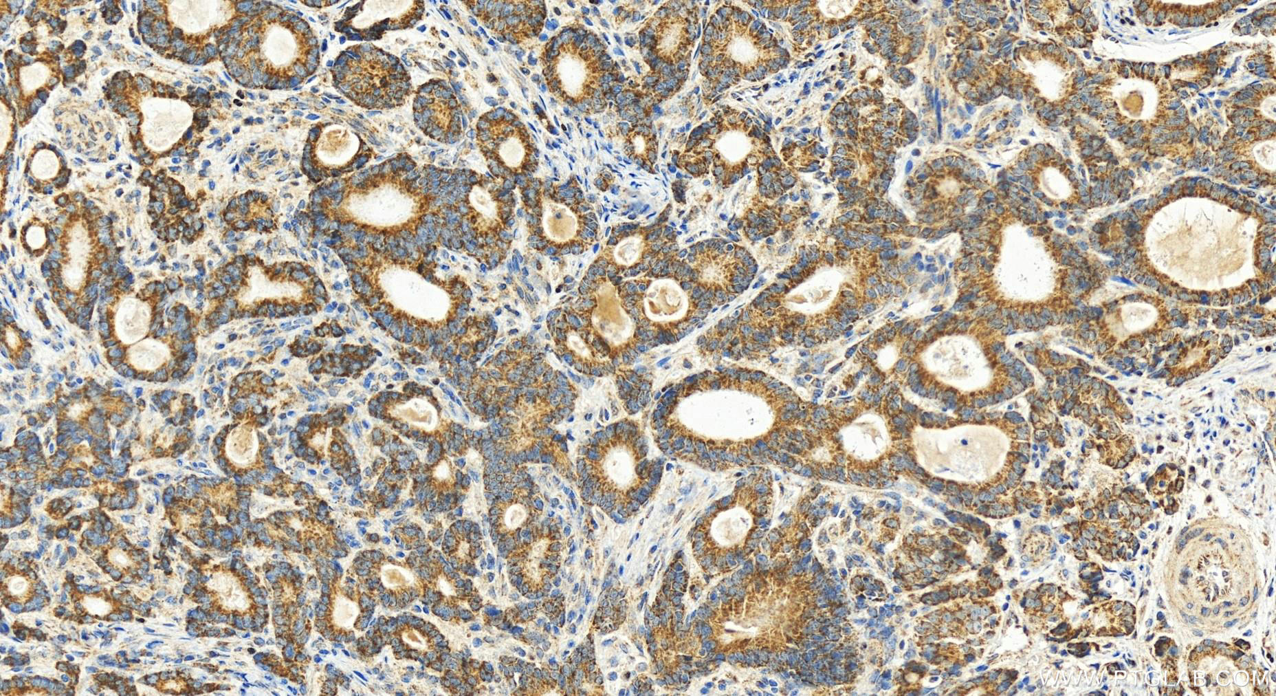
IHC staining of human stomach cancer using 15080-1-AP
Immunohistochemical analysis of paraffin-embedded human stomach cancer tissue slide using 15080-1-AP (POLDIP2 antibody) at dilution of 1:200 (under 20x lens). Heat mediated antigen retrieval with Tris-EDTA buffer (pH 9.0).
Immunohistochemical analysis of paraffin-embedded human stomach cancer tissue slide using 15080-1-AP (POLDIP2 antibody) at dilution of 1:200 (under 20x lens). Heat mediated antigen retrieval with Tris-EDTA buffer (pH 9.0).
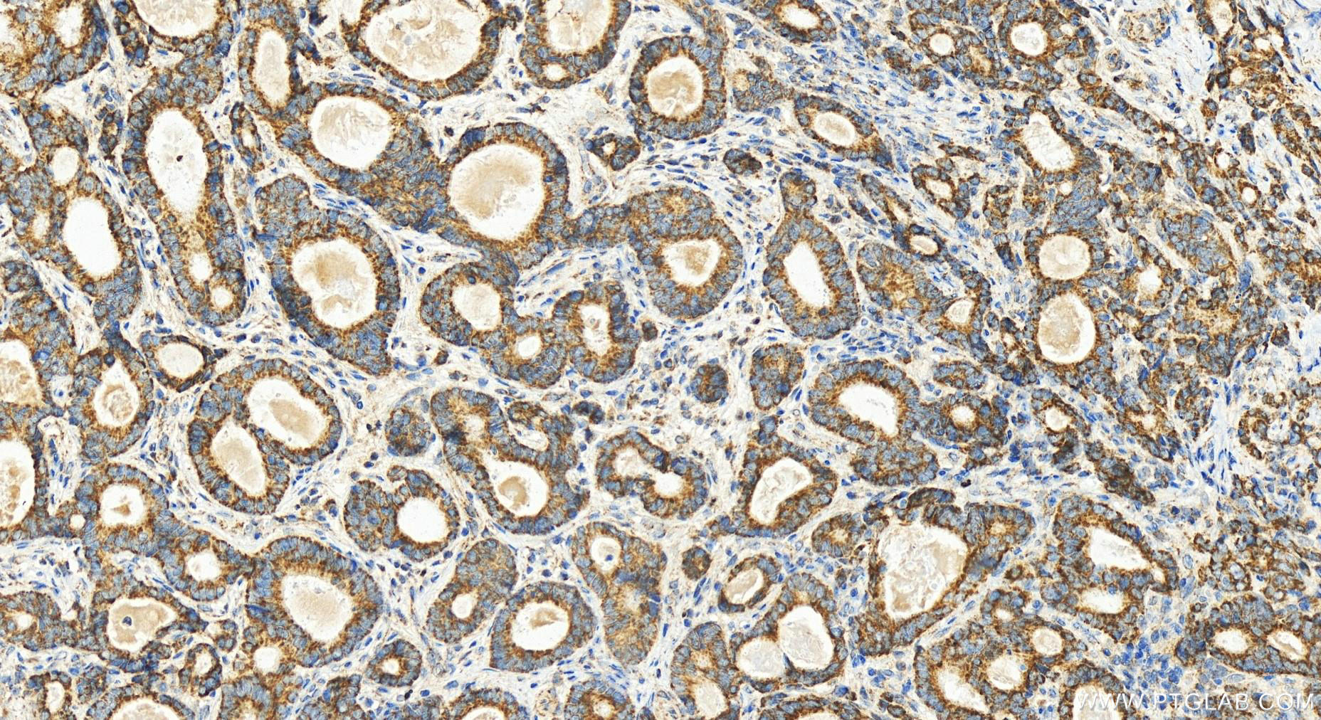
IF Staining of HeLa using 15080-1-AP
Immunofluorescent analysis of Hela cells, using POLDIP2 antibody 15080-1-AP at 1:25 dilution and Rhodamine-labeled goat anti-rabbit IgG (red).
The Proteintech guarantee covers Proteintech antibodies in any species and any application, including those not listed on the datasheet. If the antibody doesn’t perform, you can receive a hassle-free refund or credit note.
HeLa cells, HepG2 cells, Jurkat cells, K-562 cells
Positive IP detected in
HeLa cells
Positive IHC detected in
human stomach cancer tissue Note: suggested antigen retrieval with TE buffer pH 9.0; (*) Alternatively, antigen retrieval may be performed with citrate buffer pH 6.0
Positive IF/ICC detected in
Hela cells
Recommended dilution
Application
Dilution
Western Blot (WB)
WB : 1:500-1:3000
Immunoprecipitation (IP)
IP : 0.5-4.0 ug for 1.0-3.0 mg of total protein lysate
Immunohistochemistry (IHC)
IHC : 1:50-1:500
Immunofluorescence (IF)/ICC
IF/ICC : 1:10-1:100
It is recommended that this reagent should be titrated in each testing system to obtain optimal results.
Sample-dependent, Check data in validation data gallery.
PBS with 0.02% sodium azide and 50% glycerol , pH 7.3
Storage Conditions
Store at -20°C. Stable for one year after shipment. Aliquoting is unnecessary for -20oC storage. 20ul sizes contain 0.1% BSA.
Background Information
POLDIP2(Polymerase delta-interacting protein 2) is also named as PDIP38, POLD4. It is as a unique positive regulator of Nox4 via its association with p22phox and together with Nox4, has profound effects on Rhodependent cytoskeletal remodeling. It is also as a regulator of cell division. It consists of 368 amino acids and has a predicted molecular mass of 42 kDa, with a potential signal peptide cleavage site after the first N-terminal 48 residues, which would result in a protein of 37 kDa(PMID:19574552). Endogenous human and rat POLDIP2 localized to the nucleus, plasma membrane, and cytoplasm(PMID:17623671).
PDIP38 is translocated to the spliceosomes/nuclear speckles in response to UV-induced DNA damage and is required for UV-induced alternative splicing of MDM2.
HeLa cells were subjected to SDS PAGE followed by western blot with 15080-1-AP (POLDIP2 antibody) at dilution of 1:500 incubated at room temperature for 1.5 hours.
WB analysis of HepG2 using 15080-1-AP
HepG2 cells were subjected to SDS PAGE followed by western blot with 15080-1-AP (POLDIP2 antibody) at dilution of 1:500 incubated at room temperature for 1.5 hours.
WB analysis of Jurkat using 15080-1-AP
Jurkat cells were subjected to SDS PAGE followed by western blot with 15080-1-AP (POLDIP2 antibody) at dilution of 1:500 incubated at room temperature for 1.5 hours.
WB analysis of K-562 using 15080-1-AP
K-562 cells were subjected to SDS PAGE followed by western blot with 15080-1-AP (POLDIP2 antibody) at dilution of 1:500 incubated at room temperature for 1.5 hours.
IHC Figures
IHC staining of human stomach cancer using 15080-1-AP
Immunohistochemical analysis of paraffin-embedded human stomach cancer tissue slide using 15080-1-AP (POLDIP2 antibody) at dilution of 1:200 (under 20x lens). Heat mediated antigen retrieval with Tris-EDTA buffer (pH 9.0).
IHC staining of human stomach cancer using 15080-1-AP
Immunohistochemical analysis of paraffin-embedded human stomach cancer tissue slide using 15080-1-AP (POLDIP2 antibody) at dilution of 1:200 (under 20x lens). Heat mediated antigen retrieval with Tris-EDTA buffer (pH 9.0).
IP Figures
IP experiment of HeLa using 15080-1-AP
IP result of anti-POLDIP2 (IP:15080-1-AP, 4ug; Detection:15080-1-AP 1:1000) with HeLa cells lysate 1600ug.
IF/ICC Figures
IF Staining of HeLa using 15080-1-AP
Immunofluorescent analysis of Hela cells, using POLDIP2 antibody 15080-1-AP at 1:25 dilution and Rhodamine-labeled goat anti-rabbit IgG (red).
The species listed in Tested Reactivity are in-house verified and applicable species. For unlisted species, please refer to the homology analysis of the immunogen sequence and related species. For rabbit polyclonal antibodies, homology >70% is recommended. For mouse monoclonal antibodies and rabbit recombinant antibodies, homology >90% is recommended. Generally, the higher the homology, the greater the applicability. However, there will be certain differences in protein expression in different species, tissues or cells. Therefore, the homology analysis results are for reference only and do not serve as a guarantee.
At Proteintech, we pride ourselves on our antibody quality, customer service and transparency. As such, we are comparing our antibodies with other vendors, enabling easy identification and comparisons of key data to help you choose the suitable antibody for your needs.
We have selected the top cited antibodies from these vendors for you to compare.
Proteintech
POLDIP2 Polyclonal antibody
Catalog Number
15080-1-AP
Citations
3
Dilutions
WB : 1:500-1:3000 IP : 0.5-4.0 ug for IP and 0.5-4.0 ug for 1.0-3.0 mg of total protein lysate for WB IHC : 1:50-1:500 IF/ICC : 1:10-1:100
Applications
WB, IHC, IF/ICC, IP, ELISA
Reactivity
human, mouse, rat
Product Guarantee
Covers any species including not listed on datasheet
Covers any applications including not listed on datasheet
 at dilution of 1:500 incubated at room temperature for 1.5 hours.")
 at dilution of 1:500 incubated at room temperature for 1.5 hours.")
 at dilution of 1:500 incubated at room temperature for 1.5 hours.")
 at dilution of 1:500 incubated at room temperature for 1.5 hours.")
 with HeLa cells lysate 1600ug.")
 at dilution of 1:200 (under 20x lens). Heat mediated antigen retrieval with Tris-EDTA buffer (pH 9.0).")
 at dilution of 1:200 (under 20x lens). Heat mediated antigen retrieval with Tris-EDTA buffer (pH 9.0).")
.")