Tested Applications
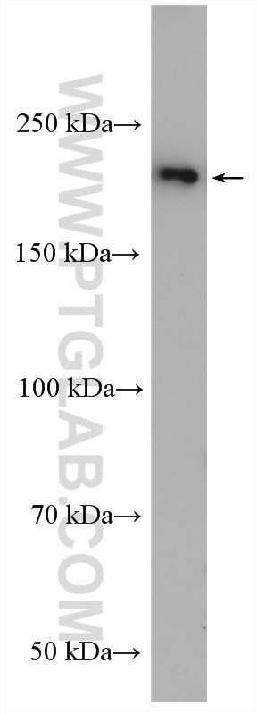
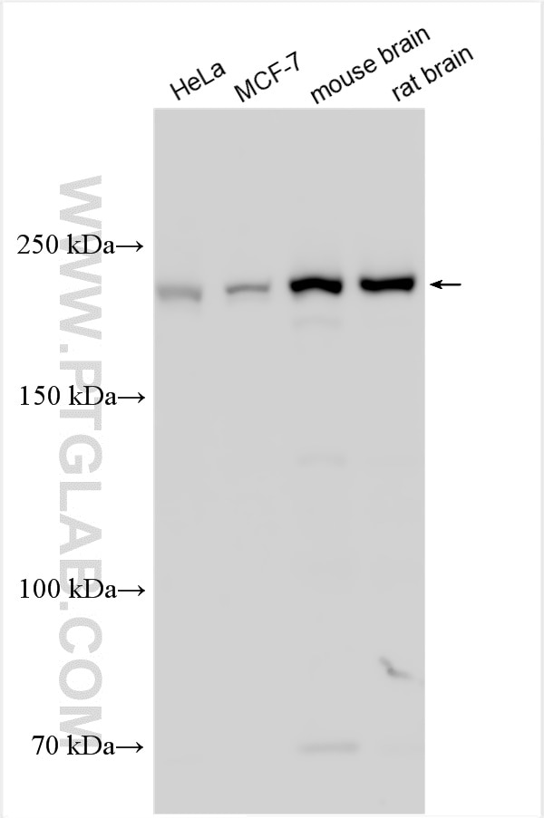
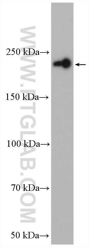
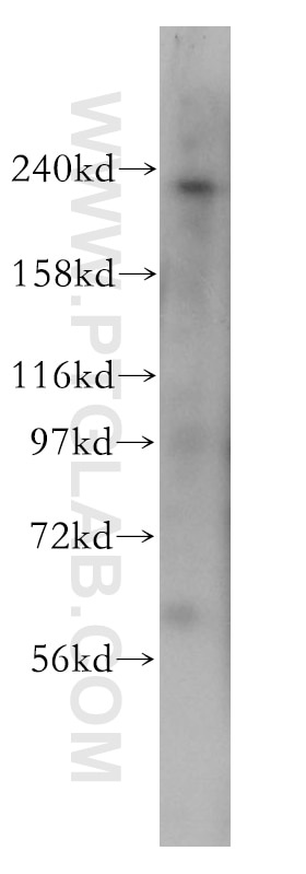
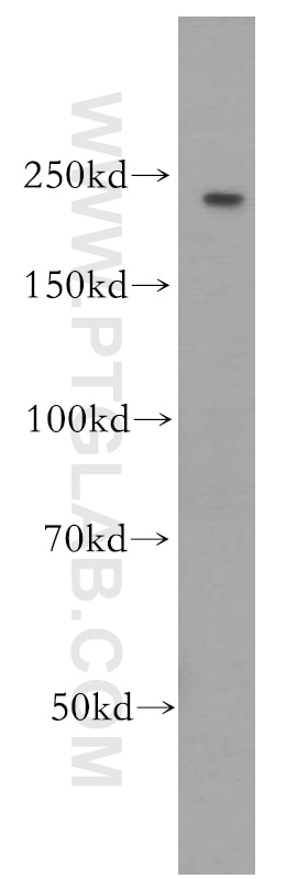
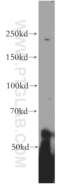
| Positive WB detected in | HeLa cells, mouse brain tissue, human brain tissue, MCF-7 cells, NIH-3T3 cells (RNAi), rat brain tissue |
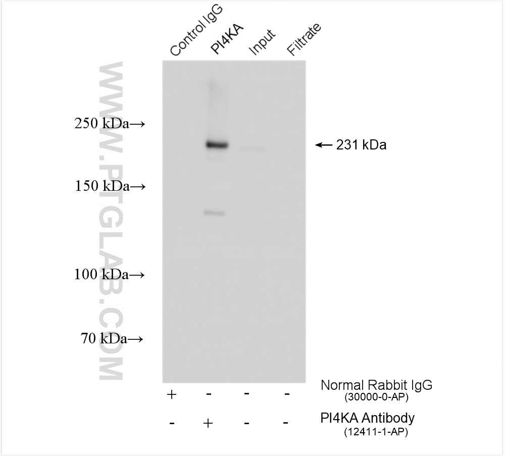
| Positive IP detected in | mouse brain tissue |
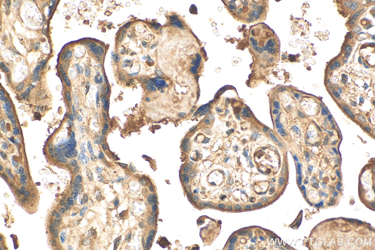
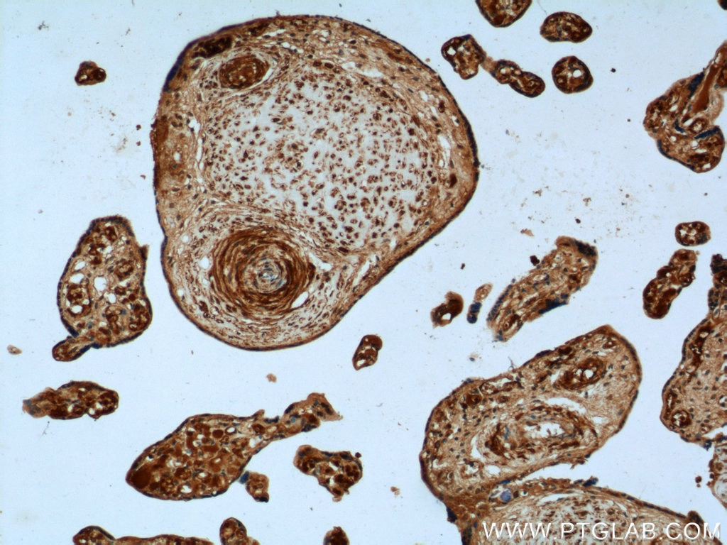
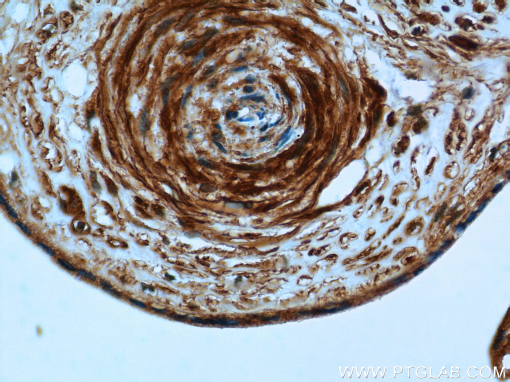
| Positive IHC detected in | human placenta tissue Note: suggested antigen retrieval with TE buffer pH 9.0; (*) Alternatively, antigen retrieval may be performed with citrate buffer pH 6.0 |
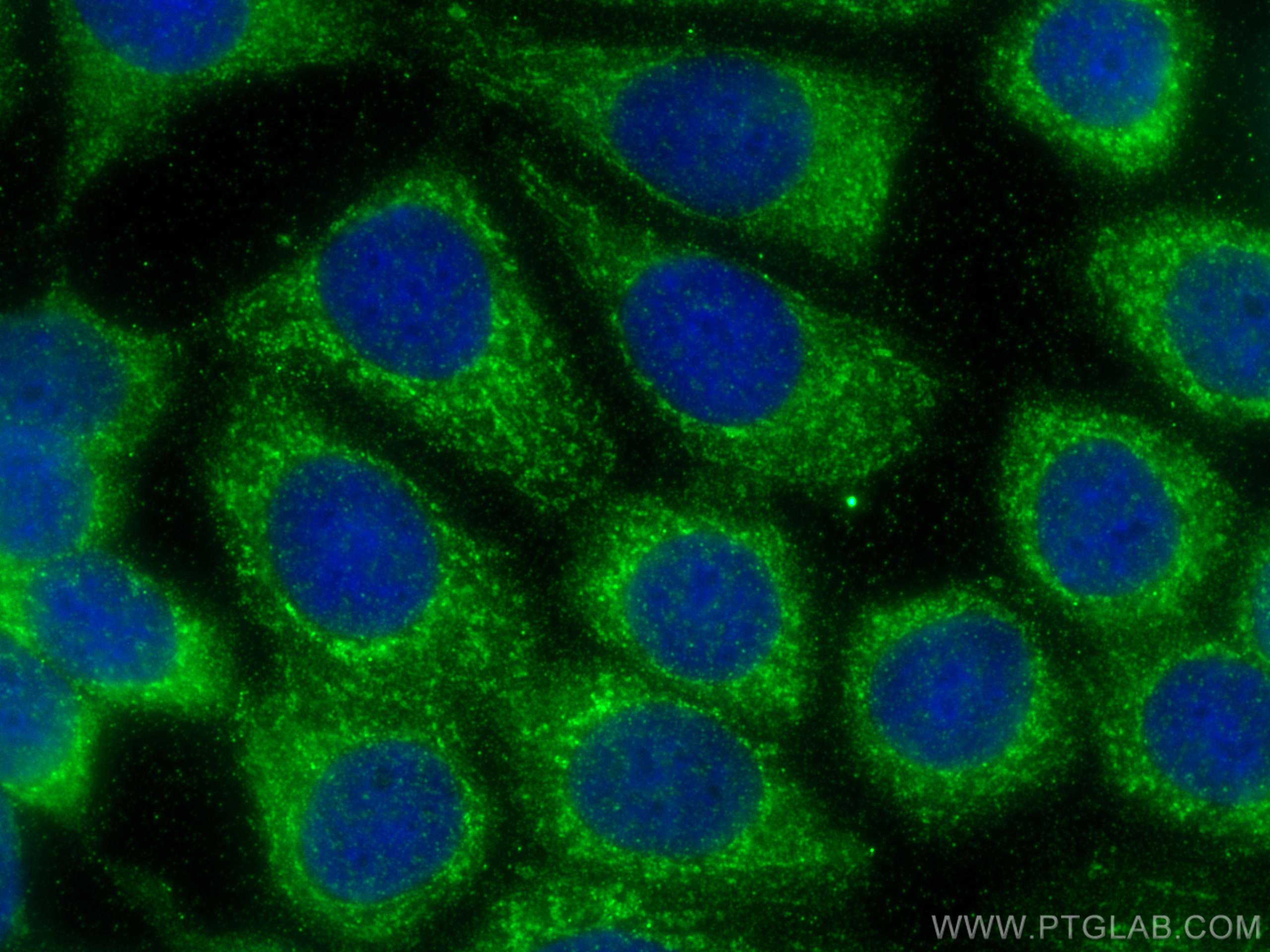
| Positive IF/ICC detected in | MCF-7 cells |
Recommended dilution
| Application | Dilution |
|---|---|
| Western Blot (WB) | WB : 1:1000-1:4000 |
| Immunoprecipitation (IP) | IP : 0.5-4.0 ug for 1.0-3.0 mg of total protein lysate |
| Immunohistochemistry (IHC) | IHC : 1:50-1:500 |
| Immunofluorescence (IF)/ICC | IF/ICC : 1:200-1:800 |
| It is recommended that this reagent should be titrated in each testing system to obtain optimal results. | |
| Sample-dependent, Check data in validation data gallery. | |
Published Applications
| KD/KO | See 8 publications below |
| WB | See 11 publications below |
| IHC | See 1 publications below |
| IF | See 4 publications below |
Product Information
12411-1-AP targets PI4KA in WB, IHC, IF/ICC, IP, ELISA applications and shows reactivity with human, mouse, rat samples.
| Tested Reactivity | human, mouse, rat |
| Cited Reactivity | human, mouse |
| Host / Isotype | Rabbit / IgG |
| Class | Polyclonal |
| Type | Antibody |
| Immunogen |
CatNo: Ag3062 Product name: Recombinant human PI4KA protein Source: e coli.-derived, PGEX-4T Tag: GST Domain: 1745-2044 aa of BC018120 Sequence: YLAKFKVKRCGVSELEKEGLRCRSDSEDECSTQEADGQKISWQAAIFKVGDDCRQDMLALQIIDLFKNIFQLVGLDLFVFPYRVVATAPGCGVIECIPDCTSRDQLGRQTDFGMYDYFTRQYGDESTLAFQQARYNFIRSMAAYSLLLFLLQIKDRHNGNIMLDKKGHIIHIDFGFMFESSPGGNLGWEPDIKLTDEMVMIMGGKMEATPFKWFMEMCVRGYLAVRPYMDAVVSLVTLMLDTGLPCFRGQTIKLLKHRFSPNMTEREAANFIMKVIQSCFLSNRSRTYDMIQYYQNDIPY Predict reactive species |
| Full Name | phosphatidylinositol 4-kinase, catalytic, alpha |
| Calculated Molecular Weight | 2044 aa, 231 kDa |
| Observed Molecular Weight | 231 kDa |
| GenBank Accession Number | BC018120 |
| Gene Symbol | PI4KA |
| Gene ID (NCBI) | 5297 |
| RRID | AB_2268237 |
| Conjugate | Unconjugated |
| Form | Liquid |
| Purification Method | Antigen affinity purification |
| UNIPROT ID | P42356 |
| Storage Buffer | PBS with 0.02% sodium azide and 50% glycerol, pH 7.3. |
| Storage Conditions | Store at -20°C. Stable for one year after shipment. Aliquoting is unnecessary for -20oC storage. 20ul sizes contain 0.1% BSA. |
Background Information
PI4KA, also named as PIK4, PIK4CA, belongs to the PI3/PI4-kinase family and Type III PI4K subfamily. It acts on phosphatidylinositol (PtdIns) in the first committed step in the production of the second messenger inositol-1,4,5,-trisphosphate. PI4KA plays a role in the formation of membrane complexes where HCV replication takes place. (PMID:19605471).
Protocols
| Product Specific Protocols | |
|---|---|
| IF protocol for PI4KA antibody 12411-1-AP | Download protocol |
| IHC protocol for PI4KA antibody 12411-1-AP | Download protocol |
| IP protocol for PI4KA antibody 12411-1-AP | Download protocol |
| WB protocol for PI4KA antibody 12411-1-AP | Download protocol |
| Standard Protocols | |
|---|---|
| Click here to view our Standard Protocols |
Publications
| Species | Application | Title |
|---|---|---|
Science Golgi-derived PI(4)P-containing vesicles drive late steps of mitochondrial division.
| ||
Brain Biallelic PI4KA variants cause a novel neurodevelopmental syndrome with hypomyelinating leukodystrophy. | ||
EMBO J Defining the proximal interaction networks of Arf GTPases reveals a mechanism for the regulation of PLD1 and PI4KB. | ||
Cell Rep A Forward Genetic Screen Targeting the Endothelium Reveals a Regulatory Role for the Lipid Kinase Pi4ka in Myelo- and Erythropoiesis.
| ||
Biomaterials Nrf-2-driven long noncoding RNA ODRUL contributes to modulating silver nanoparticle-induced effects on erythroid cells.
|
Reviews
The reviews below have been submitted by verified Proteintech customers who received an incentive for providing their feedback.
FH Patricia (Verified Customer) (06-16-2024) | Works well with 30ug of cell lysate
|


