Various lysates were subjected to SDS PAGE followed by western blot with 66372-1-Ig (PGRMC1 antibody) at dilution of 1:10000 incubated at room temperature for 1.5 hours. The membrane was stripped and reblotted with HRP-conjugated GAPDH Monoclonal antibody (HRP-60004) as loading control.
Various lysates were subjected to SDS PAGE followed by western blot with 66372-1-Ig (PGRMC1 antibody) at dilution of 1:10000 incubated at room temperature for 1.5 hours. The membrane was stripped and reblotted with HRP-conjugated GAPDH Monoclonal antibody (HRP-60004) as loading control.
WB analysis of HEK-293 using 66372-1-Ig
WB result of PGRMC1 antibody (66372-1-Ig; 1:2000; incubated at room temperature for 1.5 hours) with sh-Control and sh-PGRMC1 transfected HEK-293 cells.
WB result of PGRMC1 antibody (66372-1-Ig; 1:2000; incubated at room temperature for 1.5 hours) with sh-Control and sh-PGRMC1 transfected HEK-293 cells.
WB analysis of fetal human brain using 66372-1-Ig
fetal human brain tissue were subjected to SDS PAGE followed by western blot with 66372-1-Ig (PGRMC1 Antibody) at dilution of 1:2000 incubated at room temperature for 1.5 hours.
fetal human brain tissue were subjected to SDS PAGE followed by western blot with 66372-1-Ig (PGRMC1 Antibody) at dilution of 1:2000 incubated at room temperature for 1.5 hours.
WB analysis of MCF-7 using 66372-1-Ig
MCF-7 cells were subjected to SDS PAGE followed by western blot with 66372-1-Ig (PGRMC1 Antibody) at dilution of 1:2000 incubated at room temperature for 1.5 hours.
MCF-7 cells were subjected to SDS PAGE followed by western blot with 66372-1-Ig (PGRMC1 Antibody) at dilution of 1:2000 incubated at room temperature for 1.5 hours.
WB analysis of NIH/3T3 using 66372-1-Ig
NIH/3T3 cells were subjected to SDS PAGE followed by western blot with 66372-1-Ig (PGRMC1 Antibody) at dilution of 1:2000 incubated at room temperature for 1.5 hours.
NIH/3T3 cells were subjected to SDS PAGE followed by western blot with 66372-1-Ig (PGRMC1 Antibody) at dilution of 1:2000 incubated at room temperature for 1.5 hours.
IHC staining of human ovary tumor using 66372-1-Ig
Immunohistochemical analysis of paraffin-embedded human ovary tumor tissue slide using 66372-1-Ig (PGRMC1 Antibody) at dilution of 1:400 (under 10x lens). Heat mediated antigen retrieval with Tris-EDTA buffer (pH 9.0).
Immunohistochemical analysis of paraffin-embedded human ovary tumor tissue slide using 66372-1-Ig (PGRMC1 Antibody) at dilution of 1:400 (under 10x lens). Heat mediated antigen retrieval with Tris-EDTA buffer (pH 9.0).
IHC staining of human ovary tumor using 66372-1-Ig
Immunohistochemical analysis of paraffin-embedded human ovary tumor tissue slide using 66372-1-Ig (PGRMC1 Antibody) at dilution of 1:400 (under 40x lens). Heat mediated antigen retrieval with Tris-EDTA buffer (pH 9.0).
Immunohistochemical analysis of paraffin-embedded human ovary tumor tissue slide using 66372-1-Ig (PGRMC1 Antibody) at dilution of 1:400 (under 40x lens). Heat mediated antigen retrieval with Tris-EDTA buffer (pH 9.0).
IHC staining of human lung cancer using 66372-1-Ig
Immunohistochemical analysis of paraffin-embedded human lung cancer tissue slide using 66372-1-Ig (PGRMC1 Antibody) at dilution of 1:400 (under 10x lens). Heat mediated antigen retrieval with Tris-EDTA buffer (pH 9.0).
Immunohistochemical analysis of paraffin-embedded human lung cancer tissue slide using 66372-1-Ig (PGRMC1 Antibody) at dilution of 1:400 (under 10x lens). Heat mediated antigen retrieval with Tris-EDTA buffer (pH 9.0).
IHC staining of human lung cancer using 66372-1-Ig
Immunohistochemical analysis of paraffin-embedded human lung cancer tissue slide using 66372-1-Ig (PGRMC1 Antibody) at dilution of 1:400 (under 40x lens). Heat mediated antigen retrieval with Tris-EDTA buffer (pH 9.0).
Immunohistochemical analysis of paraffin-embedded human lung cancer tissue slide using 66372-1-Ig (PGRMC1 Antibody) at dilution of 1:400 (under 40x lens). Heat mediated antigen retrieval with Tris-EDTA buffer (pH 9.0).
IF Staining of human ovary tumor using 66372-1-Ig
Immunofluorescent analysis of (4% PFA) fixed human ovary tumor tissue using PGRMC1 antibody (66372-1-Ig, Clone: 1C10D11 ) at dilution of 1:400 and CoraLite®488-Conjugated AffiniPure Goat Anti-Mouse IgG(H+L).
Immunofluorescent analysis of (4% PFA) fixed human ovary tumor tissue using PGRMC1 antibody (66372-1-Ig, Clone: 1C10D11 ) at dilution of 1:400 and CoraLite®488-Conjugated AffiniPure Goat Anti-Mouse IgG(H+L).
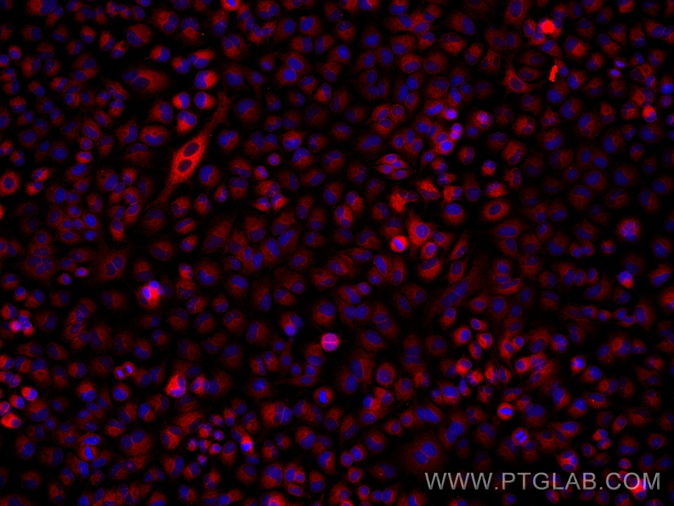
IF Staining of A549 using 66372-1-Ig
Immunofluorescent analysis of (4% PFA) fixed A549 cells using PGRMC1 antibody (66372-1-Ig, Clone: 1C10D11 ) at dilution of 1:2000 and Multi-rAb CoraLite® Plus 594-Goat Anti-Mouse Recombinant Secondary Antibody (H+L) (Cat.NO. RGAM004).
The Proteintech guarantee covers Proteintech antibodies in any species and any application, including those not listed on the datasheet. If the antibody doesn’t perform, you can receive a hassle-free refund or credit note.
A549 cells, fetal human brain tissue, MCF-7 cells, NIH/3T3 cells, HEK-293 cells, HeLa cells, Jurkat cells, K-562 cells, pig brain tissue, rat brain tissue, mouse brain tissue
Positive IHC detected in
human ovary tumor tissue, human lung cancer tissue Note: suggested antigen retrieval with TE buffer pH 9.0; (*) Alternatively, antigen retrieval may be performed with citrate buffer pH 6.0
Positive IF-P detected in
human ovary tumor tissue
Positive IF/ICC detected in
A549 cells
Recommended dilution
Application
Dilution
Western Blot (WB)
WB : 1:5000-1:50000
Immunohistochemistry (IHC)
IHC : 1:50-1:500
Immunofluorescence (IF)-P
IF-P : 1:200-1:800
Immunofluorescence (IF)/ICC
IF/ICC : 1:1000-1:4000
It is recommended that this reagent should be titrated in each testing system to obtain optimal results.
Sample-dependent, Check data in validation data gallery.
PBS with 0.02% sodium azide and 50% glycerol , pH 7.3
Storage Conditions
Store at -20°C. Stable for one year after shipment. Aliquoting is unnecessary for -20oC storage. 20ul sizes contain 0.1% BSA.
Background Information
Progesterone receptor membrane component 1 (PGRMC1) is a member of a multi-protein progesterone-binding complex. However, PGRMC1 shares homology with cytochrome b5-related proteins rather than hormone receptors (PMID: 18992768). It is a heme binding protein with biding sites for Src homology (SH2) and SH3 domain-containing proteins (PMID: 17583495). PGRMC1 is overexpressed in a variety of cancers, and thus represents an important biomarker for cancer progression and a potential target for anticancer drugs (PMID: 21730960). In nonmalignant tissues, PGRMC1 is highly expressed in the liver and kidney (PMID: 9705155; 20164297).
Various lysates were subjected to SDS PAGE followed by western blot with 66372-1-Ig (PGRMC1 antibody) at dilution of 1:10000 incubated at room temperature for 1.5 hours. The membrane was stripped and reblotted with HRP-conjugated GAPDH Monoclonal antibody (HRP-60004) as loading control.
WB analysis of HEK-293 using 66372-1-Ig
WB result of PGRMC1 antibody (66372-1-Ig; 1:2000; incubated at room temperature for 1.5 hours) with sh-Control and sh-PGRMC1 transfected HEK-293 cells.
WB analysis of fetal human brain using 66372-1-Ig
fetal human brain tissue were subjected to SDS PAGE followed by western blot with 66372-1-Ig (PGRMC1 Antibody) at dilution of 1:2000 incubated at room temperature for 1.5 hours.
WB analysis of MCF-7 using 66372-1-Ig
MCF-7 cells were subjected to SDS PAGE followed by western blot with 66372-1-Ig (PGRMC1 Antibody) at dilution of 1:2000 incubated at room temperature for 1.5 hours.
WB analysis of NIH/3T3 using 66372-1-Ig
NIH/3T3 cells were subjected to SDS PAGE followed by western blot with 66372-1-Ig (PGRMC1 Antibody) at dilution of 1:2000 incubated at room temperature for 1.5 hours.
IHC Figures
IHC staining of human ovary tumor using 66372-1-Ig
Immunohistochemical analysis of paraffin-embedded human ovary tumor tissue slide using 66372-1-Ig (PGRMC1 Antibody) at dilution of 1:400 (under 10x lens). Heat mediated antigen retrieval with Tris-EDTA buffer (pH 9.0).
IHC staining of human ovary tumor using 66372-1-Ig
Immunohistochemical analysis of paraffin-embedded human ovary tumor tissue slide using 66372-1-Ig (PGRMC1 Antibody) at dilution of 1:400 (under 40x lens). Heat mediated antigen retrieval with Tris-EDTA buffer (pH 9.0).
IHC staining of human lung cancer using 66372-1-Ig
Immunohistochemical analysis of paraffin-embedded human lung cancer tissue slide using 66372-1-Ig (PGRMC1 Antibody) at dilution of 1:400 (under 10x lens). Heat mediated antigen retrieval with Tris-EDTA buffer (pH 9.0).
IHC staining of human lung cancer using 66372-1-Ig
Immunohistochemical analysis of paraffin-embedded human lung cancer tissue slide using 66372-1-Ig (PGRMC1 Antibody) at dilution of 1:400 (under 40x lens). Heat mediated antigen retrieval with Tris-EDTA buffer (pH 9.0).
IF-P Figures
IF Staining of human ovary tumor using 66372-1-Ig
Immunofluorescent analysis of (4% PFA) fixed human ovary tumor tissue using PGRMC1 antibody (66372-1-Ig, Clone: 1C10D11 ) at dilution of 1:400 and CoraLite®488-Conjugated AffiniPure Goat Anti-Mouse IgG(H+L).
IF Staining of human ovary tumor using 66372-1-Ig
Immunofluorescent analysis of (4% PFA) fixed human ovary tumor tissue using PGRMC1 antibody (66372-1-Ig, Clone: 1C10D11 ) at dilution of 1:400 and CoraLite®488-Conjugated AffiniPure Goat Anti-Mouse IgG(H+L).
IF/ICC Figures
IF Staining of A549 using 66372-1-Ig
Immunofluorescent analysis of (4% PFA) fixed A549 cells using PGRMC1 antibody (66372-1-Ig, Clone: 1C10D11 ) at dilution of 1:2000 and Multi-rAb CoraLite® Plus 594-Goat Anti-Mouse Recombinant Secondary Antibody (H+L) (Cat.NO. RGAM004).
The species listed in Tested Reactivity are in-house verified and applicable species. For unlisted species, please refer to the homology analysis of the immunogen sequence and related species. For rabbit polyclonal antibodies, homology >70% is recommended. For mouse monoclonal antibodies and rabbit recombinant antibodies, homology >90% is recommended. Generally, the higher the homology, the greater the applicability. However, there will be certain differences in protein expression in different species, tissues or cells. Therefore, the homology analysis results are for reference only and do not serve as a guarantee.
At Proteintech, we pride ourselves on our antibody quality, customer service and transparency. As such, we are comparing our antibodies with other vendors, enabling easy identification and comparisons of key data to help you choose the suitable antibody for your needs.
We have selected the top cited antibodies from these vendors for you to compare.
 at dilution of 1:10000 incubated at room temperature for 1.5 hours. The membrane was stripped and reblotted with HRP-conjugated GAPDH Monoclonal antibody (HRP-60004) as loading control.")
 with sh-Control and sh-PGRMC1 transfected HEK-293 cells.")
 at dilution of 1:2000 incubated at room temperature for 1.5 hours.")
 at dilution of 1:2000 incubated at room temperature for 1.5 hours.")
 at dilution of 1:2000 incubated at room temperature for 1.5 hours.")
 at dilution of 1:400 (under 10x lens). Heat mediated antigen retrieval with Tris-EDTA buffer (pH 9.0).")
 at dilution of 1:400 (under 40x lens). Heat mediated antigen retrieval with Tris-EDTA buffer (pH 9.0).")
 at dilution of 1:400 (under 10x lens). Heat mediated antigen retrieval with Tris-EDTA buffer (pH 9.0).")
 at dilution of 1:400 (under 40x lens). Heat mediated antigen retrieval with Tris-EDTA buffer (pH 9.0).")
 fixed human ovary tumor tissue using PGRMC1 antibody (66372-1-Ig, Clone: 1C10D11 ) at dilution of 1:400 and CoraLite®488-Conjugated AffiniPure Goat Anti-Mouse IgG(H+L).")
 fixed human ovary tumor tissue using PGRMC1 antibody (66372-1-Ig, Clone: 1C10D11 ) at dilution of 1:400 and CoraLite®488-Conjugated AffiniPure Goat Anti-Mouse IgG(H+L).")
 fixed A549 cells using PGRMC1 antibody (66372-1-Ig, Clone: 1C10D11 ) at dilution of 1:2000 and Multi-rAb CoraLite® Plus 594-Goat Anti-Mouse Recombinant Secondary Antibody (H+L) (Cat.NO. RGAM004).")