at dilution of 1:3000 incubated at room temperature for 1.5 hours.")
 with sh-Control and sh-PFKFB3-Specific transfected HEK-293 cells.")

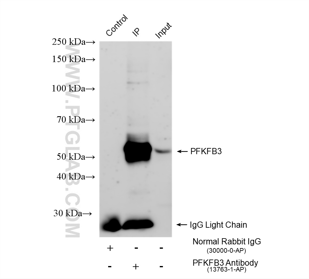
 at dilution of 1:1000 incubated at room temperature for 1.5 hours.")
 at dilution of 1:1000 incubated at room temperature for 1.5 hours.")
 with mouse spleen tissue lysate 2000 ug.")
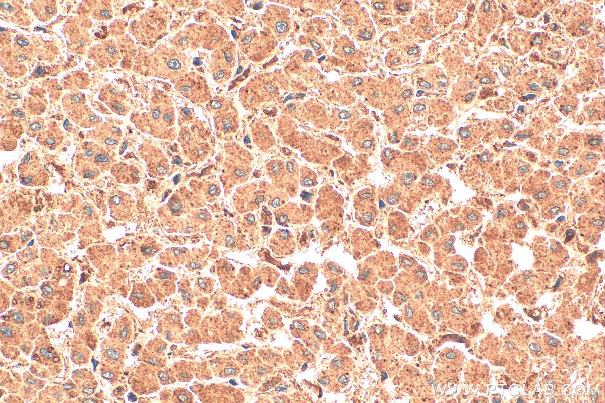
 at dilution of 1:600 (under 40x lens). Heat mediated antigen retrieval with Tris-EDTA buffer (pH 9.0).")
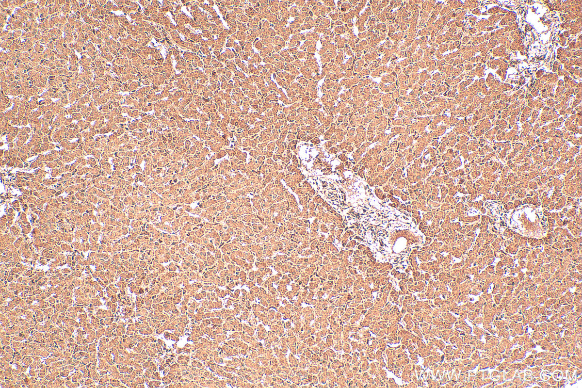
 at dilution of 1:600 (under 10x lens). Heat mediated antigen retrieval with Tris-EDTA buffer (pH 9.0).")
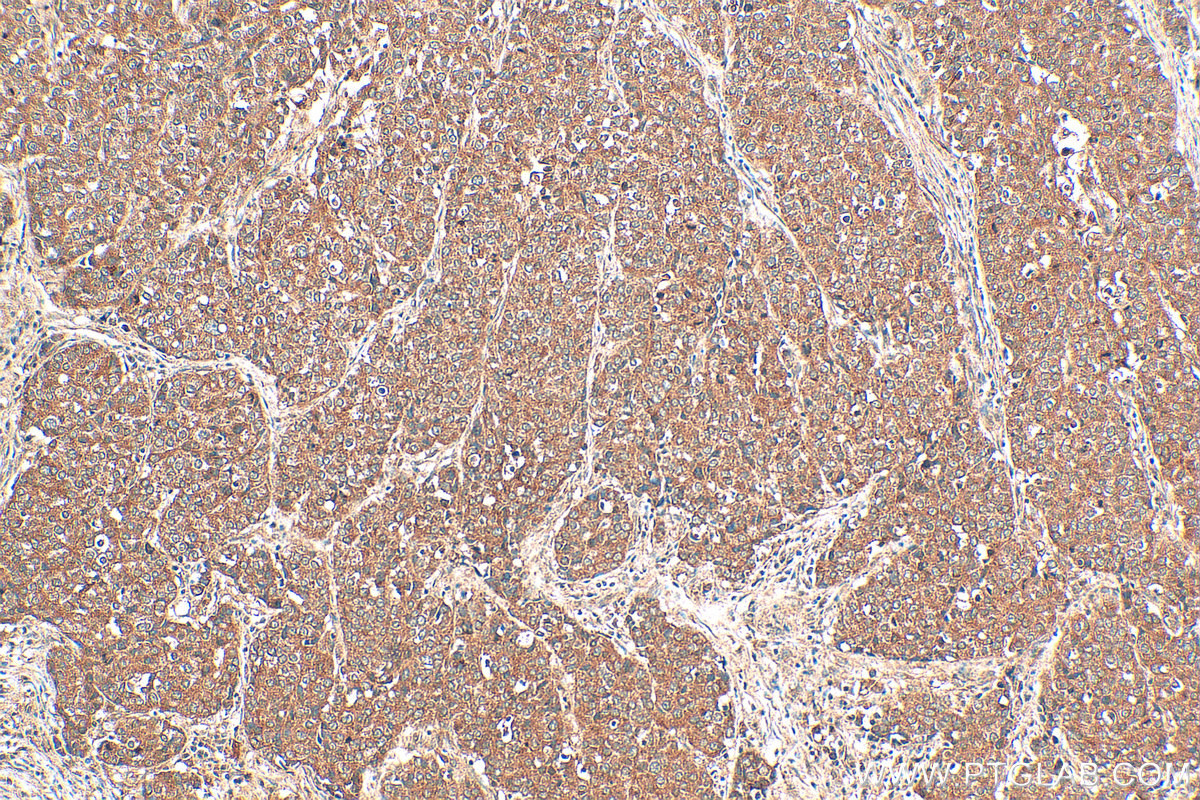
 at dilution of 1:200 (under 10x lens). Heat mediated antigen retrieval with Tris-EDTA buffer (pH 9.0).")
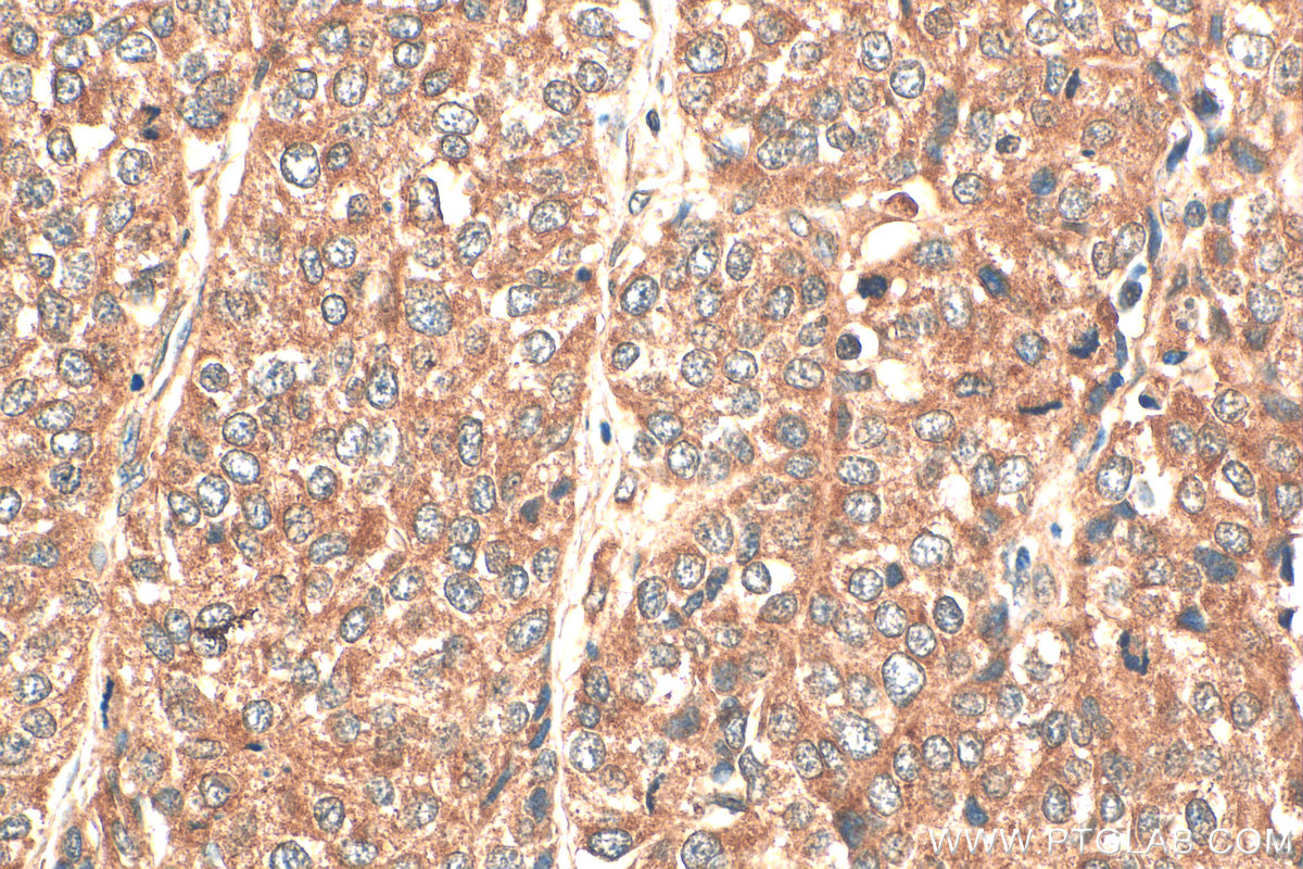
 at dilution of 1:200 (under 40x lens). Heat mediated antigen retrieval with Tris-EDTA buffer (pH 9.0).")
 at dilution of 1:50 (under 10x lens).")
 at dilution of 1:50 (under 40x lens).")
Tested Applications
| Positive WB detected in | HEK-293 cells, mouse thymus tissue, mouse heart tissue, A431 cells, HeLa cells, Jurkat cells, mouse spleen tissue |
| Positive IP detected in | mouse spleen tissue |
| Positive IHC detected in | human stomach cancer tissue, human kidney tissue, human liver tissue Note: suggested antigen retrieval with TE buffer pH 9.0; (*) Alternatively, antigen retrieval may be performed with citrate buffer pH 6.0 |
Recommended dilution
| Application | Dilution |
|---|---|
| Western Blot (WB) | WB : 1:1000-1:6000 |
| Immunoprecipitation (IP) | IP : 0.5-4.0 ug for 1.0-3.0 mg of total protein lysate |
| Immunohistochemistry (IHC) | IHC : 1:300-1:1200 |
| It is recommended that this reagent should be titrated in each testing system to obtain optimal results. | |
| Sample-dependent, Check data in validation data gallery. | |
Published Applications
| KD/KO | See 19 publications below |
| WB | See 135 publications below |
| IHC | See 31 publications below |
| IF | See 21 publications below |
| IP | See 4 publications below |
| CoIP | See 2 publications below |
Product Information
13763-1-AP targets PFKFB3 in WB, IHC, IF, IP, CoIP, ELISA applications and shows reactivity with human, mouse samples.
| Tested Reactivity | human, mouse |
| Cited Reactivity | human, mouse, rat, rabbit |
| Host / Isotype | Rabbit / IgG |
| Class | Polyclonal |
| Type | Antibody |
| Immunogen |
CatNo: Ag4744 Product name: Recombinant human PFKFB3 protein Source: e coli.-derived, PGEX-4T Tag: GST Domain: 243-520 aa of BC040482 Sequence: HVQPRTIYLCRHGENEHNLQGRIGGDSGLSSRGKKFASALSKFVEEQNLKDLRVWTSQLKSTIQTAEALRLPYEQWKALNEIDAGVCEELTYEEIRDTYPEEYALREQDKYYYRYPTGESYQDLVQRLEPVIMELERQENVLVICHQAVLRCLLAYFLDKSAEEMPYLKCPLHTVLKLTPVAYGCRVESIYLNVESVCTHRERSEDAKKGPNPLMRRNSVTPLASPEPTKKPRINSFEEHVASTSAALPSCLPPEVPTQLPGQNMKGSRSSADSSRKH Predict reactive species |
| Full Name | 6-phosphofructo-2-kinase/fructose-2,6-biphosphatase 3 |
| Calculated Molecular Weight | 520 aa, 60 kDa |
| Observed Molecular Weight | 58 kDa |
| GenBank Accession Number | BC040482 |
| Gene Symbol | PFKFB3 |
| Gene ID (NCBI) | 5209 |
| RRID | AB_2162854 |
| Conjugate | Unconjugated |
| Form | Liquid |
| Purification Method | Antigen affinity purification |
| UNIPROT ID | Q16875 |
| Storage Buffer | PBS with 0.02% sodium azide and 50% glycerol, pH 7.3. |
| Storage Conditions | Store at -20°C. Stable for one year after shipment. Aliquoting is unnecessary for -20oC storage. 20ul sizes contain 0.1% BSA. |
Background Information
PFKFB3, also named as NY-REN-56 and iPFK-2, plays a role in glucose metabolism. Its synthesis and degradation of fructose 2,6-bisphosphate. Endogenously generated adenosine cooperates with bacterial components to increase PFKFB3 isozyme activity, resulting in greater fructose 2,6-bisphosphate accumulation. PFKFB3 is required for increased growth, metabolic activity and is regulated through active JAK2 and STAT5.
Protocols
| Product Specific Protocols | |
|---|---|
| IHC protocol for PFKFB3 antibody 13763-1-AP | Download protocol |
| IP protocol for PFKFB3 antibody 13763-1-AP | Download protocol |
| WB protocol for PFKFB3 antibody 13763-1-AP | Download protocol |
| Standard Protocols | |
|---|---|
| Click here to view our Standard Protocols |
Publications
| Species | Application | Title |
|---|---|---|
Cell Metab Acetate enables metabolic fitness and cognitive performance during sleep disruption | ||
Cell Metab NEAT1 is essential for metabolic changes that promote breast cancer growth and metastasis. | ||
Cell Metab CircACC1 Regulates Assembly and Activation of AMPK Complex under Metabolic Stress. | ||
Circulation Exercise-Induced Changes in Glucose Metabolism Promote Physiologic Cardiac Growth. |
Reviews
The reviews below have been submitted by verified Proteintech customers who received an incentive for providing their feedback.
FH Balawant (Verified Customer) (08-24-2022) | this antibody is working good for westerner blot.
|
FH Balawant (Verified Customer) (05-08-2022) | tis antibody is working great at 1:2000 diluton. I have used this antibody from total cells lysate of colonic epithelial cells form human origin.
|
FH Iram (Verified Customer) (04-13-2022) | Very good signal
|
FH Susmita (Verified Customer) (03-11-2022) | This antibody works great for me.
|
FH Hsin-Sheng (Verified Customer) (08-19-2019) | Strong bend intensity at around 60 kD. However, there is a non-specific band near 65-70 kD.
|