Various lysates were subjected to SDS PAGE followed by western blot with 68238-1-Ig (PDHB antibody) at dilution of 1:10000 incubated at room temperature for 1.5 hours.
Various lysates were subjected to SDS PAGE followed by western blot with 68238-1-Ig (PDHB antibody) at dilution of 1:10000 incubated at room temperature for 1.5 hours.
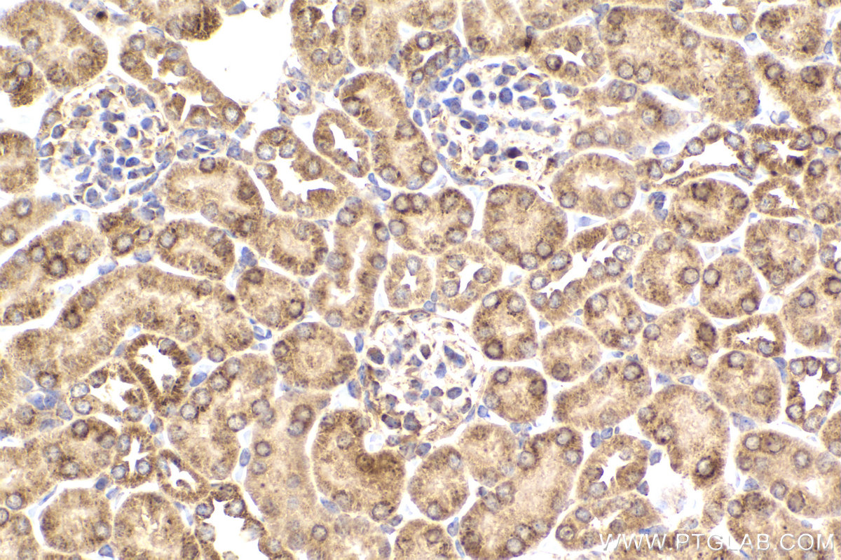
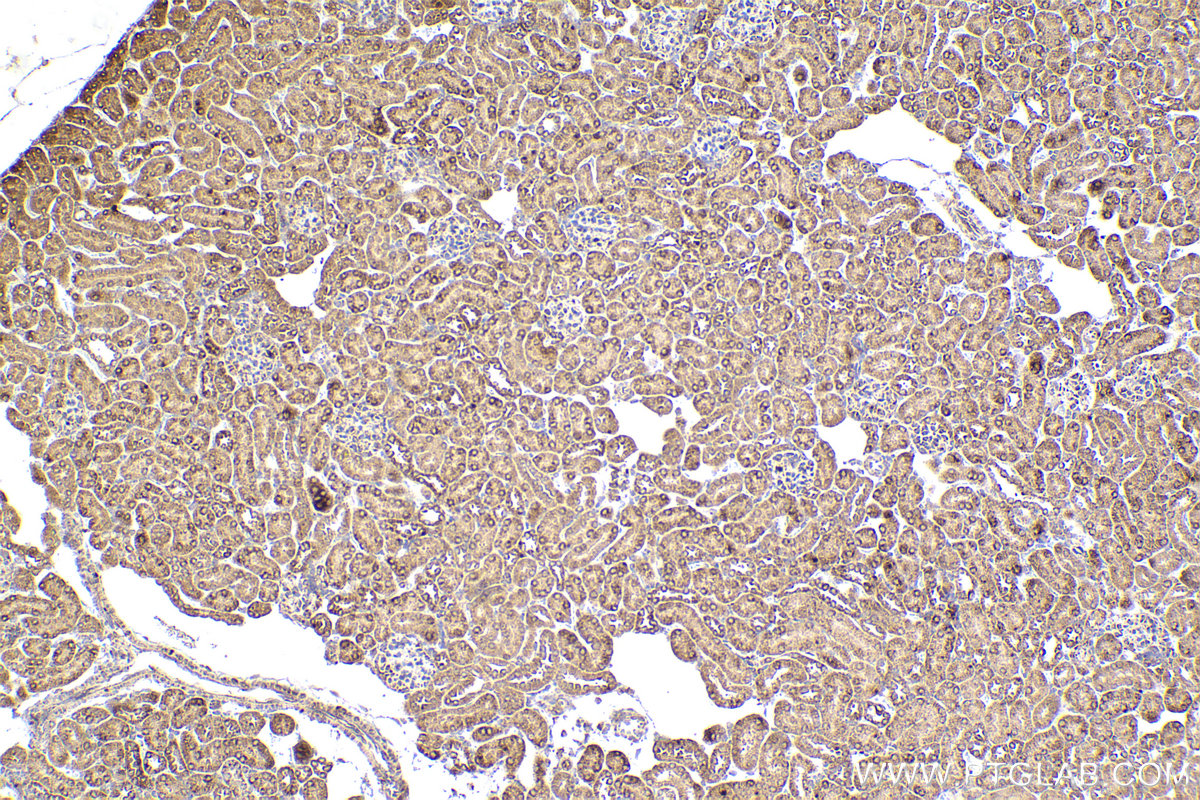
IHC staining of mouse kidney using 68238-1-Ig
Immunohistochemical analysis of paraffin-embedded mouse kidney tissue slide using 68238-1-Ig (PDHB antibody) at dilution of 1:1000 (under 40x lens). Heat mediated antigen retrieval with Tris-EDTA buffer (pH 9.0).
Immunohistochemical analysis of paraffin-embedded mouse kidney tissue slide using 68238-1-Ig (PDHB antibody) at dilution of 1:1000 (under 10x lens). Heat mediated antigen retrieval with Tris-EDTA buffer (pH 9.0).
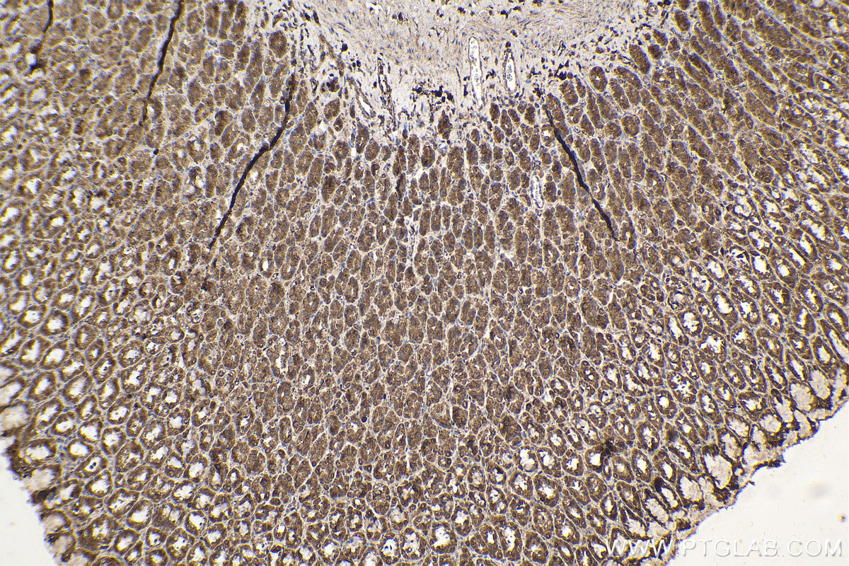
IHC staining of rat stomach using 68238-1-Ig
Immunohistochemical analysis of paraffin-embedded rat stomach tissue slide using 68238-1-Ig (PDHB antibody) at dilution of 1:500 (under 10x lens). Heat mediated antigen retrieval with Tris-EDTA buffer (pH 9.0).
Immunohistochemical analysis of paraffin-embedded rat stomach tissue slide using 68238-1-Ig (PDHB antibody) at dilution of 1:500 (under 10x lens). Heat mediated antigen retrieval with Tris-EDTA buffer (pH 9.0).
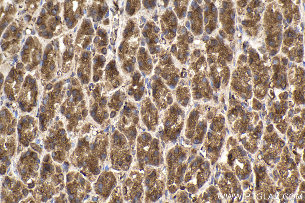
IHC staining of rat stomach using 68238-1-Ig
Immunohistochemical analysis of paraffin-embedded rat stomach tissue slide using 68238-1-Ig (PDHB antibody) at dilution of 1:500 (under 40x lens). Heat mediated antigen retrieval with Tris-EDTA buffer (pH 9.0).
Immunohistochemical analysis of paraffin-embedded rat stomach tissue slide using 68238-1-Ig (PDHB antibody) at dilution of 1:500 (under 40x lens). Heat mediated antigen retrieval with Tris-EDTA buffer (pH 9.0).
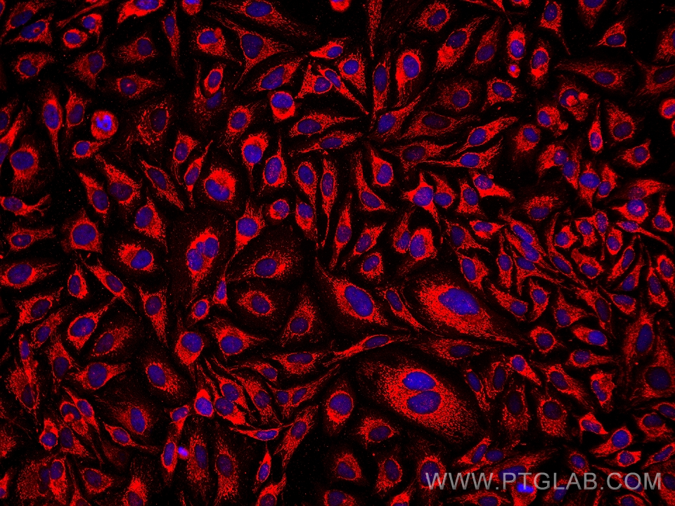
IF Staining of HeLa using 68238-1-Ig
Immunofluorescent analysis of (4% PFA) fixed HeLa cells using PDHB antibody (68238-1-Ig, Clone: 2A7C10 ) at dilution of 1:1000 and Multi-rAb CoraLite® Plus 594-Goat Anti-Mouse Recombinant Secondary Antibody (H+L) (Cat.NO. RGAM004 ).
Immunofluorescent analysis of (4% PFA) fixed HeLa cells using PDHB antibody (68238-1-Ig, Clone: 2A7C10 ) at dilution of 1:1000 and Multi-rAb CoraLite® Plus 594-Goat Anti-Mouse Recombinant Secondary Antibody (H+L) (Cat.NO. RGAM004 ).
The Proteintech guarantee covers Proteintech antibodies in any species and any application, including those not listed on the datasheet. If the antibody doesn’t perform, you can receive a hassle-free refund or credit note.
mouse kidney tissue, rat stomach tissue Note: suggested antigen retrieval with TE buffer pH 9.0; (*) Alternatively, antigen retrieval may be performed with citrate buffer pH 6.0
Positive IF/ICC detected in
HeLa cells
Recommended dilution
Application
Dilution
Western Blot (WB)
WB : 1:5000-1:50000
Immunohistochemistry (IHC)
IHC : 1:500-1:2000
Immunofluorescence (IF)/ICC
IF/ICC : 1:500-1:2000
It is recommended that this reagent should be titrated in each testing system to obtain optimal results.
Sample-dependent, Check data in validation data gallery.
Product Information
68238-1-Ig targets PDHB in WB, IHC, IF/ICC, ELISA applications and shows reactivity with human, mouse, rat, pig, rabbit samples.
PBS with 0.02% sodium azide and 50% glycerol pH 7.3.
Storage Conditions
Store at -20°C. Stable for one year after shipment. Aliquoting is unnecessary for -20oC storage. 20ul sizes contain 0.1% BSA.
Background Information
PDHB (Pyruvate dehydrogenase E1 component subunit beta, mitochondrial) is also named PHE1B. The enzyme, which is found in mitochondria, is one of the component enzymes of the pyruvate dehydrogenase multienzyme complex (PDH). It catalyzes the first reaction of an oxidative decarboxylation sequence converting pyruvate to acetyl-CoA and CO(2)(PMID:3422424). Defects in PDHB are the cause of pyruvate dehydrogenase E1-beta deficiency (PDHBD)(PMID:15138885).
Various lysates were subjected to SDS PAGE followed by western blot with 68238-1-Ig (PDHB antibody) at dilution of 1:10000 incubated at room temperature for 1.5 hours.
IHC Figures
IHC staining of mouse kidney using 68238-1-Ig
Immunohistochemical analysis of paraffin-embedded mouse kidney tissue slide using 68238-1-Ig (PDHB antibody) at dilution of 1:1000 (under 40x lens). Heat mediated antigen retrieval with Tris-EDTA buffer (pH 9.0).
IHC staining of mouse kidney using 68238-1-Ig
Immunohistochemical analysis of paraffin-embedded mouse kidney tissue slide using 68238-1-Ig (PDHB antibody) at dilution of 1:1000 (under 10x lens). Heat mediated antigen retrieval with Tris-EDTA buffer (pH 9.0).
IHC staining of rat stomach using 68238-1-Ig
Immunohistochemical analysis of paraffin-embedded rat stomach tissue slide using 68238-1-Ig (PDHB antibody) at dilution of 1:500 (under 10x lens). Heat mediated antigen retrieval with Tris-EDTA buffer (pH 9.0).
IHC staining of rat stomach using 68238-1-Ig
Immunohistochemical analysis of paraffin-embedded rat stomach tissue slide using 68238-1-Ig (PDHB antibody) at dilution of 1:500 (under 40x lens). Heat mediated antigen retrieval with Tris-EDTA buffer (pH 9.0).
IF/ICC Figures
IF Staining of HeLa using 68238-1-Ig
Immunofluorescent analysis of (4% PFA) fixed HeLa cells using PDHB antibody (68238-1-Ig, Clone: 2A7C10 ) at dilution of 1:1000 and Multi-rAb CoraLite® Plus 594-Goat Anti-Mouse Recombinant Secondary Antibody (H+L) (Cat.NO. RGAM004 ).
The species listed in Tested Reactivity are in-house verified and applicable species. For unlisted species, please refer to the homology analysis of the immunogen sequence and related species. For rabbit polyclonal antibodies, homology >70% is recommended. For mouse monoclonal antibodies and rabbit recombinant antibodies, homology >90% is recommended. Generally, the higher the homology, the greater the applicability. However, there will be certain differences in protein expression in different species, tissues or cells. Therefore, the homology analysis results are for reference only and do not serve as a guarantee.
At Proteintech, we pride ourselves on our antibody quality, customer service and transparency. As such, we are comparing our antibodies with other vendors, enabling easy identification and comparisons of key data to help you choose the suitable antibody for your needs.
We have selected the top cited antibodies from these vendors for you to compare.
 at dilution of 1:10000 incubated at room temperature for 1.5 hours.")
 at dilution of 1:1000 (under 40x lens). Heat mediated antigen retrieval with Tris-EDTA buffer (pH 9.0).")
 at dilution of 1:1000 (under 10x lens). Heat mediated antigen retrieval with Tris-EDTA buffer (pH 9.0).")
 at dilution of 1:500 (under 10x lens). Heat mediated antigen retrieval with Tris-EDTA buffer (pH 9.0).")
 at dilution of 1:500 (under 40x lens). Heat mediated antigen retrieval with Tris-EDTA buffer (pH 9.0).")
 fixed HeLa cells using PDHB antibody (68238-1-Ig, Clone: 2A7C10 ) at dilution of 1:1000 and Multi-rAb CoraLite® Plus 594-Goat Anti-Mouse Recombinant Secondary Antibody (H+L) (Cat.NO. RGAM004 ).")