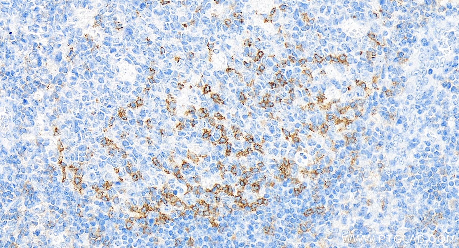
at dilution of 1:1000 (under 10x lens). Heat mediated antigen retrieval with Tris-EDTA buffer (pH 9.0).")
 at dilution of 1:1000 (under 40x lens). Heat mediated antigen retrieval with Tris-EDTA buffer (pH 9.0).")
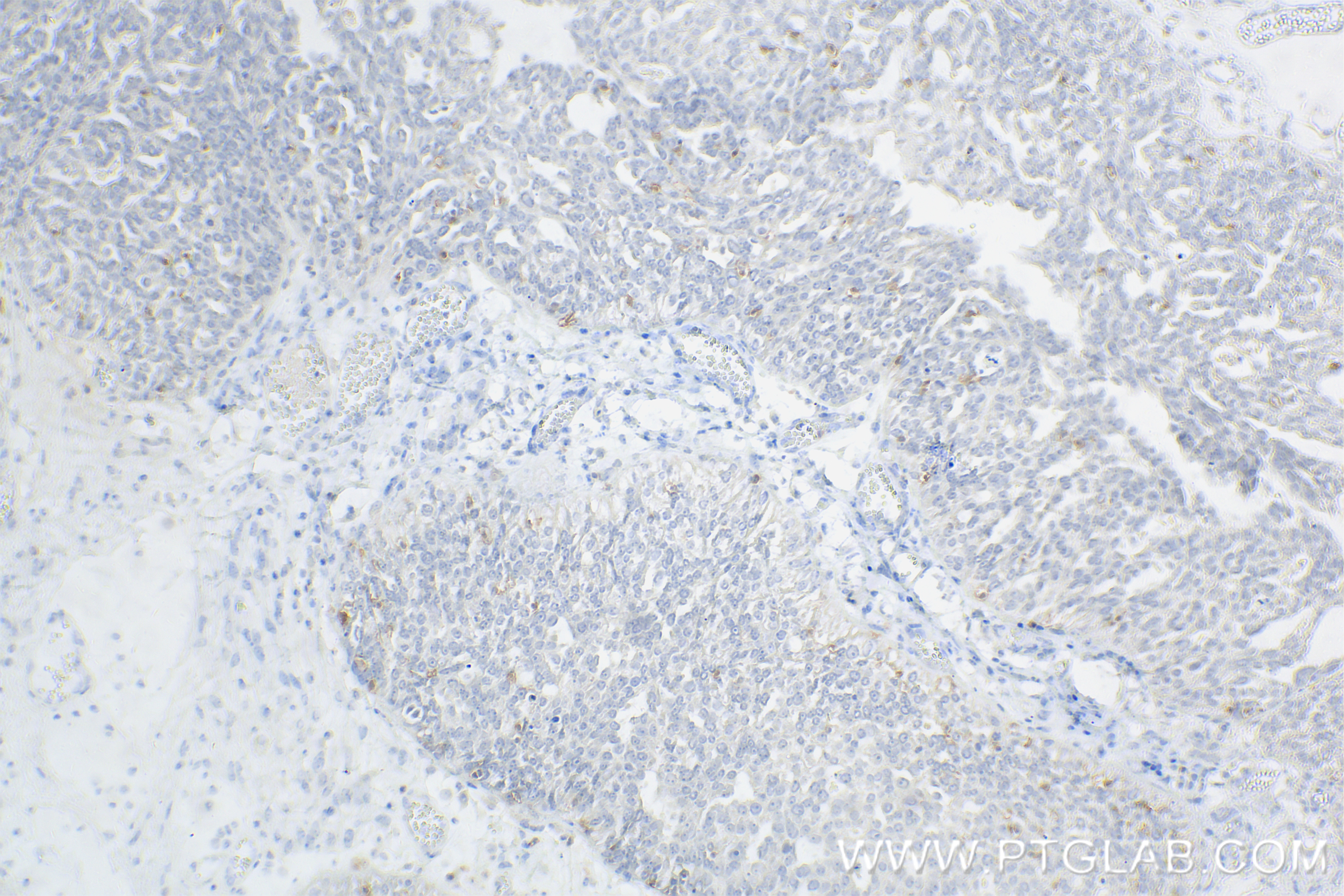
 at dilution of 1:1000 (under 10x lens). Heat mediated antigen retrieval with Tris-EDTA buffer (pH 9.0).")
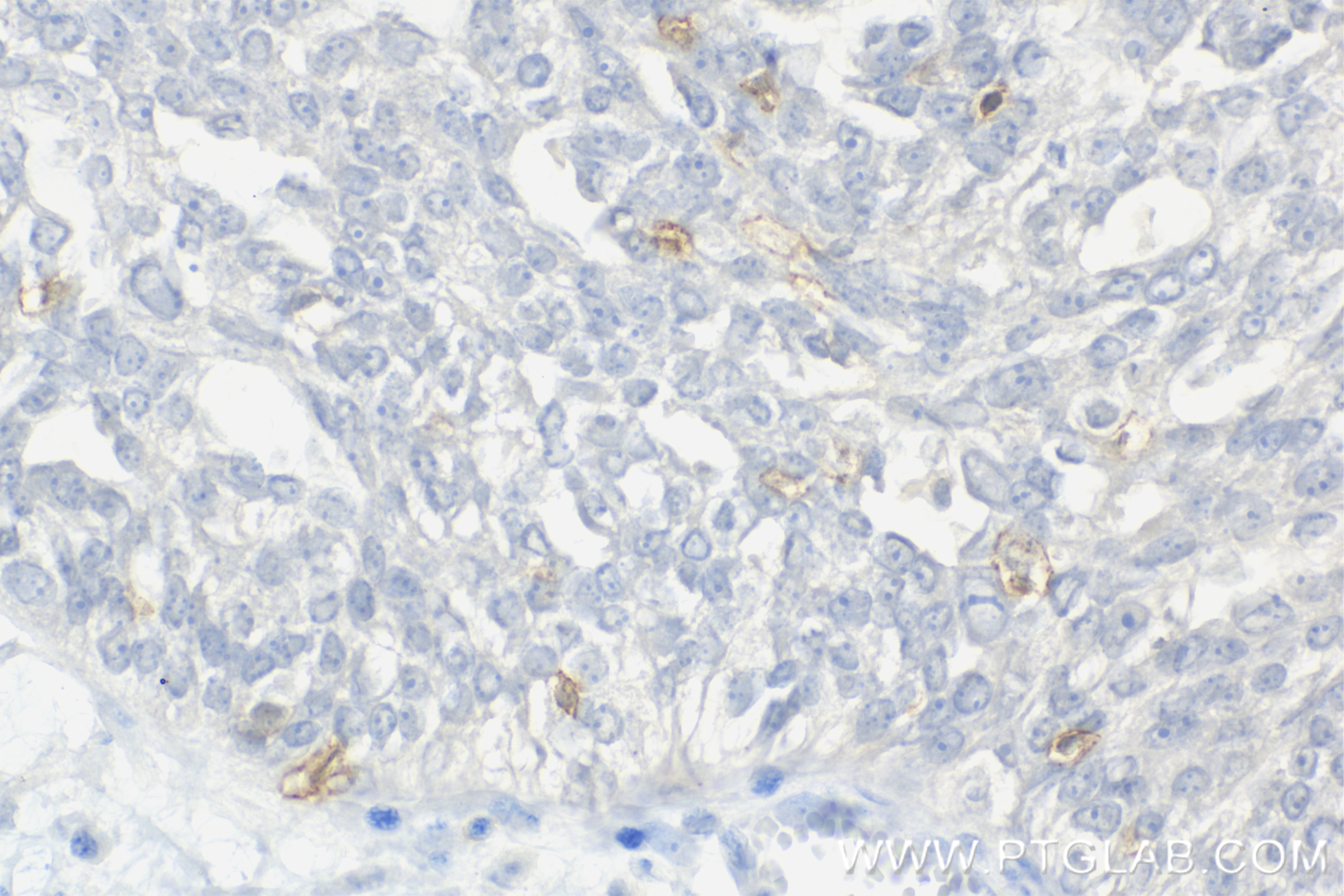
 at dilution of 1:1000 (under 40x lens). Heat mediated antigen retrieval with Tris-EDTA buffer (pH 9.0).")
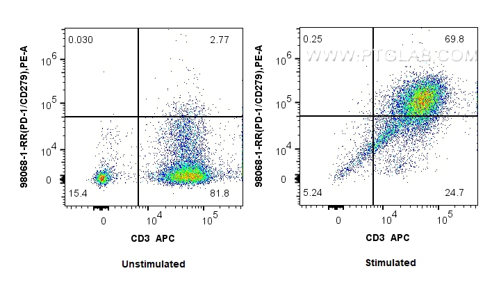
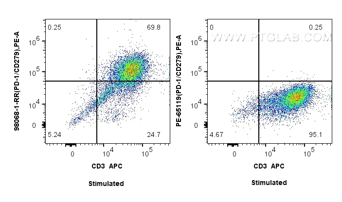
 and PE-Conjugated AffiniPure Goat Anti-Rabbit IgG(H+L). Cells were then stained with APC Anti-Human CD3. Cells were not fixed.")
(left) or 0.25 ug PE Anti-Human PD-1/CD279 (J110) (PE-65119, Clone: J110)(right) and PE-Conjugated AffiniPure Goat Anti-Rabbit IgG(H+L), Cells were then stained with APC Anti-Human CD3 (OKT3) Mouse IgG2a Recombinant Antibody (APC-65569, Clone: OKT3). Cells were not fixed.")
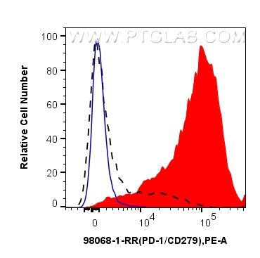
 or PHA stimulated human PBMCs (red) were stained with 0.25 ug Anti-Human PD-1/CD279 Rabbit Recombinant Antibody (98068-1-RR, Clone:240724G11) and PE-Conjugated AffiniPure Goat Anti-Rabbit IgG(H+L). 1x10^6 PHA treated human PBMCs were stained with Isotype Control(blue). Cells were not fixed.")
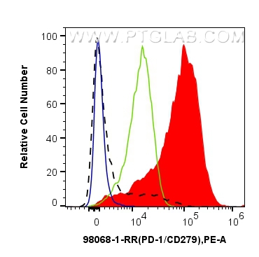
 or PHA treated human PBMCs were stained with 0.25 ug Anti-Human PD-1/CD279 Rabbit Recombinant Antibody (98068-1-RR, Clone:240724G11)(red) and PE-Conjugated AffiniPure Goat Anti-Rabbit IgG (H+L) or 0.25 ug PE Anti-Human PD-1/CD279 (J110) (PE-65119, Clone: J110)(green). 1x10^6 PHA treated human PBMCs were stained with Isotype Control(blue) . Cells were not fixed.")
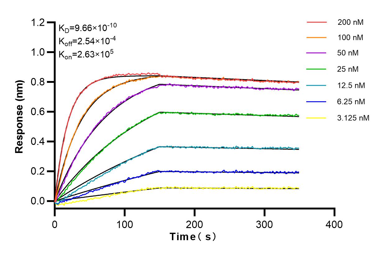
 kinetic assays of 98068-1-RR
against Human PD-1/CD279 were performed. The affinity constant is 0.966 nM.")
Tested Applications
| Positive IHC detected in | human tonsillitis tissue, human ovary cancer tissue Note: suggested antigen retrieval with TE buffer pH 9.0; (*) Alternatively, antigen retrieval may be performed with citrate buffer pH 6.0 |
| Positive FC detected in | human PBMCs |
Recommended dilution
| Application | Dilution |
|---|---|
| Immunohistochemistry (IHC) | IHC : 1:500-1:2000 |
| Flow Cytometry (FC) | FC : 0.25 ug per 10^6 cells in 100 μl suspension |
| This reagent has been tested for flow cytometric analysis. It is recommended that this reagent should be titrated in each testing system to obtain optimal results. | |
| Sample-dependent, Check data in validation data gallery. | |
Product Information
98068-1-RR targets PD-1/CD279 in IHC, FC applications and shows reactivity with human samples.
| Tested Reactivity | human |
| Host / Isotype | Rabbit / IgG |
| Class | Recombinant |
| Type | Antibody |
| Immunogen | PD-1/CD279 fusion protein Eg0974 Predict reactive species |
| Full Name | programmed cell death 1 |
| Calculated Molecular Weight | 288 aa, 32 kDa |
| GenBank Accession Number | BC074740 |
| Gene Symbol | PD-1 |
| Gene ID (NCBI) | 5133 |
| RRID | AB_3672215 |
| Conjugate | Unconjugated |
| Form | Liquid |
| Purification Method | Protein A purfication |
| UNIPROT ID | Q15116 |
| Storage Buffer | PBS with 0.09% sodium azide, pH 7.3. |
| Storage Conditions | Store at 2 - 8°C. Stable for one year after shipment. |
Background Information
Programmed cell death 1 (PD-1, also known as CD279) is an immunoinhibitory receptor that belongs to the CD28/CTLA-4 subfamily of the Ig superfamily. It is a 288 amino acid (aa) type I transmembrane protein composed of one Ig superfamily domain, a stalk, a transmembrane domain, and an intracellular domain containing an immunoreceptor tyrosine-based inhibitory motif (ITIM) as well as an immunoreceptor tyrosine-based switch motif (ITSM) (PMID: 18173375). PD-1 is expressed during thymic development and is induced in a variety of hematopoietic cells in the periphery by antigen receptor signaling and cytokines (PMID: 20636820). Engagement of PD-1 by its ligands PD-L1 or PD-L2 transduces a signal that inhibits T-cell proliferation, cytokine production, and cytolytic function (PMID: 19426218). It is critical for the regulation of T cell function during immunity and tolerance. Blockade of PD-1 can overcome immune resistance and also has been shown to have antitumor activity (PMID: 22658127; 23169436). It has been reported that PD-1 is heavily glycosylated and migrates with an apparent molecular mass of 47-55 kDa on SDS-PAGE , which is larger than its predicted mass of 32 kDa (PMID: 8671665; 17640856; 17003438).
Protocols
| Product Specific Protocols | |
|---|---|
| IHC protocol for PD-1/CD279 antibody 98068-1-RR | Download protocol |
| FC protocol for PD-1/CD279 antibody 98068-1-RR | Download protocol |
| Standard Protocols | |
|---|---|
| Click here to view our Standard Protocols |