Filter:
 at dilution of 1:1000 incubated at room temperature for 1.5 hours.")
 at dilution of 1:500 incubated at room temperature for 1.5 hours.")
 at dilution of 1:500 incubated at room temperature for 1.5 hours.")
 with mouse testis tissue lysate 4000 ug.")
 at dilution of 1:200 (under 10x lens. Heat mediated antigen retrieval with Tris-EDTA buffer (pH 9.0).")
 at dilution of 1:200 (under 40x lens. Heat mediated antigen retrieval with Tris-EDTA buffer (pH 9.0).")
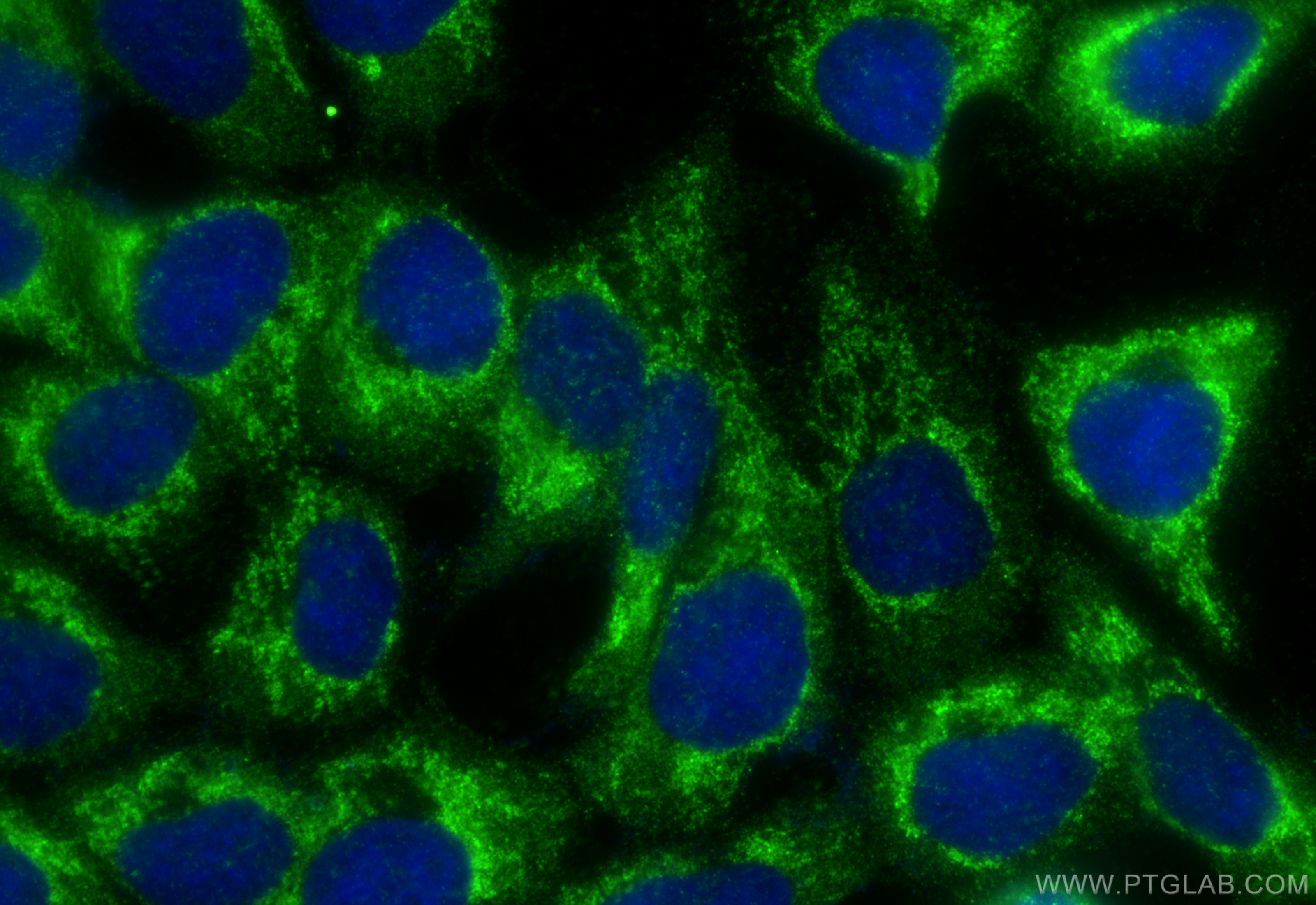
 fixed A431 cells using PAK6 antibody (13539-1-AP) at dilution of 1:200 and CoraLite®488-Conjugated Goat Anti-Rabbit IgG(H+L) (SA00013-2).")
Tested Applications
| Positive WB detected in | mouse testis tissue, Raji cells, rat testis tissue |
| Positive IP detected in | mouse testis tissue |
| Positive IHC detected in | human prostate cancer tissue Note: suggested antigen retrieval with TE buffer pH 9.0; (*) Alternatively, antigen retrieval may be performed with citrate buffer pH 6.0 |
| Positive IF/ICC detected in | A431 cells |
Recommended dilution
| Application | Dilution |
|---|---|
| Western Blot (WB) | WB : 1:500-1:2000 |
| Immunoprecipitation (IP) | IP : 0.5-4.0 ug for 1.0-3.0 mg of total protein lysate |
| Immunohistochemistry (IHC) | IHC : 1:50-1:500 |
| Immunofluorescence (IF)/ICC | IF/ICC : 1:50-1:500 |
| It is recommended that this reagent should be titrated in each testing system to obtain optimal results. | |
| Sample-dependent, Check data in validation data gallery. | |
Published Applications
| KD/KO | See 1 publications below |
| WB | See 4 publications below |
| IHC | See 2 publications below |
Product Information
13539-1-AP targets PAK6 in WB, IHC, IF/ICC, IP, ELISA applications and shows reactivity with human, mouse, rat samples.
| Tested Reactivity | human, mouse, rat |
| Cited Reactivity | human, rat |
| Host / Isotype | Rabbit / IgG |
| Class | Polyclonal |
| Type | Antibody |
| Immunogen | PAK6 fusion protein Ag4467 Predict reactive species |
| Full Name | p21 protein (Cdc42/Rac)-activated kinase 6 |
| Calculated Molecular Weight | 681 aa, 75 kDa |
| Observed Molecular Weight | 70-75 kDa |
| GenBank Accession Number | BC035596 |
| Gene Symbol | PAK6 |
| Gene ID (NCBI) | 56924 |
| RRID | AB_2158467 |
| Conjugate | Unconjugated |
| Form | Liquid |
| Purification Method | Antigen affinity purification |
| UNIPROT ID | Q9NQU5 |
| Storage Buffer | PBS with 0.02% sodium azide and 50% glycerol , pH 7.3 |
| Storage Conditions | Store at -20°C. Stable for one year after shipment. Aliquoting is unnecessary for -20oC storage. 20ul sizes contain 0.1% BSA. |
Background Information
PAK6(p21-activated kinase 6) is a serine/threonine kinase belonging to the p21-activated kinase (PAK) family. .This protein plays a role in the regulation of gene transcription and may protect cells from apoptosis through phosphorylation of BAD. Northern blot analysis revealed that PAK6 is highly expressed in testis and prostate tissues (PMID:11278661). The immunogen of this antibody is the C-terminal of PAK6. This antibody has weak cross-reaction with other PAK members.
Protocols
| Product Specific Protocols | |
|---|---|
| WB protocol for PAK6 antibody 13539-1-AP | Download protocol |
| IHC protocol for PAK6 antibody 13539-1-AP | Download protocol |
| IF protocol for PAK6 antibody 13539-1-AP | Download protocol |
| IP protocol for PAK6 antibody 13539-1-AP | Download protocol |
| Standard Protocols | |
|---|---|
| Click here to view our Standard Protocols |
Publications
| Species | Application | Title |
|---|---|---|
Oncol Lett PAK6 promotes cervical cancer progression through activation of the Wnt/β-catenin signaling pathway.
| ||
J Pain Res Proteomics analysis of the amygdala in rats with CFA-induced pain aversion with electro-acupuncture stimulation. | ||
BMC Nephrol Urinary exosome proteins PAK6 and EGFR as noninvasive diagnostic biomarkers of diabetic nephropathy |