Filter:

 with si-Control and si-NUMBL transfected HeLa cells.")
 at dilution of 1:1000 incubated at room temperature for 1.5 hours.")
 at dilution of 1:1000 incubated at room temperature for 1.5 hours.")
 at dilution of 1:1000 incubated at room temperature for 1.5 hours.")
 at dilution of 1:1000 incubated at room temperature for 1.5 hours.")
 with HeLa cells lysate 2800 ug.")
 at dilution of 1:200 (under 10x lens. Heat mediated antigen retrieval with Tris-EDTA buffer (pH 9.0).")
 at dilution of 1:200 (under 40x lens. Heat mediated antigen retrieval with Tris-EDTA buffer (pH 9.0).")
.")
.")
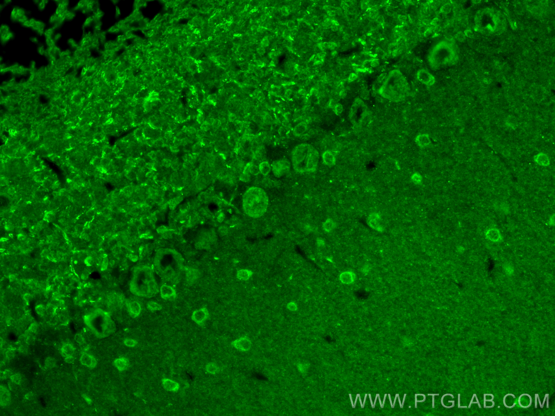
 fixed paraffin-embedded mouse cerebellum tissue using NUMBL antibody (10111-1-AP) at dilution of 1:200 and CoraLite®488-Conjugated Goat Anti-Rabbit IgG(H+L). Heat mediated antigen retrieval with Tris-EDTA buffer (pH 9.0).")
 fixed PC-12 cells using 10111-1-AP (NUMBL antibody) at dilution of 1:50 and Alexa Fluor 488-Conjugated Goat Anti-Rabbit IgG(H+L).")
 fixed HepG2 cells using 10111-1-AP (NUMBL antibody) at dilution of 1:50 and Alexa Fluor 488-conjugated Goat Anti-Rabbit IgG(H+L).")
Tested Applications
| Positive WB detected in | mouse brain tissue, mouse spleen tissue, HeLa cells, mouse lung tissue, mouse liver tissue |
| Positive IP detected in | HeLa cells |
| Positive IHC detected in | human brain tissue, human gliomas tissue Note: suggested antigen retrieval with TE buffer pH 9.0; (*) Alternatively, antigen retrieval may be performed with citrate buffer pH 6.0 |
| Positive IF-P detected in | mouse cerebellum tissue |
| Positive IF/ICC detected in | PC-12 cells, HepG2 cells |
Recommended dilution
| Application | Dilution |
|---|---|
| Western Blot (WB) | WB : 1:1000-1:4000 |
| Immunoprecipitation (IP) | IP : 0.5-4.0 ug for 1.0-3.0 mg of total protein lysate |
| Immunohistochemistry (IHC) | IHC : 1:50-1:500 |
| Immunofluorescence (IF)-P | IF-P : 1:50-1:500 |
| Immunofluorescence (IF)/ICC | IF/ICC : 1:50-1:500 |
| It is recommended that this reagent should be titrated in each testing system to obtain optimal results. | |
| Sample-dependent, Check data in validation data gallery. | |
Published Applications
| KD/KO | See 1 publications below |
| WB | See 4 publications below |
| IHC | See 3 publications below |
Product Information
10111-1-AP targets NUMBL in WB, IHC, IF/ICC, IF-P, IP, ELISA applications and shows reactivity with human, mouse, rat samples.
| Tested Reactivity | human, mouse, rat |
| Cited Reactivity | human, mouse |
| Host / Isotype | Rabbit / IgG |
| Class | Polyclonal |
| Type | Antibody |
| Immunogen | NUMBL fusion protein Ag0156 Predict reactive species |
| Full Name | numb homolog (Drosophila)-like |
| Calculated Molecular Weight | 65 kDa |
| Observed Molecular Weight | 65-70 kDa |
| GenBank Accession Number | BC001794 |
| Gene Symbol | NUMBL |
| Gene ID (NCBI) | 9253 |
| RRID | AB_2154440 |
| Conjugate | Unconjugated |
| Form | Liquid |
| Purification Method | Antigen affinity purification |
| UNIPROT ID | Q9Y6R0 |
| Storage Buffer | PBS with 0.02% sodium azide and 50% glycerol , pH 7.3 |
| Storage Conditions | Store at -20°C. Stable for one year after shipment. Aliquoting is unnecessary for -20oC storage. 20ul sizes contain 0.1% BSA. |
Background Information
Numbl (also known as numblike or Nbl), as a conserved homolog of Drosophila Numb, is specifically expressed in the brain. Numbl is a cytoplasm protein and has redundant functions in embryonic neurogenesis. It plays an important role in ependymal wall integrity and subventricular zone neuroblast survival. However, its expression and function in the central nervous system lesion are still unclear.
Protocols
| Product Specific Protocols | |
|---|---|
| WB protocol for NUMBL antibody 10111-1-AP | Download protocol |
| IHC protocol for NUMBL antibody 10111-1-AP | Download protocol |
| IF protocol for NUMBL antibody 10111-1-AP | Download protocol |
| IP protocol for NUMBL antibody 10111-1-AP | Download protocol |
| Standard Protocols | |
|---|---|
| Click here to view our Standard Protocols |
Publications
| Species | Application | Title |
|---|---|---|
Nat Neurosci Numb and Numbl are required for maintenance of cadherin-based adhesion and polarity of neural progenitors. | ||
Development Cleaved Delta like 1 intracellular domain regulates neural development via Notch signal-dependent and -independent pathways. | ||
Mol Endocrinol Numb deletion in POMC-expressing cells impairs pituitary intermediate lobe cell adhesion, progenitor cell localization, and neuro-intermediate lobe boundary formation. |