Various lysates were subjected to SDS PAGE followed by western blot with 66999-1-Ig (NFIA antibody) at dilution of 1:20000 incubated at room temperature for 1.5 hours. The membrane was stripped and reblotted with HRP-conjugated Beta Actin Monoclonal antibody (HRP-66009) as loading control.
Various lysates were subjected to SDS PAGE followed by western blot with 66999-1-Ig (NFIA antibody) at dilution of 1:20000 incubated at room temperature for 1.5 hours. The membrane was stripped and reblotted with HRP-conjugated Beta Actin Monoclonal antibody (HRP-66009) as loading control.
WB analysis of MCF-7 using 66999-1-Ig
WB result of NFIA antibody (66999-1-Ig; 1:12000; incubated at room temperature for 1.5 hours) with sh-Control and sh-NFIA transfected MCF-7 cells.
WB result of NFIA antibody (66999-1-Ig; 1:12000; incubated at room temperature for 1.5 hours) with sh-Control and sh-NFIA transfected MCF-7 cells.
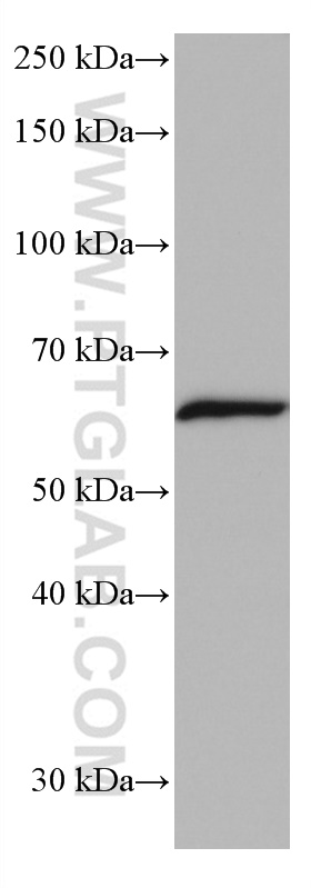
WB analysis of mouse cerebellum using 66999-1-Ig
mouse cerebellum tissue were subjected to SDS PAGE followed by western blot with 66999-1-Ig (NFIA antibody) at dilution of 1:20000 incubated at room temperature for 1.5 hours.
mouse cerebellum tissue were subjected to SDS PAGE followed by western blot with 66999-1-Ig (NFIA antibody) at dilution of 1:20000 incubated at room temperature for 1.5 hours.
WB analysis of HeLa using 66999-1-Ig
HeLa cells were subjected to SDS PAGE followed by western blot with 66999-1-Ig (NFIA antibody) at dilution of 1:3000 incubated at room temperature for 1.5 hours.
HeLa cells were subjected to SDS PAGE followed by western blot with 66999-1-Ig (NFIA antibody) at dilution of 1:3000 incubated at room temperature for 1.5 hours.
WB analysis using 66999-1-Ig
Various lysates were subjected to SDS PAGE followed by western blot with 66999-1-Ig (NFIA antibody) at dilution of 1:3000 incubated at room temperature for 1.5 hours.
Various lysates were subjected to SDS PAGE followed by western blot with 66999-1-Ig (NFIA antibody) at dilution of 1:3000 incubated at room temperature for 1.5 hours.
IP experiment of HeLa using 66999-1-Ig
IP result of anti-NFIA (IP:66999-1-Ig, 5ug; Detection:66999-1-Ig 1:500) with HeLa cells lysate 1600 ug.
The Proteintech guarantee covers Proteintech antibodies in any species and any application, including those not listed on the datasheet. If the antibody doesn’t perform, you can receive a hassle-free refund or credit note.
PBS with 0.02% sodium azide and 50% glycerol , pH 7.3
Storage Conditions
Store at -20°C. Stable for one year after shipment. Aliquoting is unnecessary for -20oC storage. 20ul sizes contain 0.1% BSA.
Background Information
The NFI (nuclear factor I) family consists of four members in vertebrates (NFI-A, NFI-B, NFI-C and NFI-X), and the four NFI genes are expressed in unique patterns during mouse embryogenesis and in the adult. Four isoforms of NFIA were found in human and they play various roles in DNA replication, DNA-dependent transcritpion via their DNA binding property. Multiple residues of NFIA can be phosphorylated resulting in mild shifts of its practical molecular weight. Recent finding also revealed its neuroprotective function in NMDA-induced neuronal damage. The calculated molecular weight of NFIA is 55 kDa, However the size of the proteins cross-linked to the adenoviral NF-I element ranged from 60 to 80 kDa . The larger size observed by us could be due to the oligo protein complex, which would increase the size by 15-20 kDa. (PMID: 11447215)
Various lysates were subjected to SDS PAGE followed by western blot with 66999-1-Ig (NFIA antibody) at dilution of 1:20000 incubated at room temperature for 1.5 hours. The membrane was stripped and reblotted with HRP-conjugated Beta Actin Monoclonal antibody (HRP-66009) as loading control.
WB analysis of MCF-7 using 66999-1-Ig
WB result of NFIA antibody (66999-1-Ig; 1:12000; incubated at room temperature for 1.5 hours) with sh-Control and sh-NFIA transfected MCF-7 cells.
WB analysis of mouse cerebellum using 66999-1-Ig
mouse cerebellum tissue were subjected to SDS PAGE followed by western blot with 66999-1-Ig (NFIA antibody) at dilution of 1:20000 incubated at room temperature for 1.5 hours.
WB analysis of HeLa using 66999-1-Ig
HeLa cells were subjected to SDS PAGE followed by western blot with 66999-1-Ig (NFIA antibody) at dilution of 1:3000 incubated at room temperature for 1.5 hours.
WB analysis using 66999-1-Ig
Various lysates were subjected to SDS PAGE followed by western blot with 66999-1-Ig (NFIA antibody) at dilution of 1:3000 incubated at room temperature for 1.5 hours.
IP Figures
IP experiment of HeLa using 66999-1-Ig
IP result of anti-NFIA (IP:66999-1-Ig, 5ug; Detection:66999-1-Ig 1:500) with HeLa cells lysate 1600 ug.
The species listed in Tested Reactivity are in-house verified and applicable species. For unlisted species, please refer to the homology analysis of the immunogen sequence and related species. For rabbit polyclonal antibodies, homology >70% is recommended. For mouse monoclonal antibodies and rabbit recombinant antibodies, homology >90% is recommended. Generally, the higher the homology, the greater the applicability. However, there will be certain differences in protein expression in different species, tissues or cells. Therefore, the homology analysis results are for reference only and do not serve as a guarantee.
At Proteintech, we pride ourselves on our antibody quality, customer service and transparency. As such, we are comparing our antibodies with other vendors, enabling easy identification and comparisons of key data to help you choose the suitable antibody for your needs.
We have selected the top cited antibodies from these vendors for you to compare.
Proteintech
KD/KO VALIDATED
NFIA Monoclonal antibody
Catalog Number
66999-1-Ig
Citations
1
Dilutions
WB : 1:5000-1:50000 IP : 0.5-4.0 ug for IP and 0.5-4.0 ug for 1.0-3.0 mg of total protein lysate for WB
Applications
WB, IP, ELISA
Reactivity
human, rat, mouse, pig, rabbit
Product Guarantee
Covers any species including not listed on datasheet
Covers any applications including not listed on datasheet
 at dilution of 1:20000 incubated at room temperature for 1.5 hours. The membrane was stripped and reblotted with HRP-conjugated Beta Actin Monoclonal antibody (HRP-66009) as loading control.")
 with sh-Control and sh-NFIA transfected MCF-7 cells.")
 at dilution of 1:20000 incubated at room temperature for 1.5 hours.")
 at dilution of 1:3000 incubated at room temperature for 1.5 hours.")
 at dilution of 1:3000 incubated at room temperature for 1.5 hours.")
 with HeLa cells lysate 1600 ug.")