Tested Applications
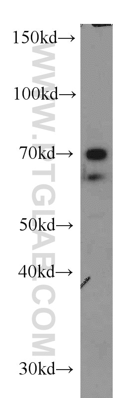
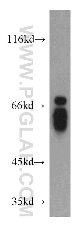
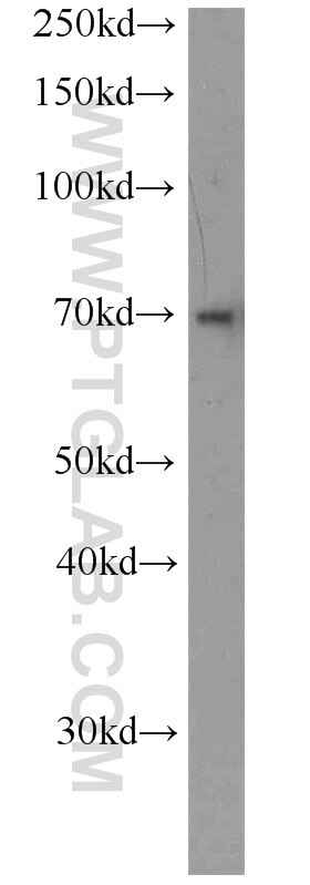
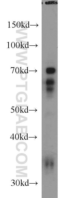
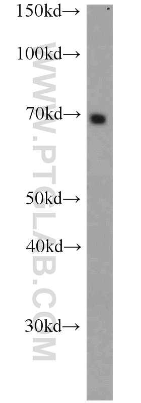
| Positive WB detected in | A431 cells, HeLa cells, Jurkat cells, L02 cells, mouse liver tissue |
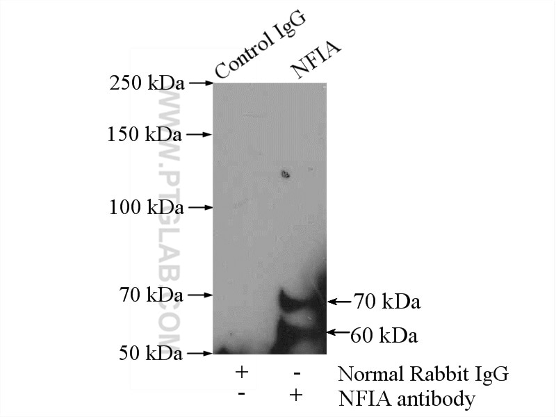
| Positive IP detected in | A431 cells |
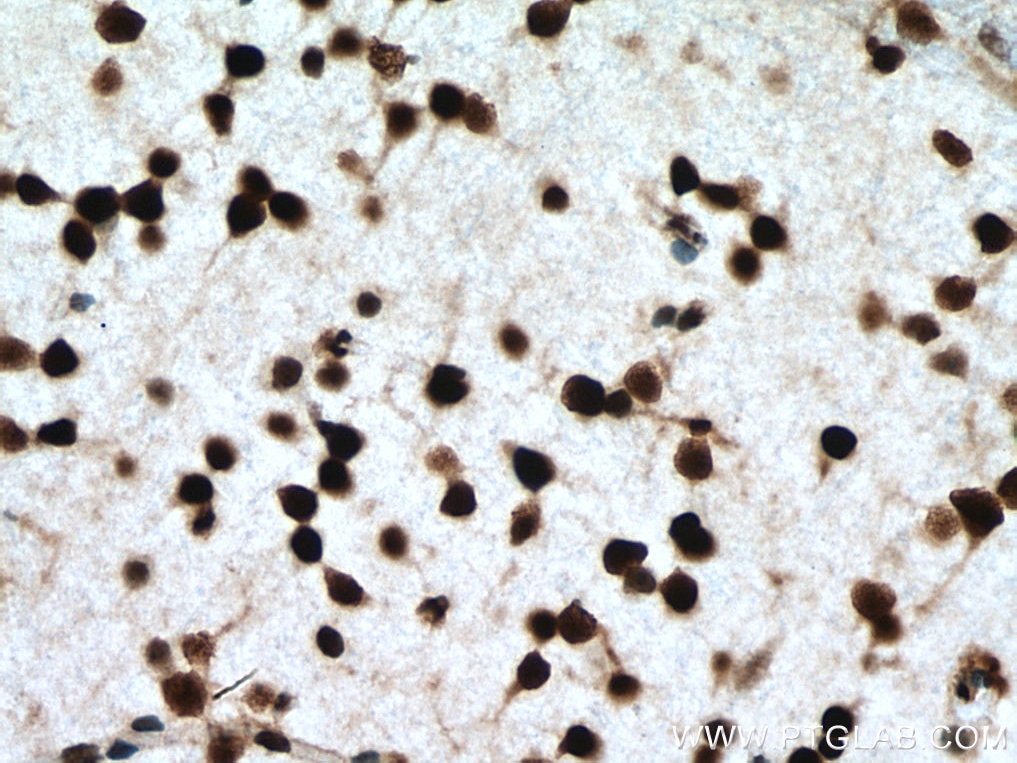
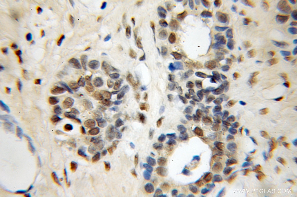
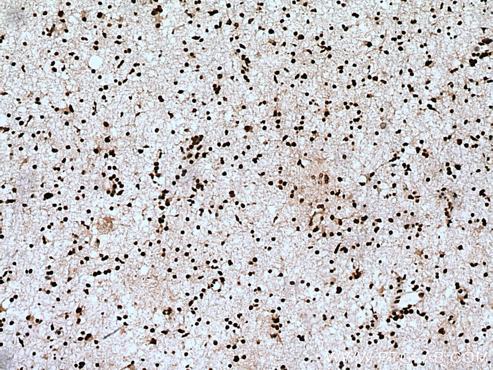
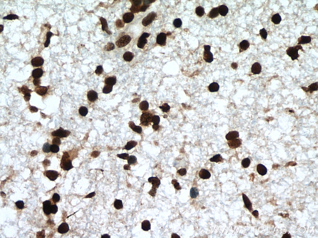
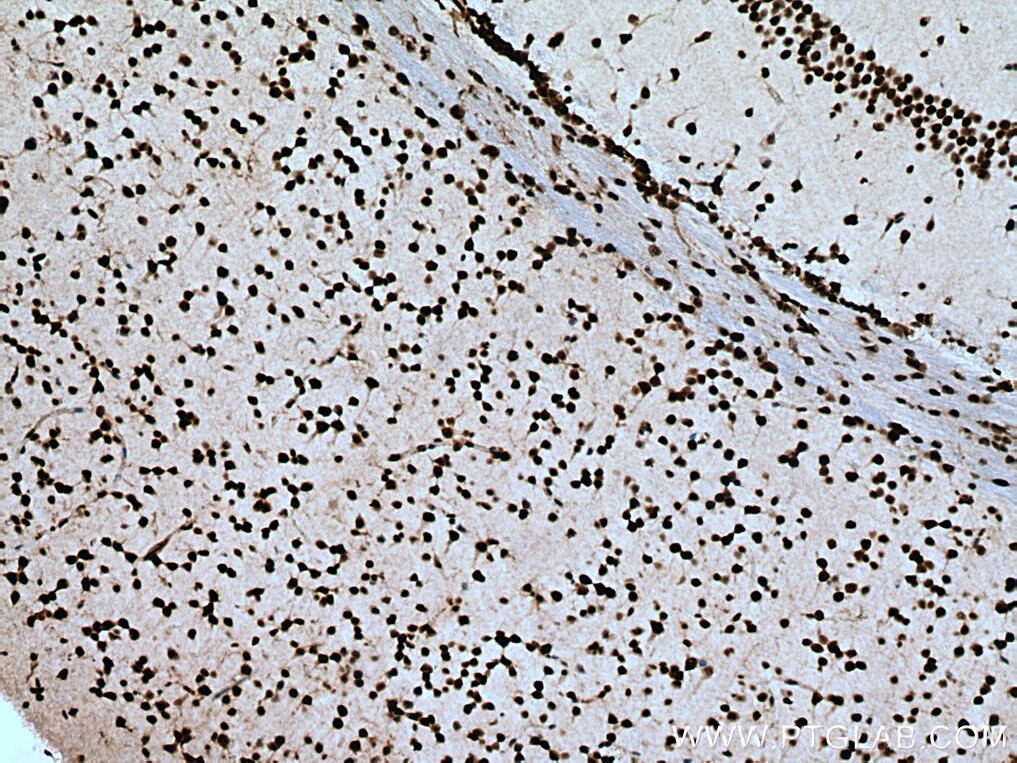
| Positive IHC detected in | mouse brain tissue, human prostate cancer tissue, human gliomas tissue Note: suggested antigen retrieval with TE buffer pH 9.0; (*) Alternatively, antigen retrieval may be performed with citrate buffer pH 6.0 |
Recommended dilution
| Application | Dilution |
|---|---|
| Western Blot (WB) | WB : 1:500-1:1000 |
| Immunoprecipitation (IP) | IP : 0.5-4.0 ug for 1.0-3.0 mg of total protein lysate |
| Immunohistochemistry (IHC) | IHC : 1:50-1:500 |
| It is recommended that this reagent should be titrated in each testing system to obtain optimal results. | |
| Sample-dependent, Check data in validation data gallery. | |
Published Applications
| WB | See 3 publications below |
| IHC | See 1 publications below |
| CoIP | See 1 publications below |
Product Information
11750-1-AP targets NFIA in WB, IP, IHC, CoIP, ELISA applications and shows reactivity with human, mouse, rat samples.
| Tested Reactivity | human, mouse, rat |
| Cited Reactivity | human, mouse |
| Host / Isotype | Rabbit / IgG |
| Class | Polyclonal |
| Type | Antibody |
| Immunogen | NFIA fusion protein Ag2346 Predict reactive species |
| Full Name | nuclear factor I/A |
| Calculated Molecular Weight | 498 aa, 55 kDa |
| Observed Molecular Weight | 60-70 kDa |
| GenBank Accession Number | BC022264 |
| Gene Symbol | NFIA |
| Gene ID (NCBI) | 4774 |
| RRID | AB_2153214 |
| Conjugate | Unconjugated |
| Form | Liquid |
| Purification Method | Antigen affinity purification |
| UNIPROT ID | Q12857 |
| Storage Buffer | PBS with 0.02% sodium azide and 50% glycerol , pH 7.3 |
| Storage Conditions | Store at -20°C. Stable for one year after shipment. Aliquoting is unnecessary for -20oC storage. 20ul sizes contain 0.1% BSA. |
Background Information
The NFI (nuclear factor I) family consists of four members in vertebrates (NFI-A, NFI-B, NFI-C and NFI-X), and the four NFI genes are expressed in unique patterns during mouse embryogenesis and in the adult. Four isoforms of NFIA were found in human and they play various roles in DNA replication, DNA-dependent transcritpion via their DNA binding property. Multiple residues of NFIA can be phosphorylated resulting in mild shifts of its practical molecular weight. Recent finding also revealed its neuroprotective function in NMDA-induced neuronal damage. Catalog# 11750-1-AP is a rabbit polyclonal antibody raised against N-terminal of human original NFIA. The calculated molecular weight of NFIA is 55 kDa, However the size of the proteins cross-linked to the adenoviral NF-I element ranged from 60 to 80 kDa . The larger size observed by us could be due to the oligo protein complex, which would increase the size by 15-20 kDa. (PMID: 11447215)
Protocols
| Product Specific Protocols | |
|---|---|
| WB protocol for NFIA antibody 11750-1-AP | Download protocol |
| IHC protocol for NFIA antibody 11750-1-AP | Download protocol |
| IP protocol for NFIA antibody 11750-1-AP | Download protocol |
| Standard Protocols | |
|---|---|
| Click here to view our Standard Protocols |
Publications
| Species | Application | Title |
|---|---|---|
Cell Cycle TRIM71 binds to IMP1 and is capable of positive and negative regulation of target RNAs. | ||
Cancer Cell Int Macrophages-derived exosomal lncRNA LIFR-AS1 promotes osteosarcoma cell progression via miR-29a/NFIA axis. | ||
FEBS Lett STAT3 transcriptionally regulates the expression of genes related to glycogen metabolism in developing motor neurons | ||
Pathol Res Pract SOX9/NFIA promotes human ovarian cancer metastasis through the Wnt/β-catenin signaling pathway |
Reviews
The reviews below have been submitted by verified Proteintech customers who received an incentive for providing their feedback.
FH KUN (Verified Customer) (01-13-2021) | Works for western blot, recogizning both NFIA and also other NFI family proteins
|
