Tested Applications
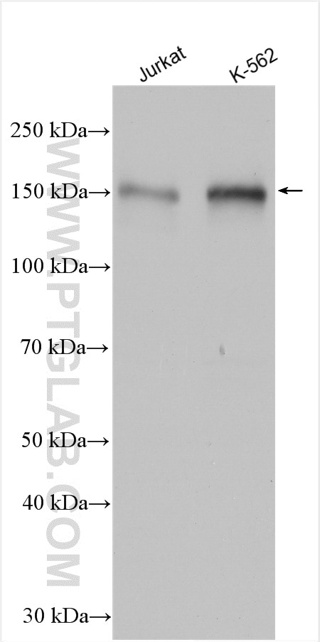
| Positive WB detected in | Jurkat cells, K-562 cells |
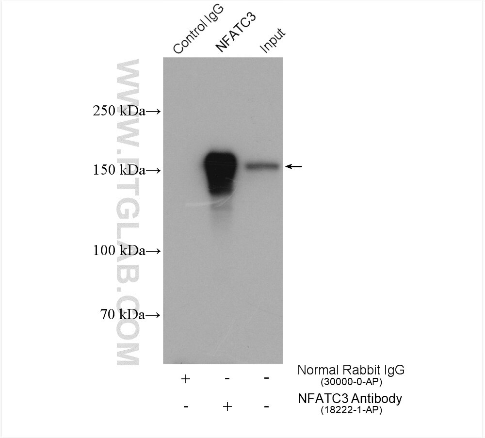
| Positive IP detected in | K-562 cells |
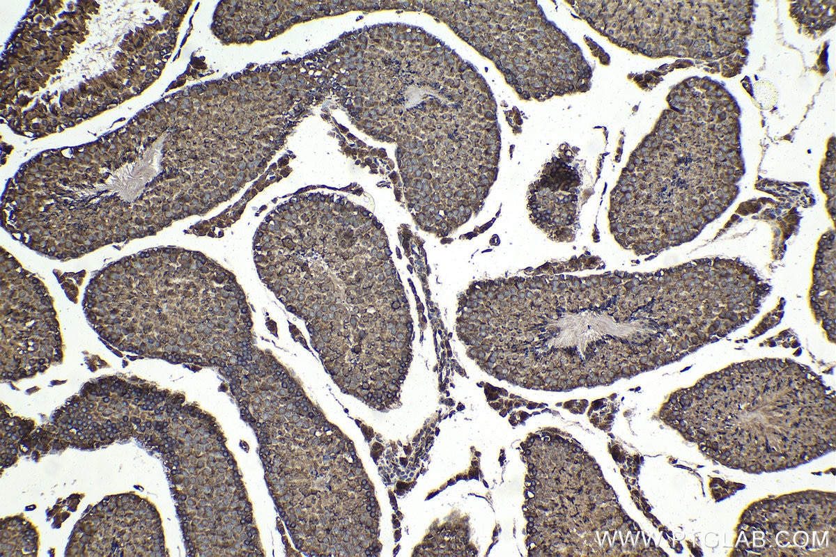
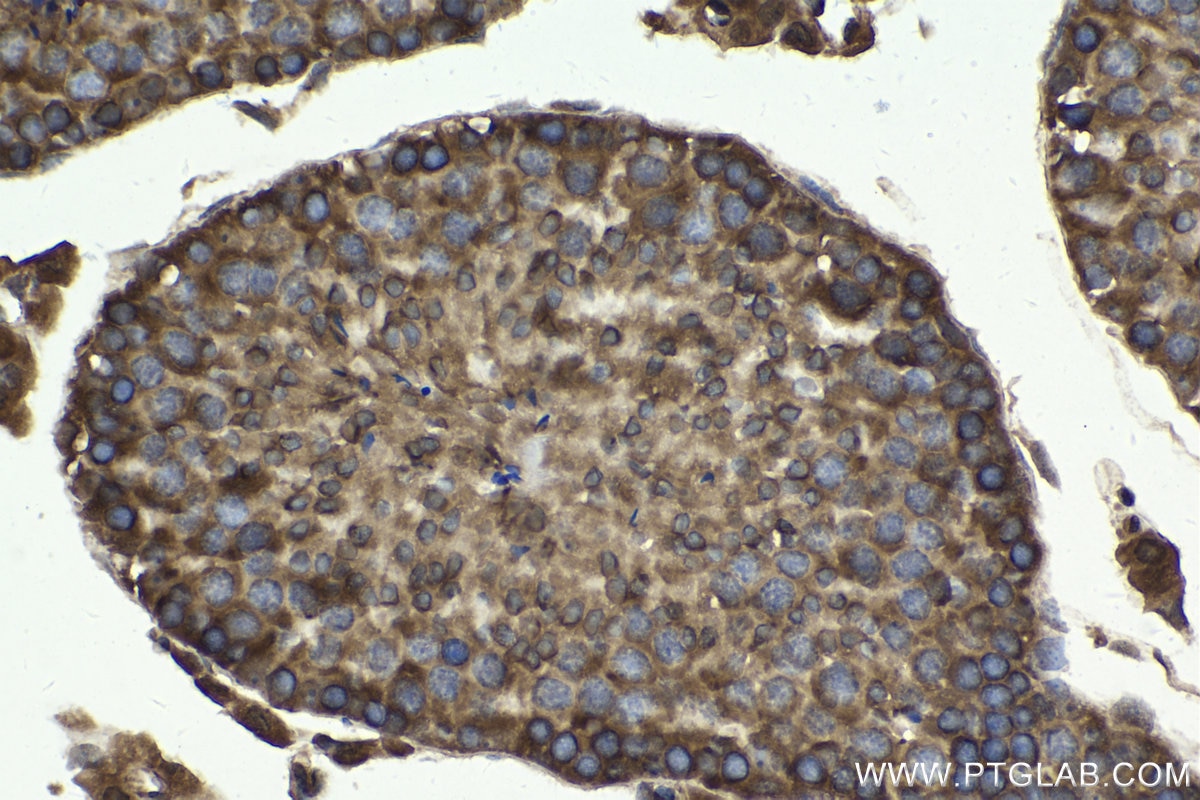
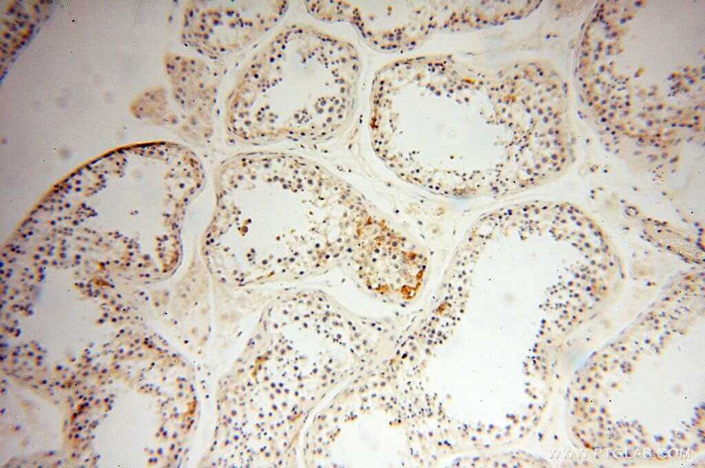
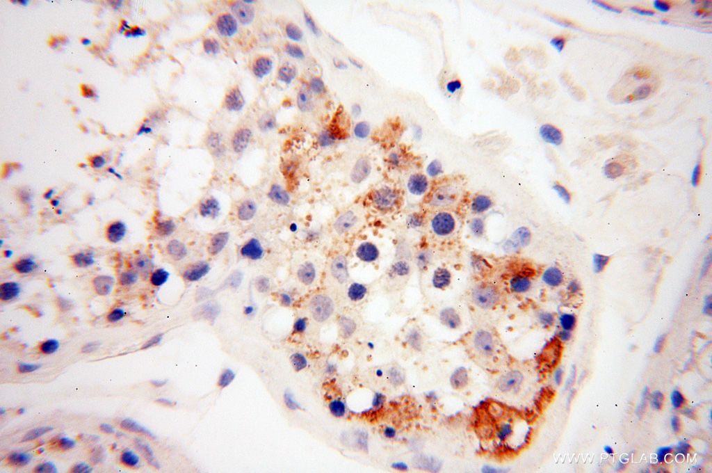
| Positive IHC detected in | mouse testis tissue, human testis tissue Note: suggested antigen retrieval with TE buffer pH 9.0; (*) Alternatively, antigen retrieval may be performed with citrate buffer pH 6.0 |
Recommended dilution
| Application | Dilution |
|---|---|
| Western Blot (WB) | WB : 1:2000-1:12000 |
| Immunoprecipitation (IP) | IP : 0.5-4.0 ug for 1.0-3.0 mg of total protein lysate |
| Immunohistochemistry (IHC) | IHC : 1:500-1:2000 |
| It is recommended that this reagent should be titrated in each testing system to obtain optimal results. | |
| Sample-dependent, Check data in validation data gallery. | |
Published Applications
| KD/KO | See 2 publications below |
| WB | See 24 publications below |
| IHC | See 2 publications below |
| IF | See 9 publications below |
| IP | See 1 publications below |
| CoIP | See 1 publications below |
| ChIP | See 4 publications below |
Product Information
18222-1-AP targets NFATC3 in WB, IHC, IF, IP, CoIP, chIP, ELISA applications and shows reactivity with human samples.
| Tested Reactivity | human |
| Cited Reactivity | human, mouse, rat |
| Host / Isotype | Rabbit / IgG |
| Class | Polyclonal |
| Type | Antibody |
| Immunogen |
CatNo: Ag12985 Product name: Recombinant human NFATC3 protein Source: e coli.-derived, PGEX-4T Tag: GST Domain: 1-205 aa of BC001050 Sequence: MTTANCGAHDELDFKLVFGEDGAPAPPPPGSRPADLEPDDCASIYIFNVDPPPSTLTTPLCLPHHGLPSHSSVLSPSFQLQSHKNYEGTCEIPESKYSPLGGPKPFECPSIQITSISPNCHQELDAHEDDLQINDPEREFLERPSRDHLYLPLEPSYRESSLSPSPASSISSRSWFSDASSCESLSHIYDDVDSELNEAAARFTL Predict reactive species |
| Full Name | nuclear factor of activated T-cells, cytoplasmic, calcineurin-dependent 3 |
| Calculated Molecular Weight | 116 kDa |
| Observed Molecular Weight | 130-170 kDa |
| GenBank Accession Number | BC001050 |
| Gene Symbol | NFATC3 |
| Gene ID (NCBI) | 4775 |
| RRID | AB_2152773 |
| Conjugate | Unconjugated |
| Form | Liquid |
| Purification Method | Antigen affinity purification |
| UNIPROT ID | Q12968 |
| Storage Buffer | PBS with 0.02% sodium azide and 50% glycerol, pH 7.3. |
| Storage Conditions | Store at -20°C. Stable for one year after shipment. Aliquoting is unnecessary for -20oC storage. 20ul sizes contain 0.1% BSA. |
Background Information
NFAT (nuclear factors of activated T cells) proteins are a family of transcription factors originally identified as mediators of activation of cytokine genes in response to antigenic stimulation of T cells. NFAT proteins also play varied roles in cells outside of the immune system [PMID:11877454]. NFATc3 has specifically been implicated in vasculature development, regulation of smooth muscle contractile phenotype, and modulation of vascular smooth muscle contractility [PMID:11439183,17148444]. The calculated molecular weight of NFATC3 is 115 kDa, but the post-modified protein is about 130-170 kDa (PMID: 15728531, PMID: 15857835)
Protocols
| Product Specific Protocols | |
|---|---|
| IHC protocol for NFATC3 antibody 18222-1-AP | Download protocol |
| IP protocol for NFATC3 antibody 18222-1-AP | Download protocol |
| WB protocol for NFATC3 antibody 18222-1-AP | Download protocol |
| Standard Protocols | |
|---|---|
| Click here to view our Standard Protocols |
Publications
| Species | Application | Title |
|---|---|---|
Immunity Metformin inhibition of mitochondrial ATP and DNA synthesis abrogates NLRP3 inflammasome activation and pulmonary inflammation. | ||
Gastroenterology Regulator of Calcineurin 1 Gene Isoform 4, Downregulated in Hepatocellular Carcinoma, Prevents Proliferation, Migration, and Invasive Activity of Cancer Cells and Growth of Orthotopic Tumors by Inhibiting Nuclear Translocation of NFAT1. | ||
Acta Pharm Sin B Cytoplasmic and nuclear NFATc3 cooperatively contributes to vascular smooth muscle cell dysfunction and drives aortic aneurysm and dissection | ||
Cell Death Differ Trim39 regulates neuronal apoptosis by acting as a SUMO-targeted E3 ubiquitin-ligase for the transcription factor NFATc3. | ||
Oncogene Regulator of calcineurin 1 gene isoform 4 in pancreatic ductal adenocarcinoma regulates the progression of tumor cells. | ||
Cancer Lett TRPC3 promotes tumorigenesis of gastric cancer via the CNB2/GSK3β/NFATc2 signaling pathway. |

