Tested Applications
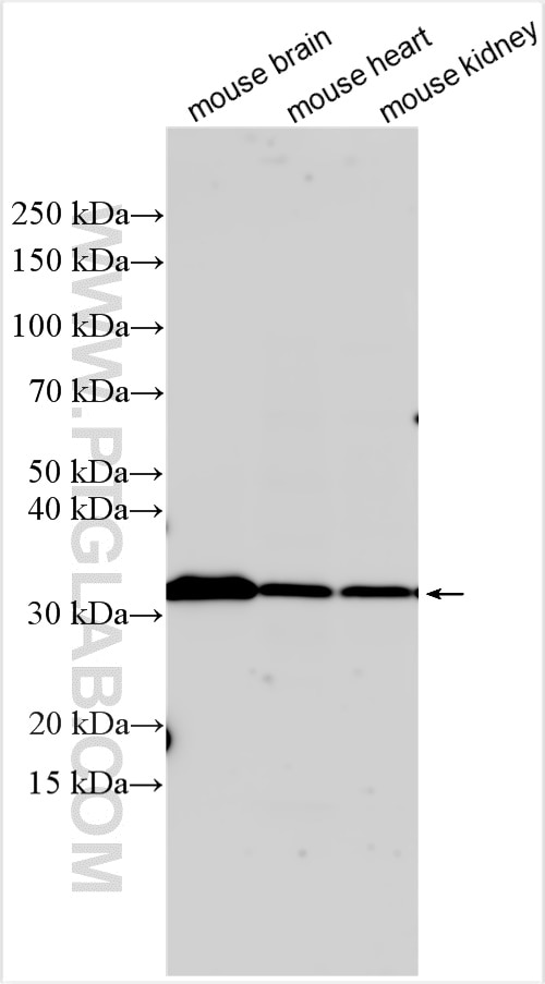
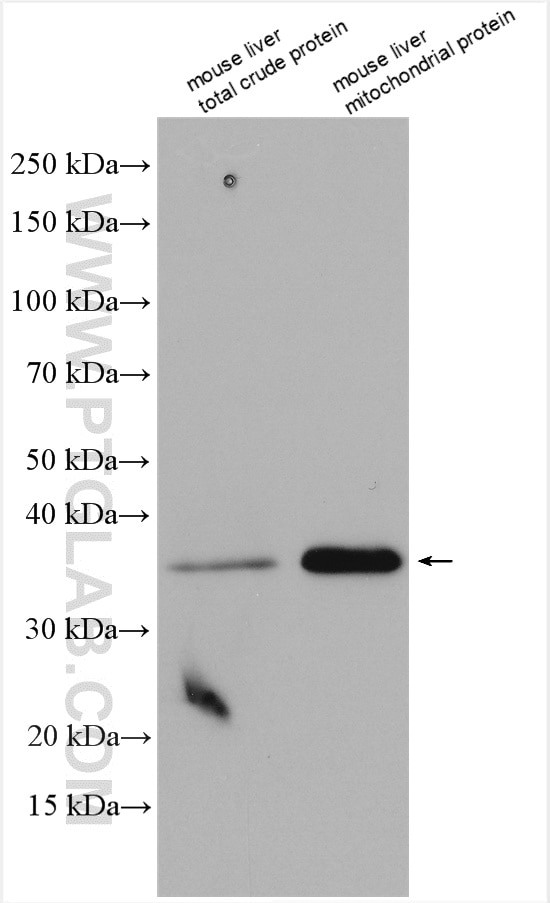
| Positive WB detected in | mouse brain tissue, mouse liver tissue, mouse heart tissue, mouse kidney tissue, mouse liver mitochondria |
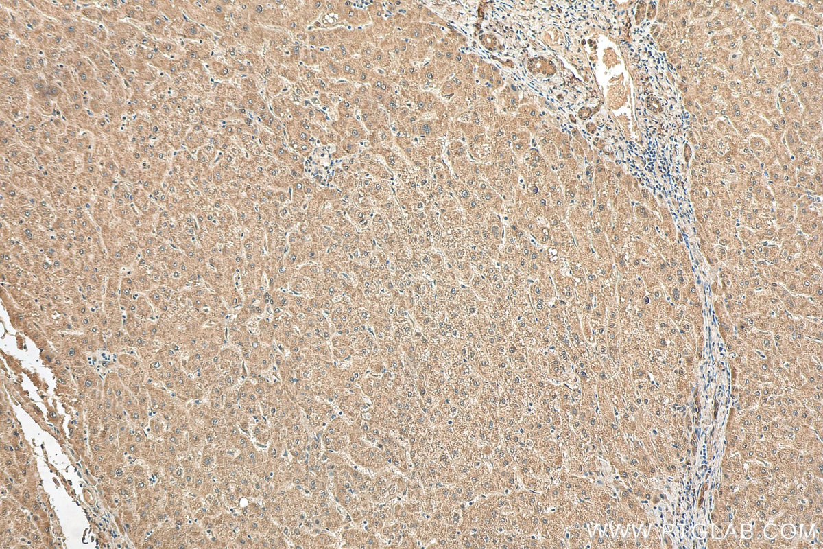
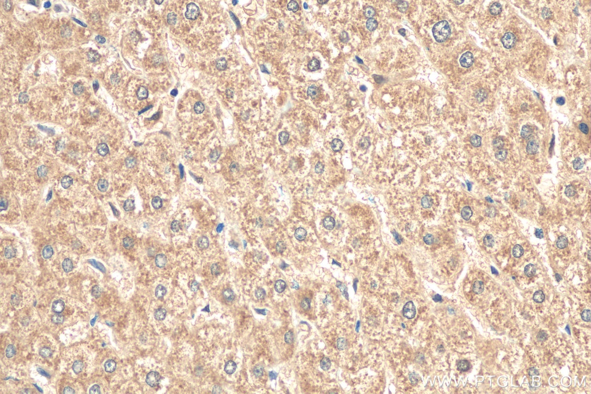
| Positive IHC detected in | human liver tissue Note: suggested antigen retrieval with TE buffer pH 9.0; (*) Alternatively, antigen retrieval may be performed with citrate buffer pH 6.0 |
Recommended dilution
| Application | Dilution |
|---|---|
| Western Blot (WB) | WB : 1:1000-1:8000 |
| Immunohistochemistry (IHC) | IHC : 1:50-1:500 |
| It is recommended that this reagent should be titrated in each testing system to obtain optimal results. | |
| Sample-dependent, Check data in validation data gallery. | |
Published Applications
| KD/KO | See 2 publications below |
| WB | See 85 publications below |
| IHC | See 5 publications below |
| IF | See 2 publications below |
Product Information
19703-1-AP targets ND1 in WB, IHC, IF, ELISA applications and shows reactivity with human, mouse samples.
| Tested Reactivity | human, mouse |
| Cited Reactivity | human, mouse, rat, pig, hamster |
| Host / Isotype | Rabbit / IgG |
| Class | Polyclonal |
| Type | Antibody |
| Immunogen |
Peptide Predict reactive species |
| Full Name | NADH dehydrogenase, subunit 1 (complex I) |
| Calculated Molecular Weight | 36 kDa |
| Observed Molecular Weight | 28-38 kDa |
| GenBank Accession Number | NC_012920 |
| Gene Symbol | ND1 |
| Gene ID (NCBI) | 4535 |
| RRID | AB_10637853 |
| Conjugate | Unconjugated |
| Form | Liquid |
| Purification Method | Antigen affinity purification |
| UNIPROT ID | P03886 |
| Storage Buffer | PBS with 0.02% sodium azide and 50% glycerol, pH 7.3. |
| Storage Conditions | Store at -20°C. Stable for one year after shipment. Aliquoting is unnecessary for -20oC storage. 20ul sizes contain 0.1% BSA. |
Background Information
ND1 belongs to the complex I subunit 1 family. ND1 is a core subunit of the mitochondrial membrane respiratory chain NADH dehydrogenase (Complex I) that is believed to belong to the minimal assembly required for catalysis. Complex I functions in the transfer of electrons from NADH to the respiratory chain. The immediate electron acceptor for the enzyme is believed to be ubiquinone.
Protocols
| Product Specific Protocols | |
|---|---|
| IHC protocol for ND1 antibody 19703-1-AP | Download protocol |
| WB protocol for ND1 antibody 19703-1-AP | Download protocol |
| Standard Protocols | |
|---|---|
| Click here to view our Standard Protocols |
Publications
| Species | Application | Title |
|---|---|---|
Cell MicroRNA Directly Enhances Mitochondrial Translation during Muscle Differentiation.
| ||
Mol Cell Filamentous GLS1 promotes ROS-induced apoptosis upon glutamine deprivation via insufficient asparagine synthesis. | ||
Nat Commun Disuse-associated loss of the protease LONP1 in muscle impairs mitochondrial function and causes reduced skeletal muscle mass and strength. |
Reviews
The reviews below have been submitted by verified Proteintech customers who received an incentive for providing their feedback.
FH Lenie (Verified Customer) (10-30-2025) | Both WB and IF no result on zebrafish even though protocol was followed.
|
FH Tania (Verified Customer) (03-05-2022) | a lot of background. difficult to say each band is the correct one.
|
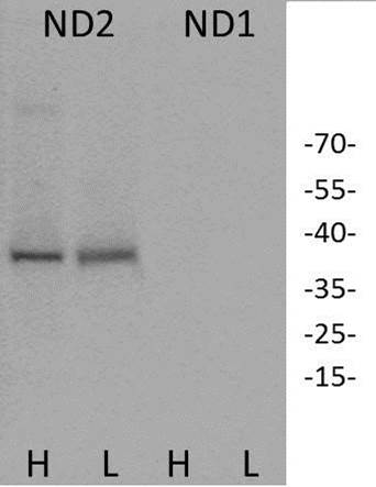
FH Daniel (Verified Customer) (10-01-2019) | ND2 was detected in both liver (L) and heart (H) lysate from a wildtype mouse. No ND1 was detected in samples processed in parallel.
 |
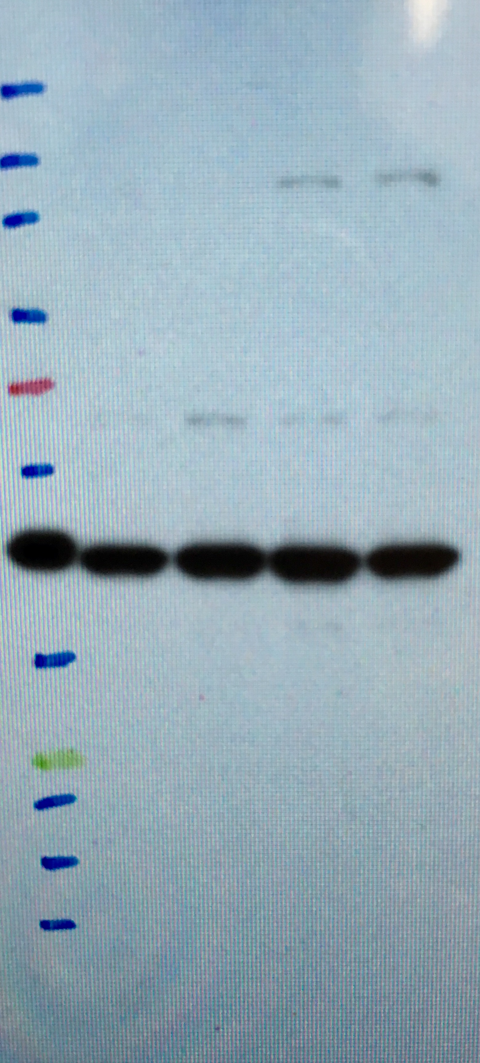
FH Loukmane (Verified Customer) (08-10-2018) | It works very well with A549 and lung tissue, although with the former the background is higherThe image shows ND1 in whole A549 lysate
 |

