


Tested Applications
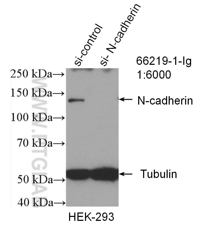
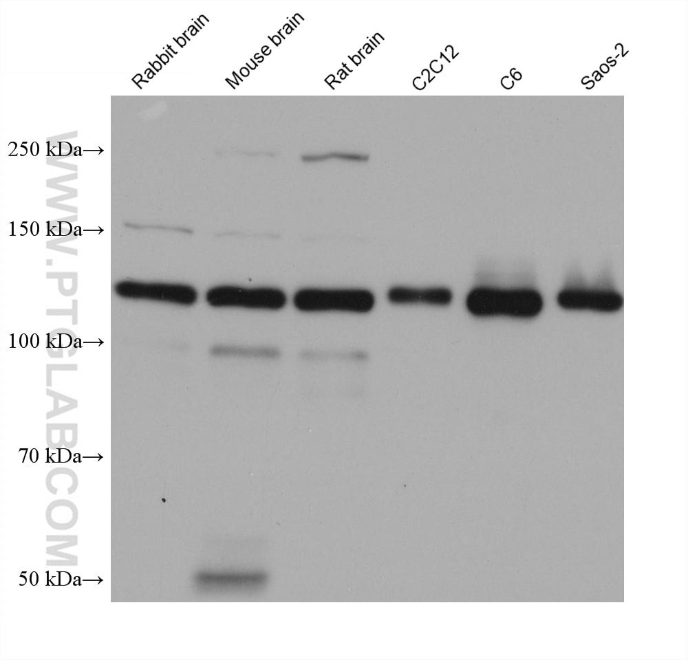
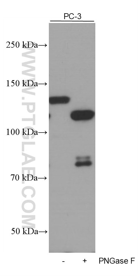
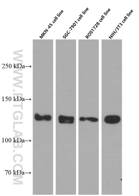
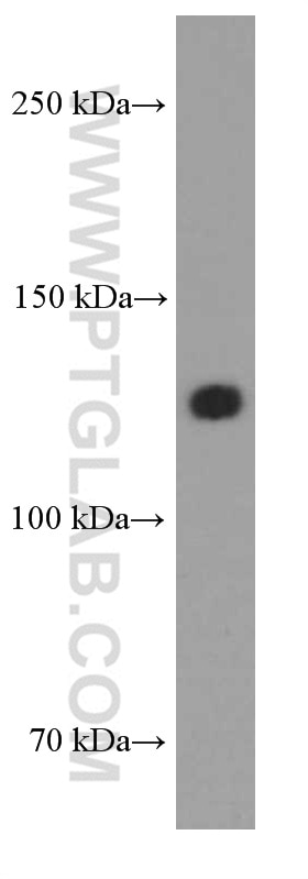
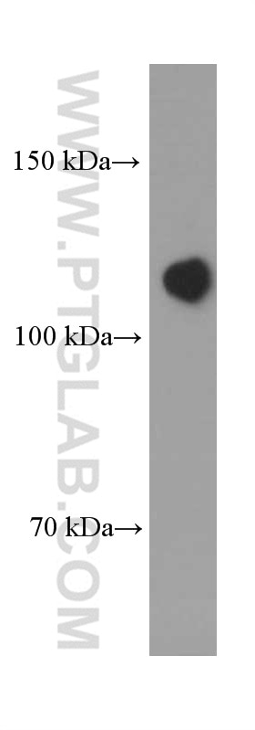
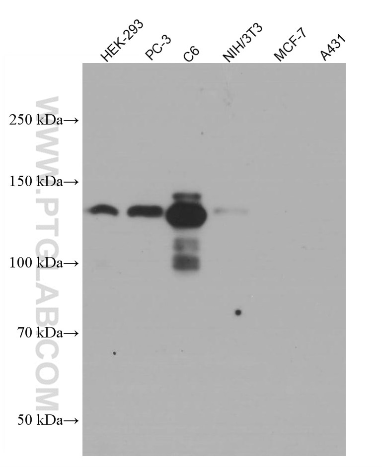
| Positive WB detected in | rabbit brain tissue, pig brain tissue, human heart tissue, MKN-45 cells, HEK-293 cells, PC-3 cells, mouse brain tissue, rat brain tissue, C2C12 cells, C6 cells, Saos-2 cells, SGC-7901 cells, ROS1728 cells, NIH/3T3 cells |
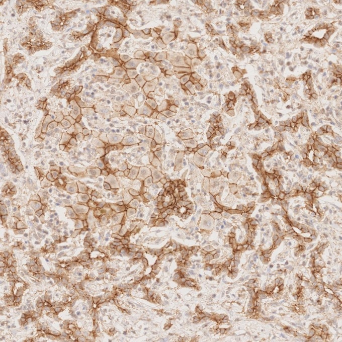
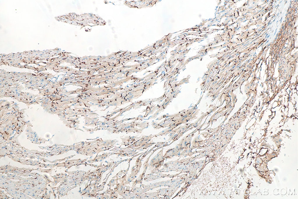
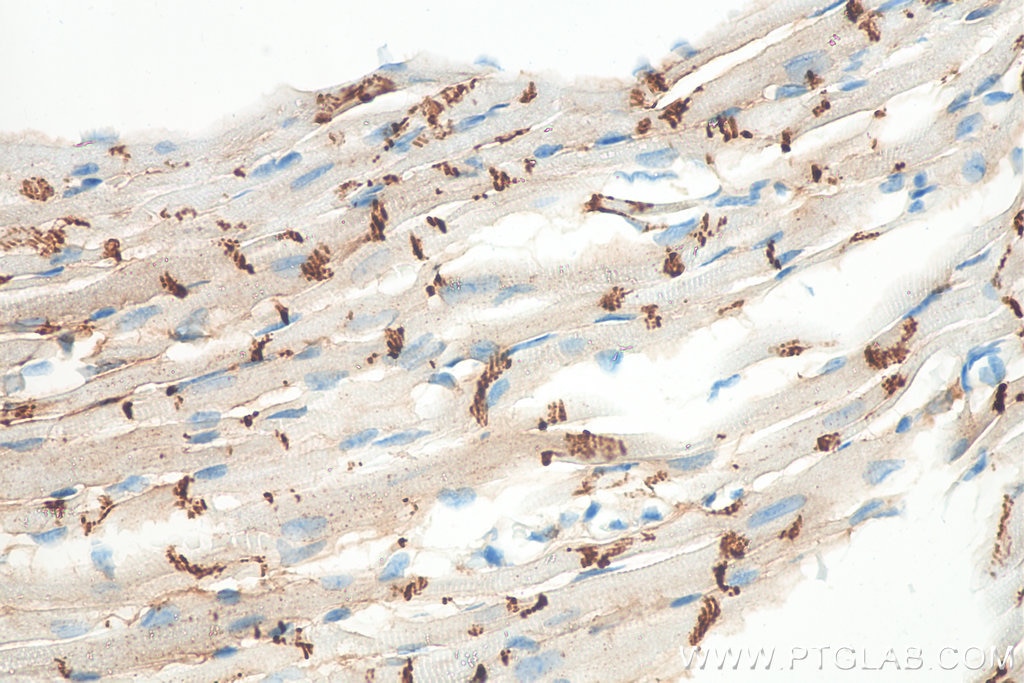
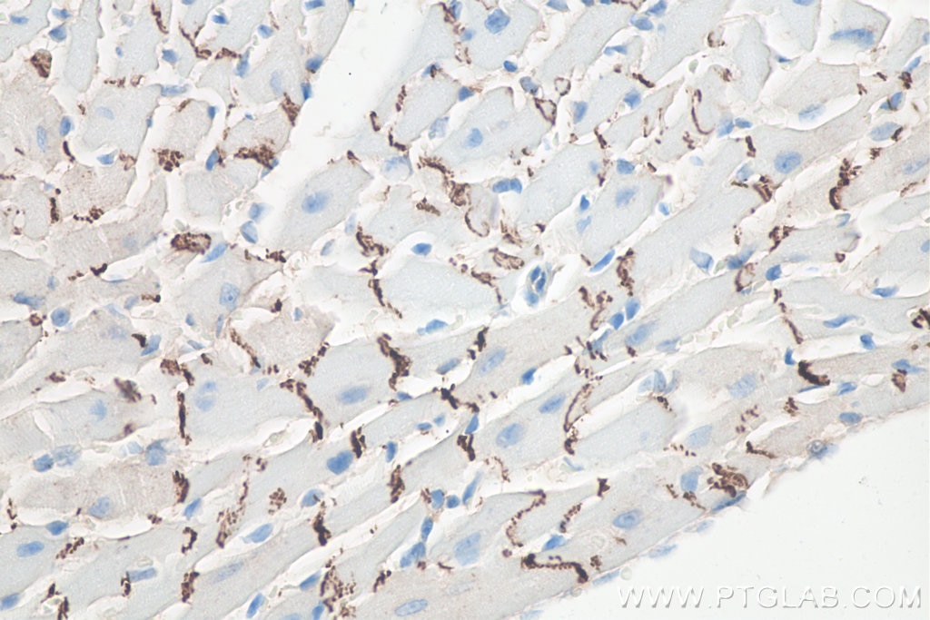
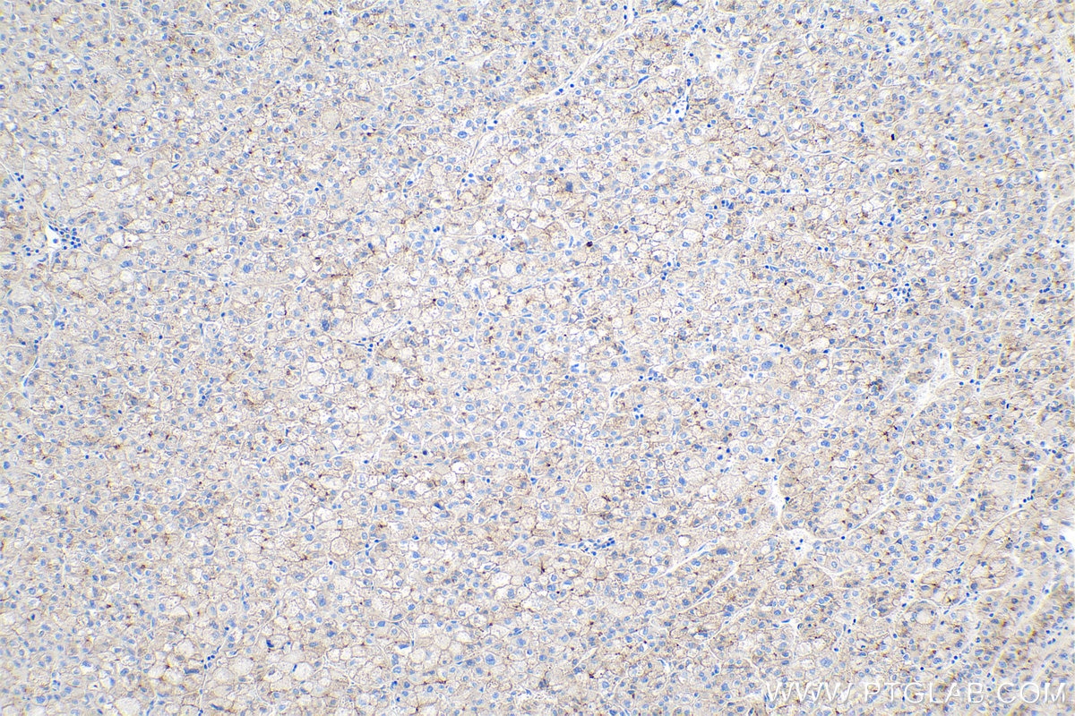
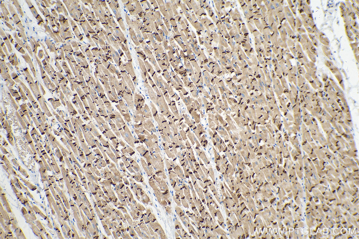
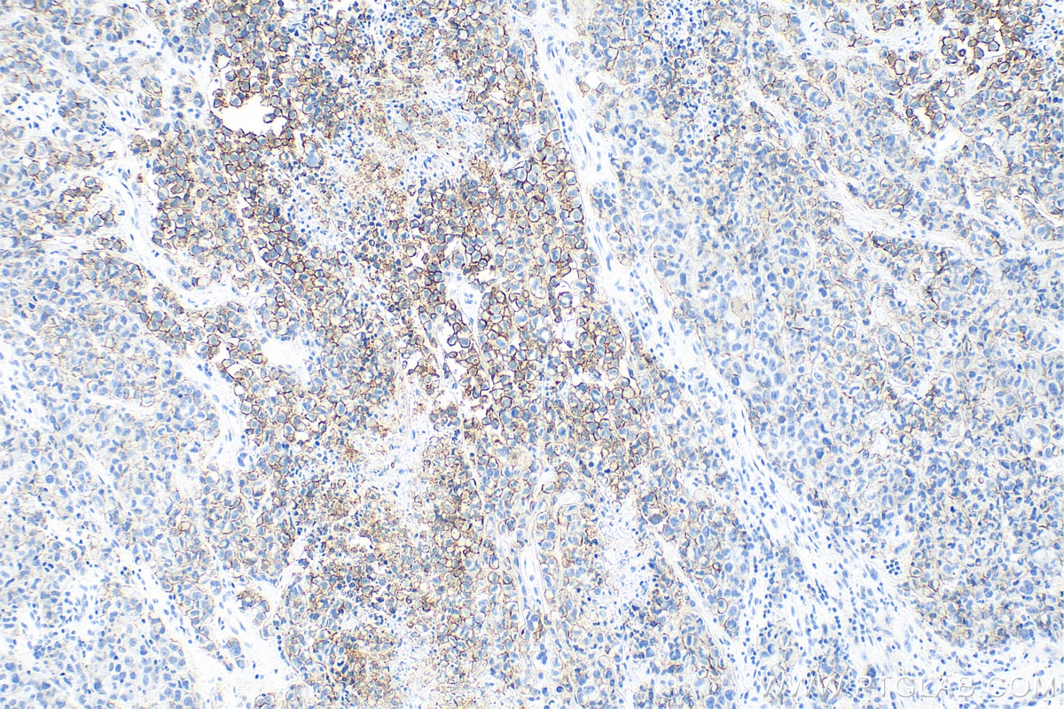
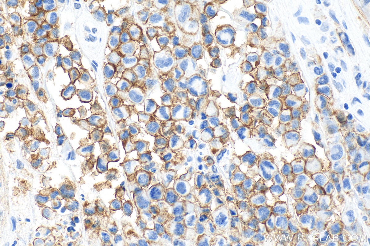
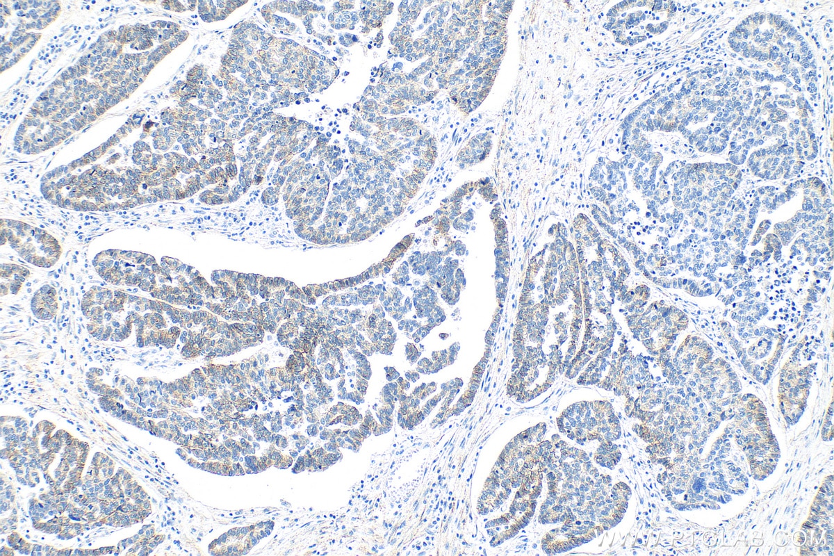
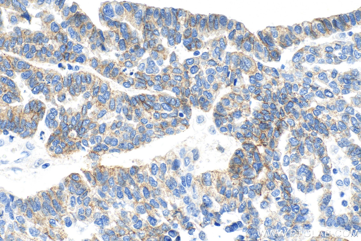
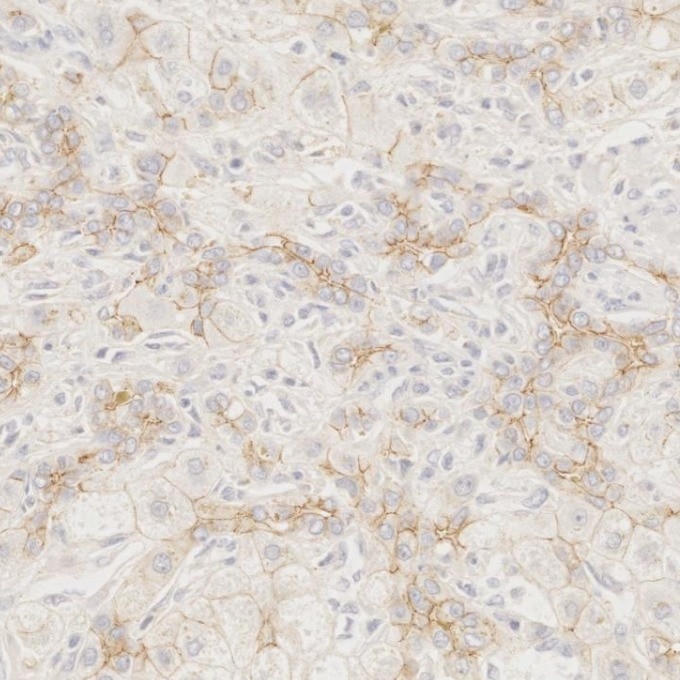
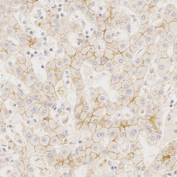
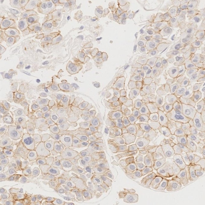
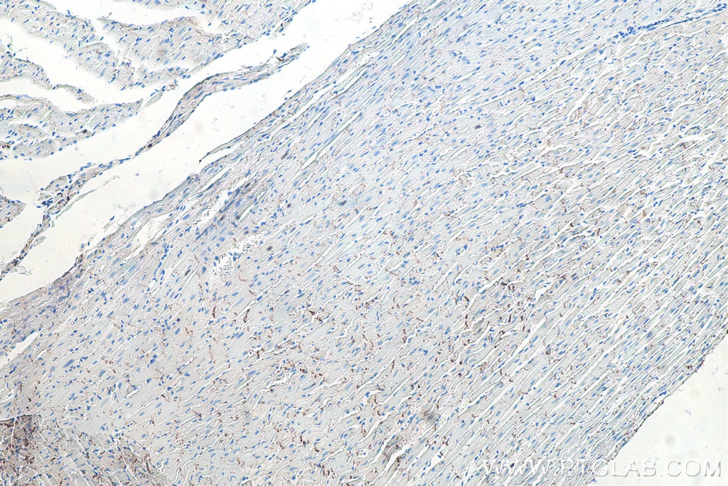
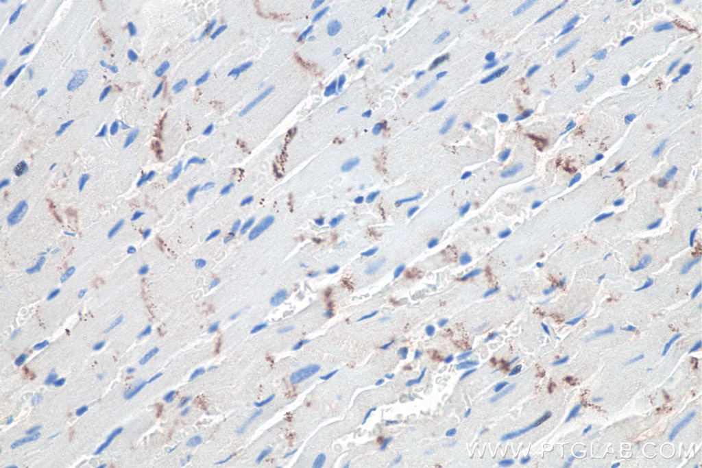
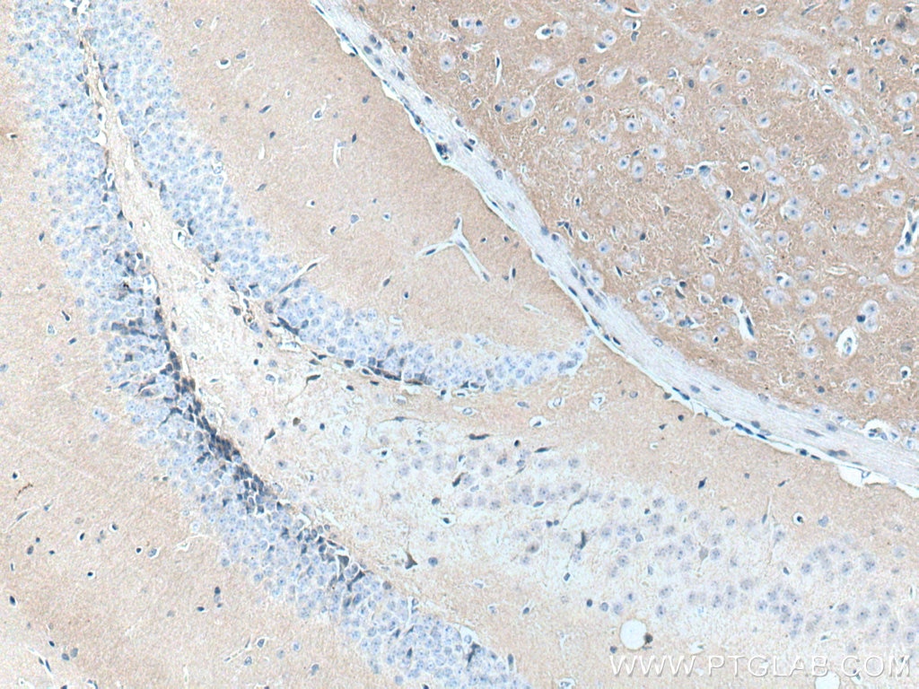
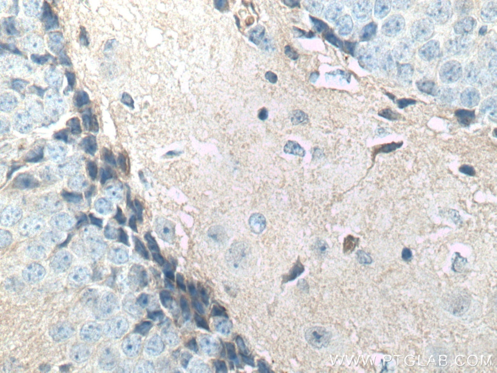
| Positive IHC detected in | mouse heart tissue, mouse brain tissue, human malignant melanoma tissue, human ovary tumor tissue, rat heart tissue Note: suggested antigen retrieval with TE buffer pH 9.0; (*) Alternatively, antigen retrieval may be performed with citrate buffer pH 6.0 |
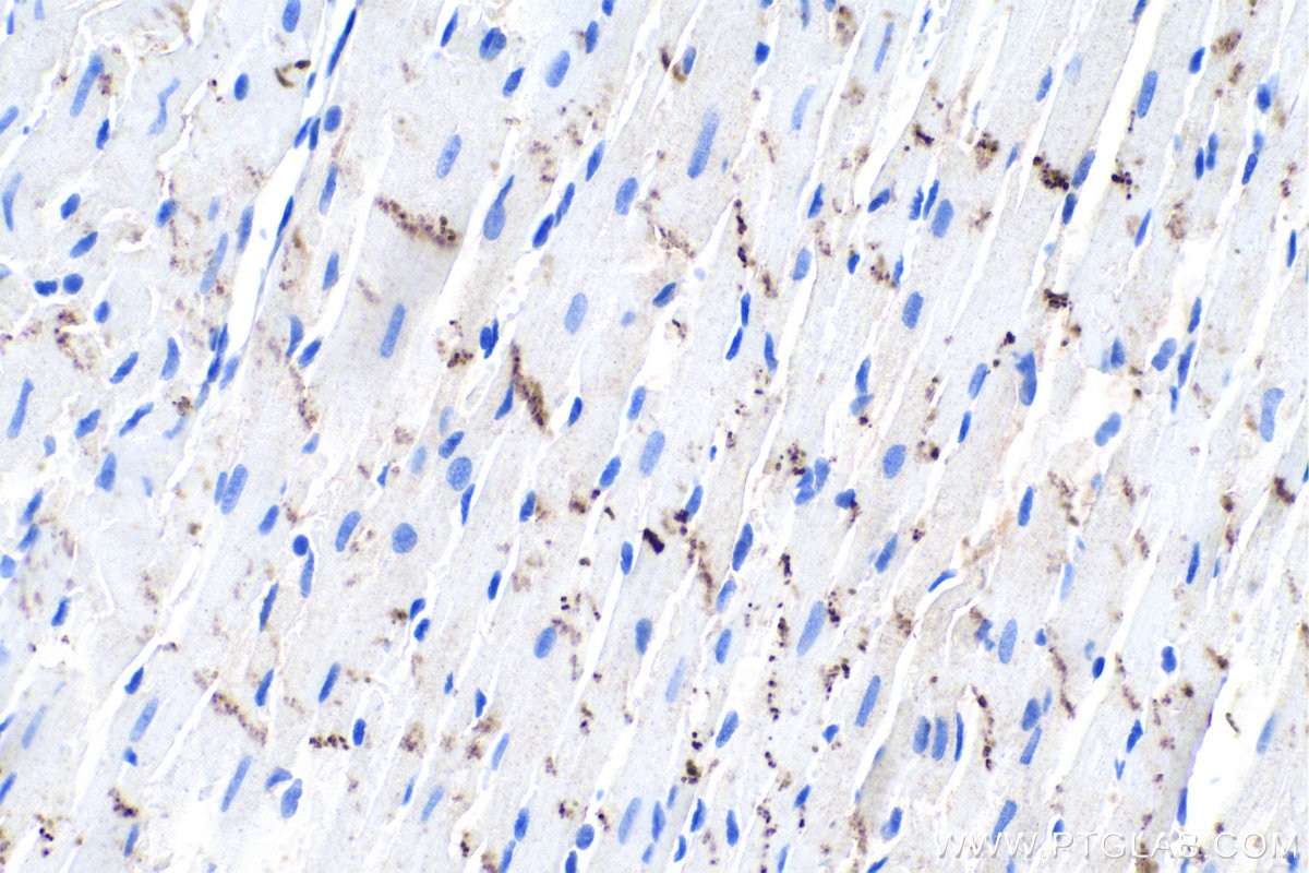
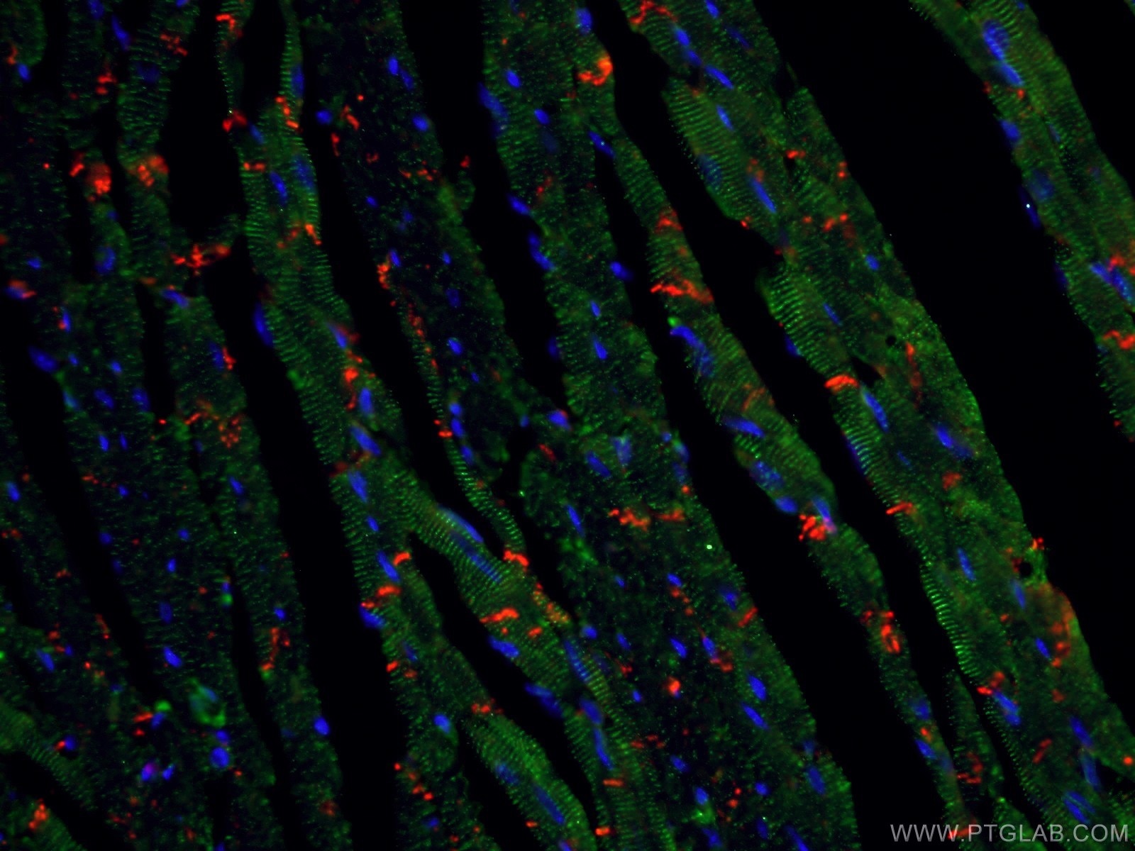
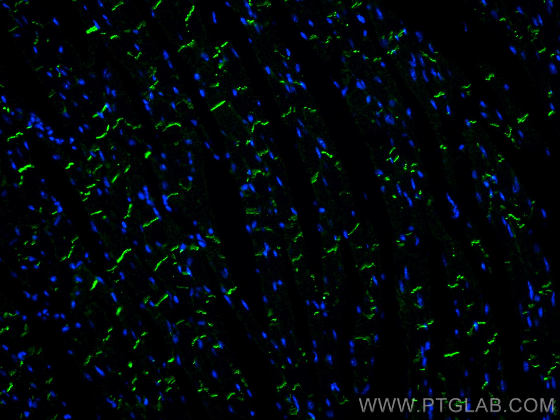
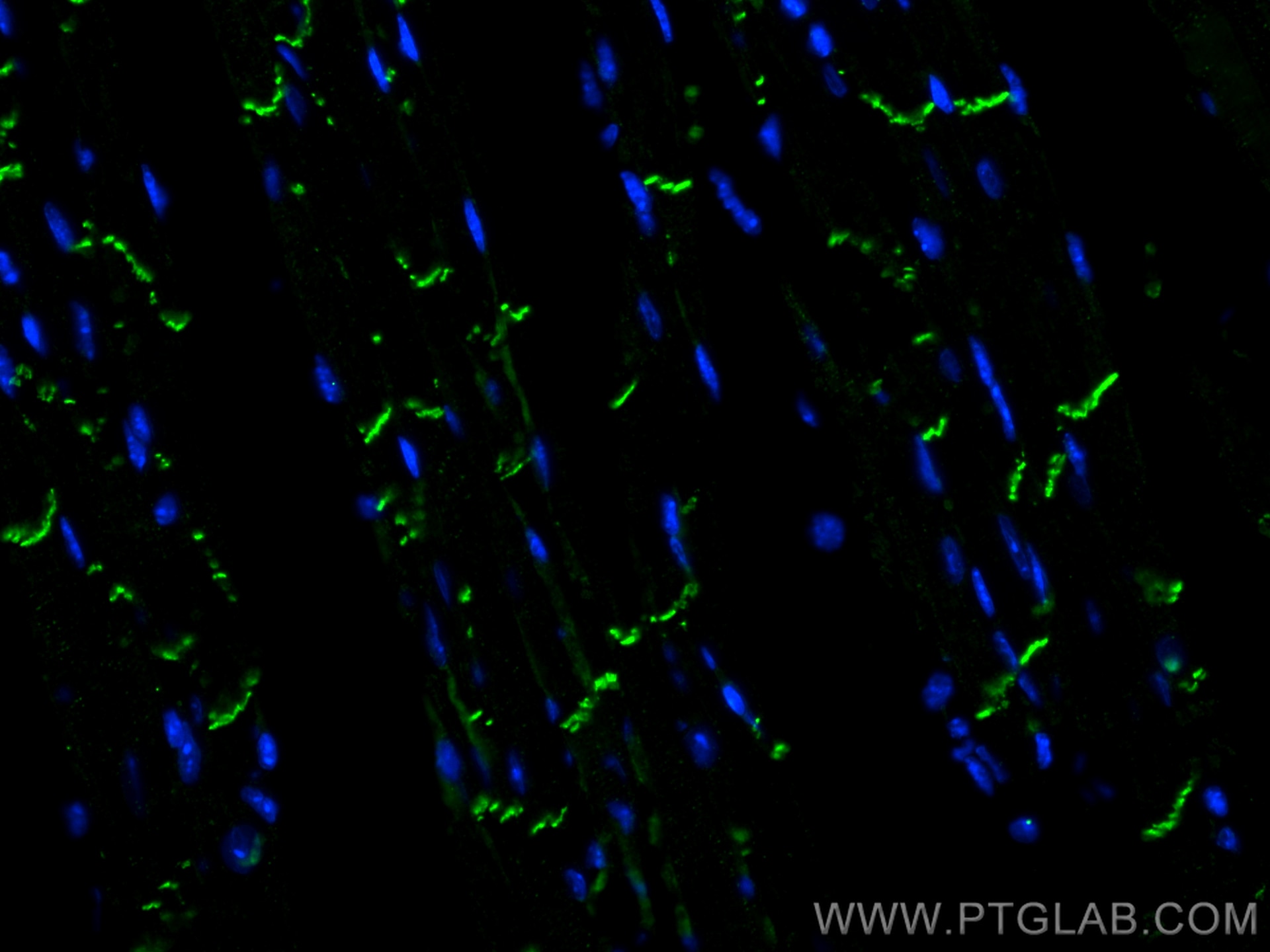
| Positive IF-P detected in | mouse heart tissue |
Recommended dilution
| Application | Dilution |
|---|---|
| Western Blot (WB) | WB : 1:5000-1:50000 |
| Immunohistochemistry (IHC) | IHC : 1:7500-1:30000 |
| Immunofluorescence (IF)-P | IF-P : 1:200-1:800 |
| It is recommended that this reagent should be titrated in each testing system to obtain optimal results. | |
| Sample-dependent, Check data in validation data gallery. | |
Product Information
66219-1-Ig targets N-cadherin in WB, IHC, IF-P, ELISA applications and shows reactivity with human, mouse, rat, pig, rabbit samples.
| Tested Reactivity | human, mouse, rat, pig, rabbit |
| Cited Reactivity | human, mouse, rat, pig, canine, monkey |
| Host / Isotype | Mouse / IgG1 |
| Class | Monoclonal |
| Type | Antibody |
| Immunogen |
CatNo: Ag4996 Product name: Recombinant human N-cadherin protein Source: e coli.-derived, PET28a Tag: 6*His Domain: 33-350 aa of BC036470 Sequence: KTGFPEDVYSAVLSKDVHEGQPLLNVKFSNCNGKRKVQYESSEPADFKVDEDGMVYAVRSFPLSSEHAKFLIYAQDKETQEKWQVAVKLSLKPTLTEESVKESAEVEEIVFPRQFSKHSGHLQRQKRDWVISPINLPENSRGPFPQELVRIRSDRDKNLSLRYSVTGPGADQPPTGIFIINPISGQLSVTKPLDREQIARFHLRAHAVDINGNQVENPIDIVINVIDMNDNRPEFLHQVWNGTVPEGSKPGTYVMTVTAIDADDPNALNGMLRYRIVSQAPSTPSPNMFTINNETGDIITVAAGLDREKVQQYTLIIQ Predict reactive species |
| Full Name | cadherin 2, type 1, N-cadherin (neuronal) |
| Calculated Molecular Weight | 906 aa, 100 kDa |
| Observed Molecular Weight | 130 kDa |
| GenBank Accession Number | BC036470 |
| Gene Symbol | N-cadherin |
| Gene ID (NCBI) | 1000 |
| RRID | AB_2881610 |
| Conjugate | Unconjugated |
| Form | Liquid |
| Purification Method | Protein G purification |
| UNIPROT ID | P19022 |
| Storage Buffer | PBS with 0.02% sodium azide and 50% glycerol, pH 7.3. |
| Storage Conditions | Store at -20°C. Stable for one year after shipment. Aliquoting is unnecessary for -20oC storage. 20ul sizes contain 0.1% BSA. |
Background Information
Cadherins are a family of transmembrane glycoproteins that mediate calcium-dependent cell-cell adhesion and play an important role in the maintenance of normal tissue architecture. N-cadherin (neural cadherin), also known as CDH2 (cadherin 2), is a 130-kDa transmembrane protein and a classical member of the cadherin superfamily which also include E-, P-, R-, and B-cadherins. Expression of N-cadherin has been reported on various cell types including neurons, endothelial cells and cardiac myocytes (PMID: 11282032; 9508779; 8125202). N-cadherin has functions in early brain morphogenesis, synaptogenesis and synaptic plasticity (PMID: 23321619). The N-cadherin ectodomain can be cleaved, leading to the generation of a 90 kD N-terminal fragment (PMID: 16998833; 17028923).
Protocols
| Product Specific Protocols | |
|---|---|
| IF protocol for N-cadherin antibody 66219-1-Ig | Download protocol |
| IHC protocol for N-cadherin antibody 66219-1-Ig | Download protocol |
| WB protocol for N-cadherin antibody 66219-1-Ig | Download protocol |
| Standard Protocols | |
|---|---|
| Click here to view our Standard Protocols |
Publications
What published studies show
| Species | Application | Title |
|---|---|---|
Mol Cancer YTHDF2 mediates the mRNA degradation of the tumor suppressors to induce AKT phosphorylation in N6-methyladenosine-dependent way in prostate cancer. | ||
Cell Stem Cell Long-term engraftment and maturation of autologous iPSC-derived cardiomyocytes in two rhesus macaques | ||
Cancer Res Proteomic Profiling of Serum Extracellular Vesicles Identifies Diagnostic Signatures and Therapeutic Targets in Breast Cancer | ||
Cancer Res SRSF9 Forms Phase-separated Condensates to Promote Ovarian Cancer Progression by Inducing RNA Alternative Splicing that is Inhibited by m6A Modification | ||
J Extracell Vesicles Extracellular vesicles derived from Akkermansia muciniphila promote placentation and mitigate preeclampsia in a mouse model | ||
Autophagy Dual roles of CXCR4 (C-X-C motif chemokine receptor 4) in promoting entry of ebolavirus and targeting excessive glycoprotein for reticulophagic degradation to facilitate viral fitness |
Reviews
What customers say
The reviews below have been submitted by verified Proteintech customers who received an incentive for providing their feedback.
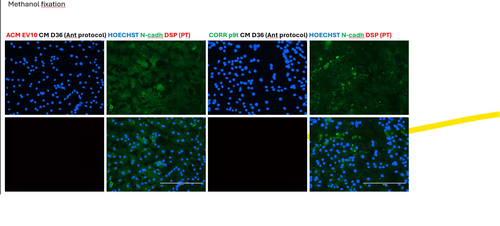
Kiara (Verified Customer) (07-28-2025) | This primary antibody was not internally validated for IF, but there was a paper published that it worked on 4% PFA-fixed H9 ESCs, so I gave it a shot anyway. However, the staining did not show a nice membrane localization that our lab usually sees when using a different rabbit pan-cadherin primary antibody.
 |
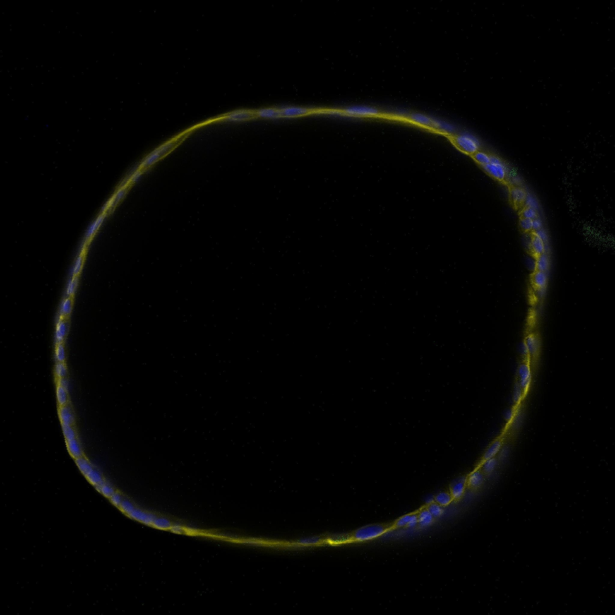
Davide (Verified Customer) (06-13-2023) | The antibody didn't produce any signal in 3d primary human cholangiocyte organoids. I would recommend either to try other antibody concentration or to try another Ab. It was tried on a cell condition which was recently reported to stimulate the CDH2 protein expression. However it should also be present in control condition at low levels.
 |
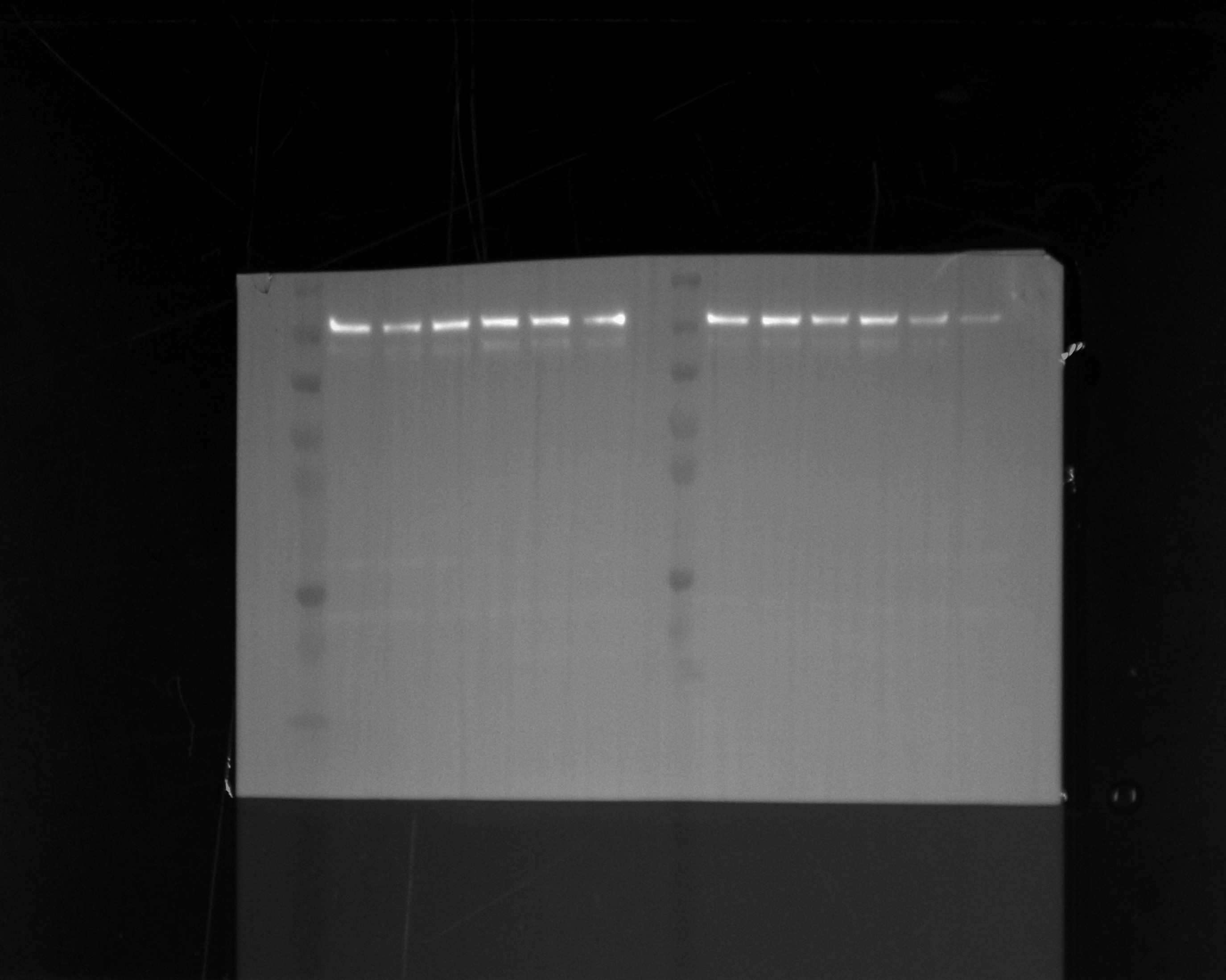
Udesh (Verified Customer) (10-13-2022) | Used the Ab at 1:2000 dilution in 5% NFDM and observed a band at 140 kD. Blocked in 5% NFDM for 1H at RT.
 |


