

Tested Applications
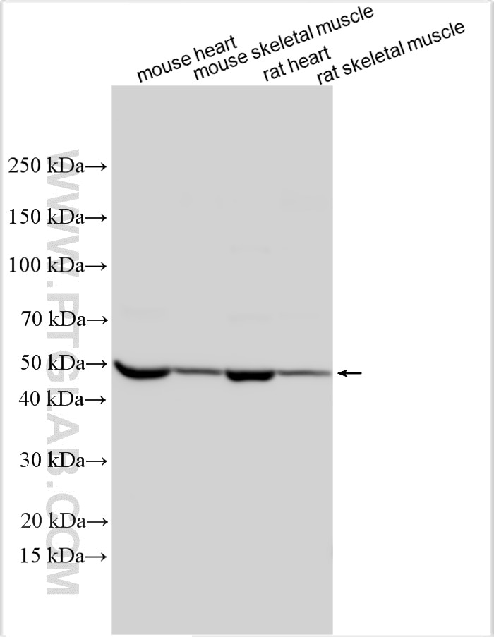
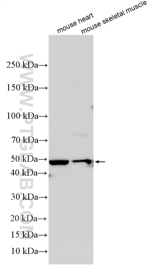
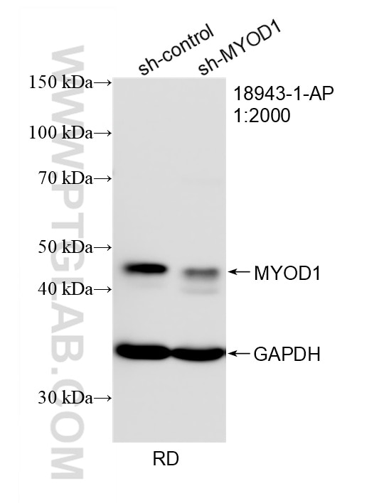
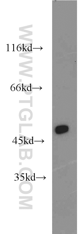
| Positive WB detected in | mouse heart tissue, human heart tissue, RD cells |
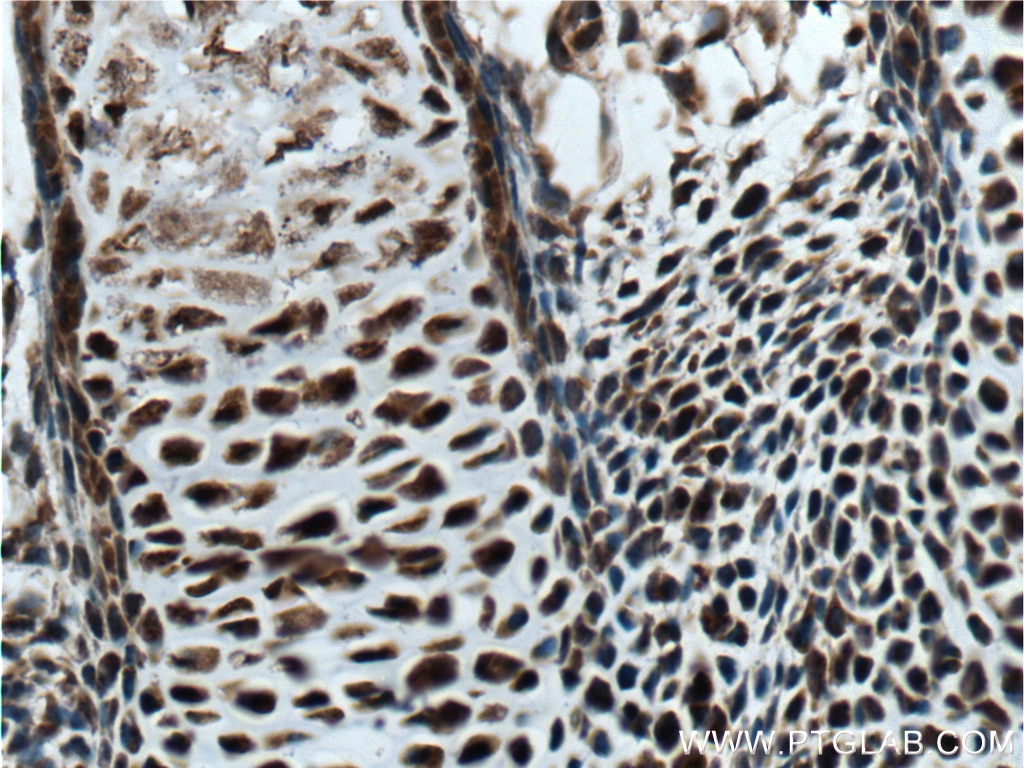
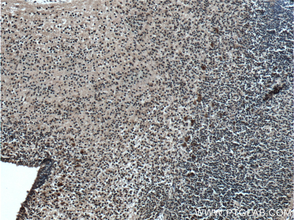
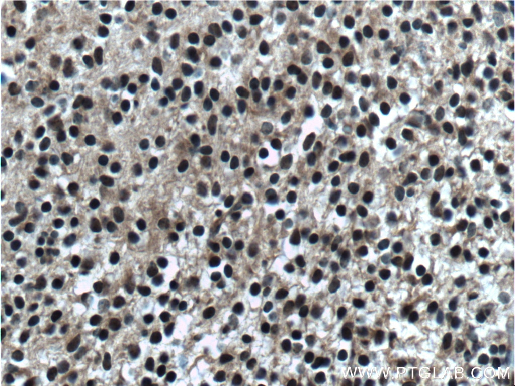
| Positive IHC detected in | mouse embryo tissue Note: suggested antigen retrieval with TE buffer pH 9.0; (*) Alternatively, antigen retrieval may be performed with citrate buffer pH 6.0 |
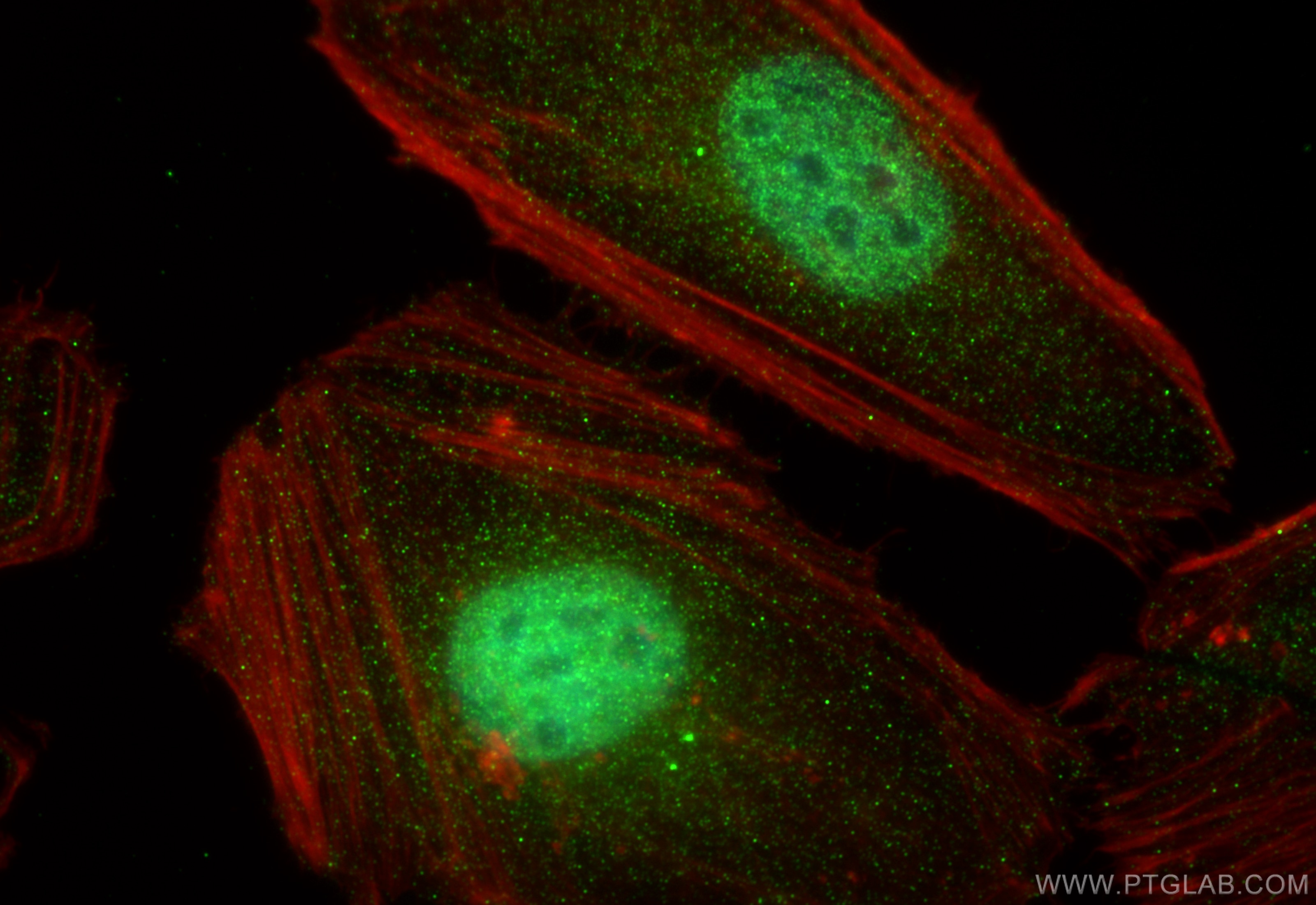
| Positive IF/ICC detected in | HeLa cells |
Recommended dilution
| Application | Dilution |
|---|---|
| Western Blot (WB) | WB : 1:2000-1:12000 |
| Immunohistochemistry (IHC) | IHC : 1:50-1:500 |
| Immunofluorescence (IF)/ICC | IF/ICC : 1:400-1:1600 |
| It is recommended that this reagent should be titrated in each testing system to obtain optimal results. | |
| Sample-dependent, Check data in validation data gallery. | |
Product Information
18943-1-AP targets MYOD1 in WB, IHC, IF/ICC, CoIP, ChIP, ELISA applications and shows reactivity with human, mouse, rat samples.
| Tested Reactivity | human, mouse, rat |
| Cited Reactivity | human, mouse, rat, pig, rabbit, chicken, zebrafish, sheep, goat |
| Host / Isotype | Rabbit / IgG |
| Class | Polyclonal |
| Type | Antibody |
| Immunogen |
CatNo: Ag13512 Product name: Recombinant human MYOD1 protein Source: e coli.-derived, PGEX-4T Tag: GST Domain: 1-320 aa of BC064493 Sequence: MELLSPPLRDVDLTAPDGSLCSFATTDDFYDDPCFDSPDLRFFEDLDPRLMHVGALLKPEEHSHFPAAVHPAPGAREDEHVRAPSGHHQAGRCLLWACKACKRKTTNADRRKAATMRERRRLSKVNEAFETLKRCTSSNPNQRLPKVEILRNAIRYIEGLQALLRDQDAAPPGAAAAFYAPGPLPPGRGGEHYSGDSDASSPRSNCSDGMMDYSGPPSGARRRNCYEGAYYNEAPSEPRPGKSAAVSSLDCLSSIVERISTESPAAPALLLADVPSESPPRRQEAAAPSEGESSGDPTQSPDAAPQCPAGANPNPIYQVL Predict reactive species |
| Full Name | myogenic differentiation 1 |
| Calculated Molecular Weight | 320 aa, 35 kDa |
| Observed Molecular Weight | 35-45 kDa |
| GenBank Accession Number | BC064493 |
| Gene Symbol | MYOD1 |
| Gene ID (NCBI) | 4654 |
| RRID | AB_10603467 |
| Conjugate | Unconjugated |
| Form | Liquid |
| Purification Method | Antigen affinity purification |
| UNIPROT ID | P15172 |
| Storage Buffer | PBS with 0.02% sodium azide and 50% glycerol, pH 7.3. |
| Storage Conditions | Store at -20°C. Stable for one year after shipment. Aliquoting is unnecessary for -20oC storage. 20ul sizes contain 0.1% BSA. |
Background Information
MYOD1, also named as BHLHC1 or MYF3, is a 320 amino acid protein, which promotes the transcriptional activity of MYOD1 through its CDK9-mediated phosphorylation. This phosphorylation promotes its function in muscle differentiation. MYOD1 acts as a transcriptional activator that promotes transcription of muscle-specific target genes and plays a role in muscle differentiation. MYOD1 together with MYF5 and MYOG, co-occupies muscle-specific gene promoter core region during myogenesis. Induces fibroblasts to differentiate into myoblasts. The calcualted molecular weight of MYOD1 is 34 kDa, but modified MYOD1 is about 45 kDa. (PMID: 12037670)
Protocols
| Product Specific Protocols | |
|---|---|
| IF protocol for MYOD1 antibody 18943-1-AP | Download protocol |
| IHC protocol for MYOD1 antibody 18943-1-AP | Download protocol |
| WB protocol for MYOD1 antibody 18943-1-AP | Download protocol |
| Standard Protocols | |
|---|---|
| Click here to view our Standard Protocols |
Publications
What published studies show
MYOD1 Polyclonal antibody (Cat# 18943-1-AP) has accumulated 192 citations across journals including Nature, Cell Stem Cell, Nature Communications, Science Advances, Advanced Science, FASEB Journal, Cell Reports, Developmental Cell, and Autophagy. Research fields span skeletal muscle biology, myoblast differentiation, muscle regeneration, sarcopenia, cancer cachexia, ALS, Duchenne muscular dystrophy, tissue engineering, cultured meat production, and stem cell biology, with primary applications in WB, IHC, IF, CoIP, and ChIP.
| Species | Application | Title |
|---|---|---|
Bioact Mater Injectable chondroitin sulfate-glycosylated decellularized extracellular matrix microgels activate Wnt/β-Catenin signaling to promote functional muscle regeneration in VML. | ||
Cell Stem Cell A reference human induced pluripotent stem cell line for large-scale collaborative studies | ||
Cancer Res YAP1-mediated suppression of USP31 enhances NF-κB activity to promote sarcomagenesis. |
Reviews
What customers say
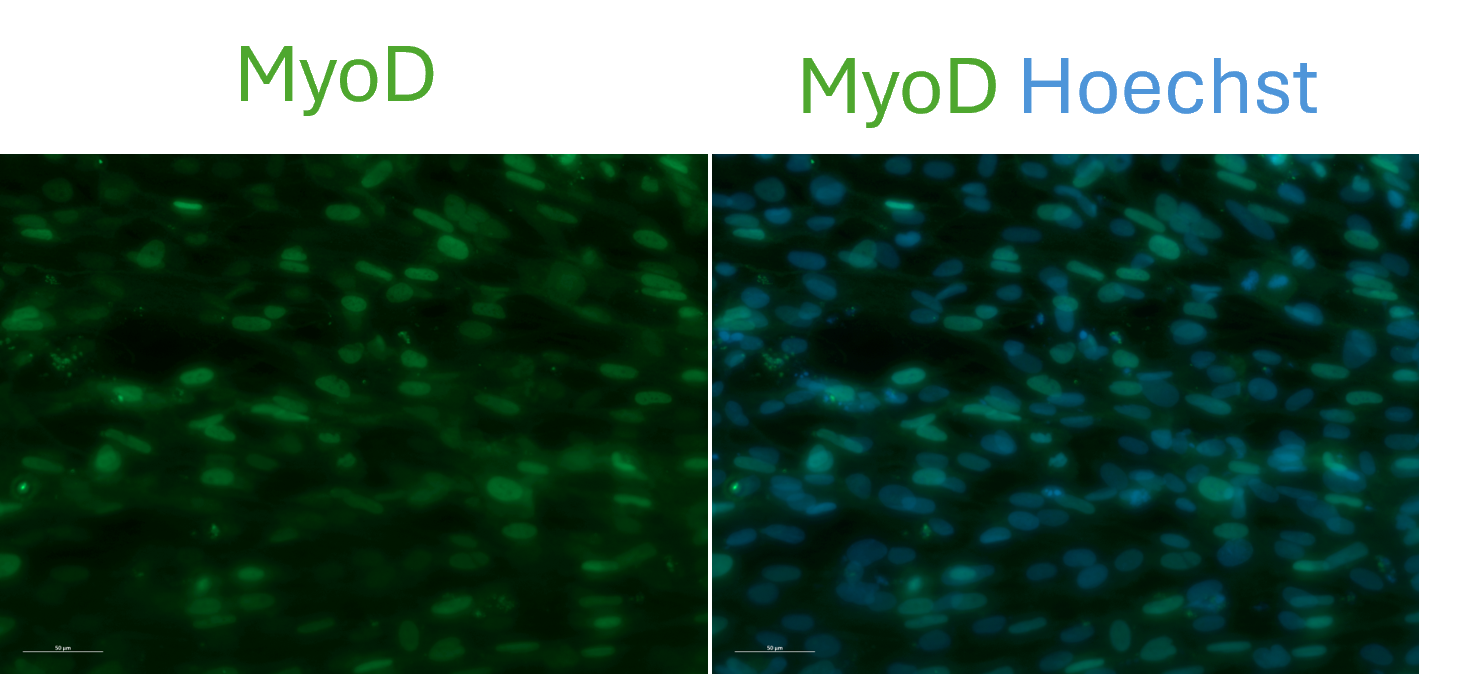
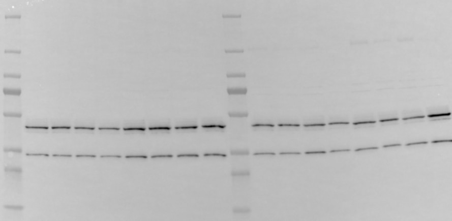
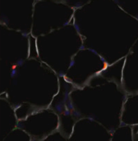
Users report consistent performance across applications. Two immunofluorescence reviews (5/5 and 5/5) confirm reliable staining at 1:100 dilution in myogenic progenitors, nascent myotubes, and satellite cells. One Western blot review (4/5) using 1:1000 dilution in C2C12 cells detected the expected MyoD1 band, though a lower non-specific band below 35 kDa was noted. Overall, reviewers find the antibody specific and effective for its intended targets in both IF and WB.
The reviews below have been submitted by verified Proteintech customers who received an incentive for providing their feedback.
Pavithra (Verified Customer) (08-21-2026) | Immunofluorescence as expected using 1:100 dilution
 |
Charlotte (Verified Customer) (07-23-2024) | I reveal MyoD1 in C2C12 cells that differentiate into myoblast lineage upon stimulation. Antibodies were diluted in 5% milk TBST. The upper band is the expected one. I guess the lower one (< 35kDa) is aspecific
 |
Cassandra (Verified Customer) (02-04-2021) | This antibody worked quite well in my IF staining. I used 1 to 100 antibody dilution.
 |


