Tested Applications
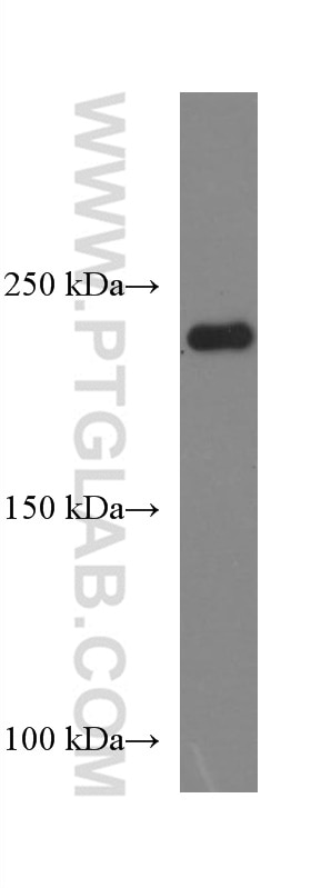
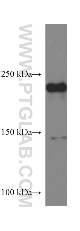
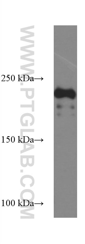
| Positive WB detected in | human skeletal muscle tissue, pig skeletal muscle tissue, mouse skeletal muscle tissue |
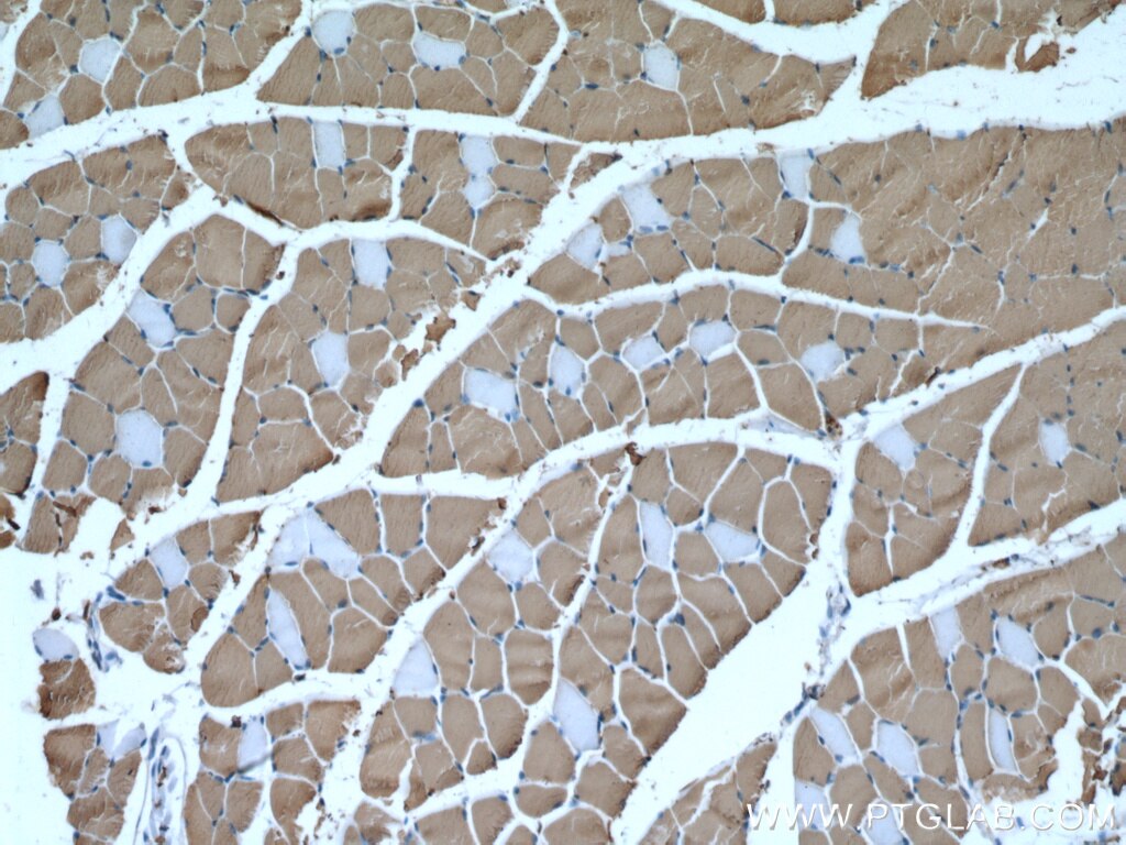
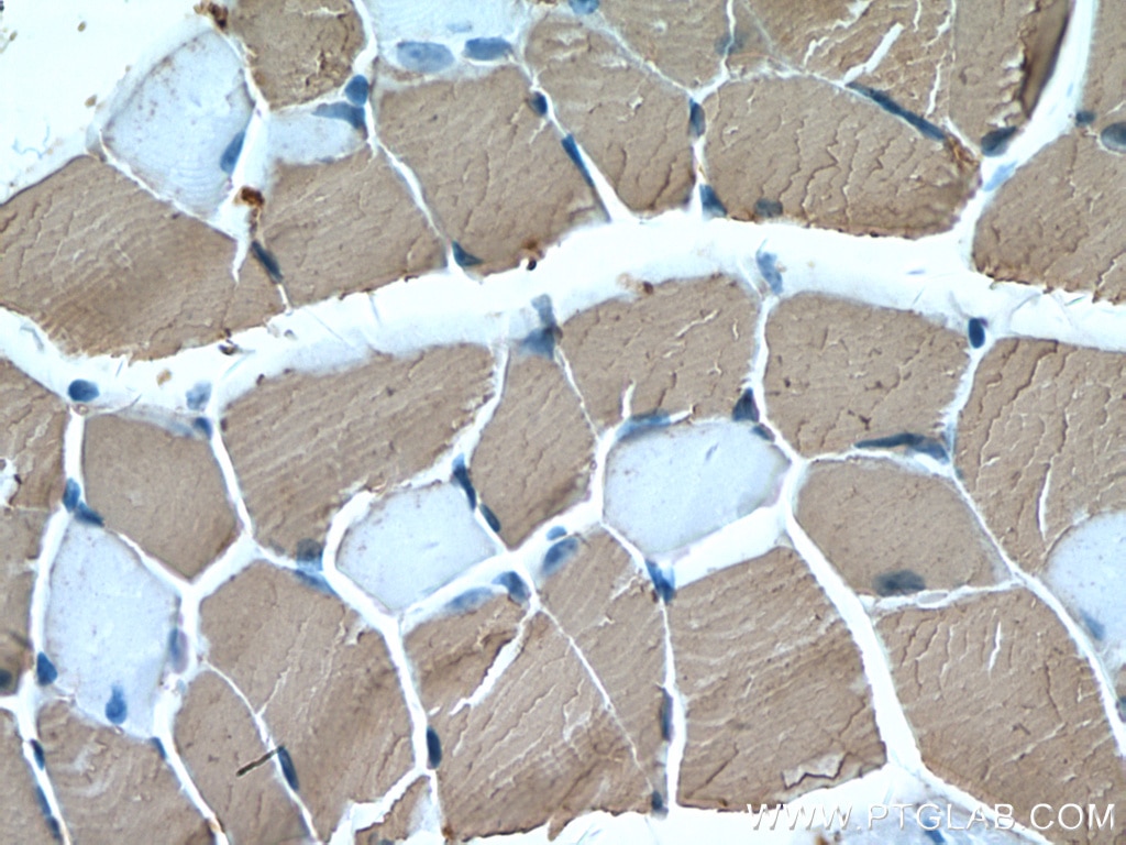
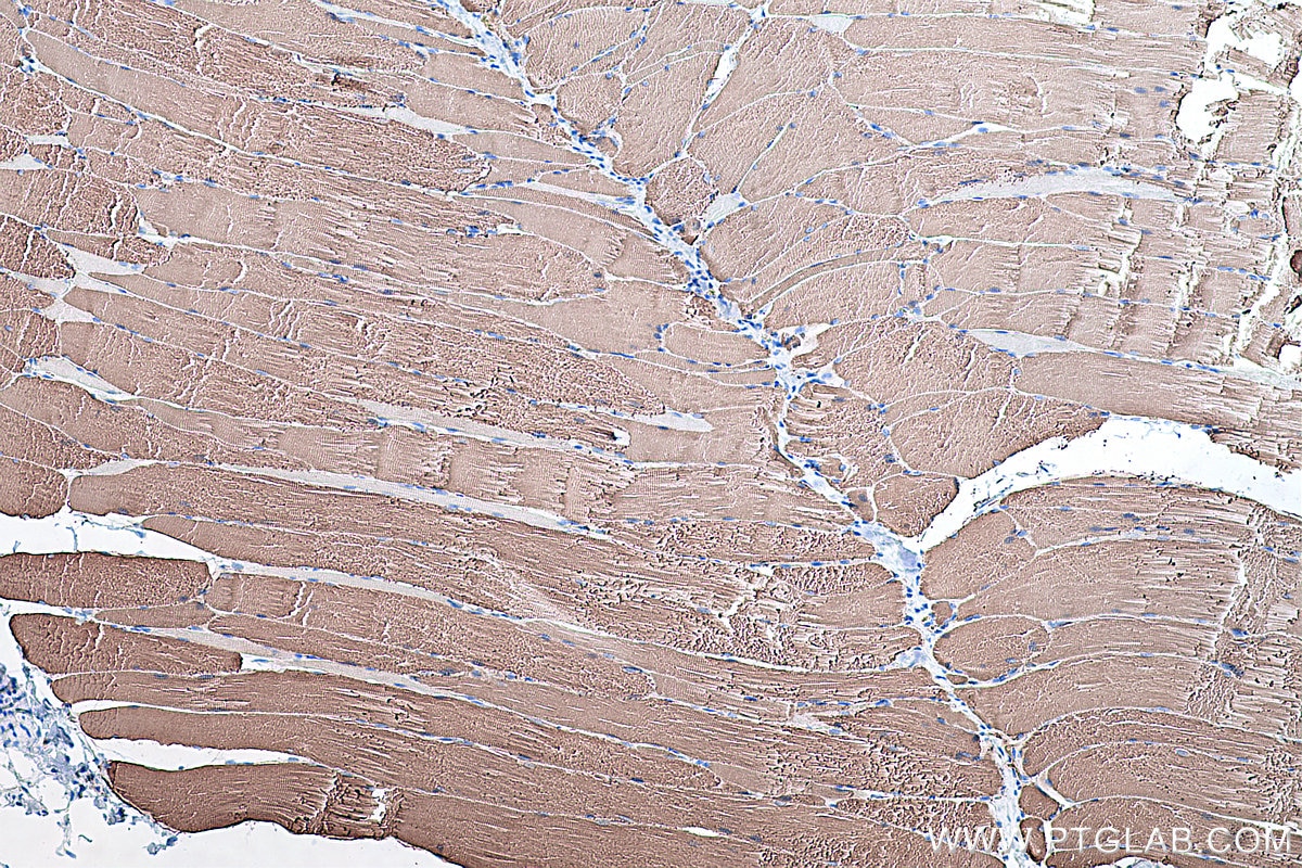
| Positive IHC detected in | mouse skeletal muscle tissue, rat skeletal muscle tissue Note: suggested antigen retrieval with TE buffer pH 9.0; (*) Alternatively, antigen retrieval may be performed with citrate buffer pH 6.0 |
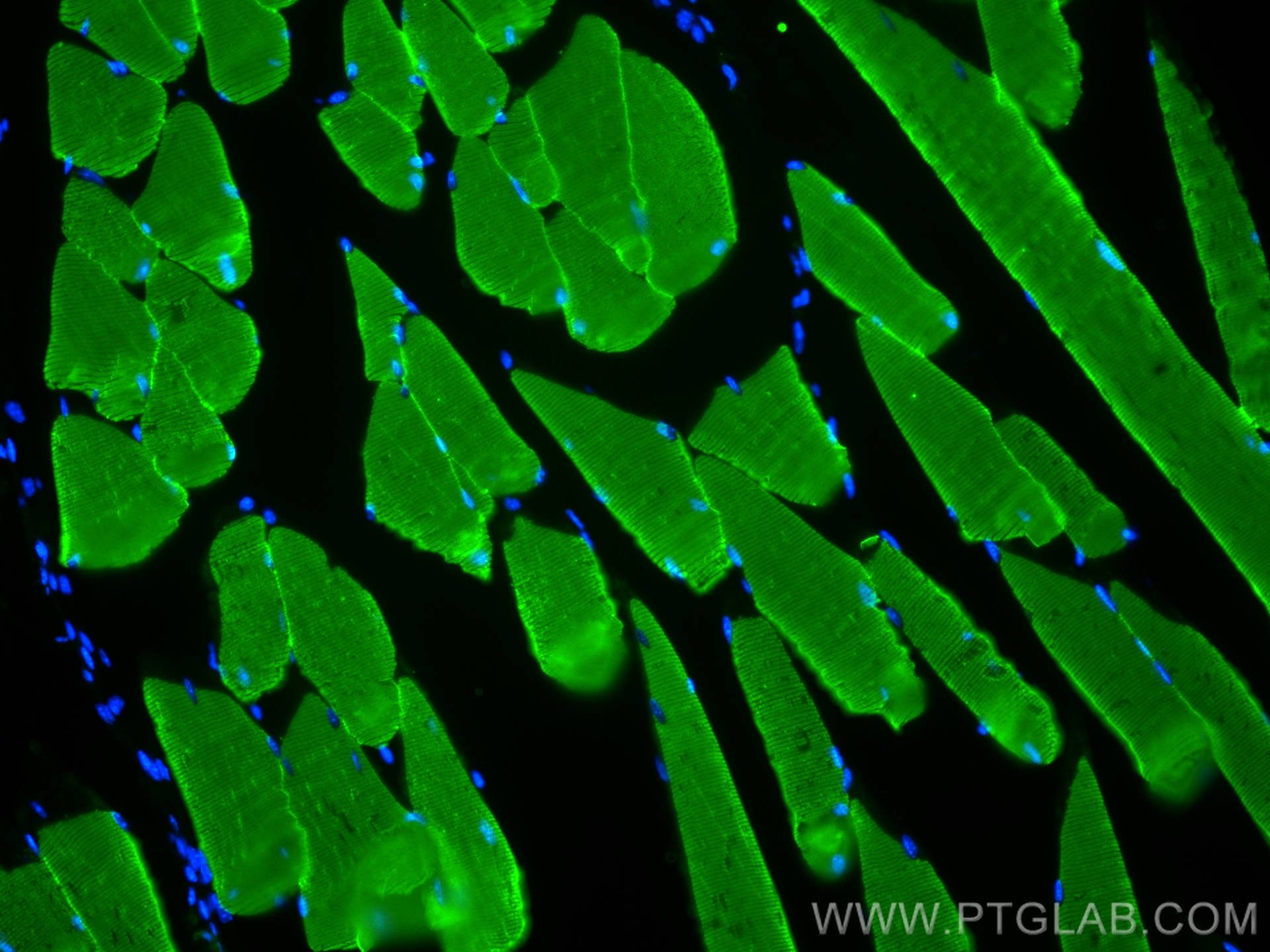
| Positive IF-P detected in | mouse skeletal muscle tissue |
Recommended dilution
| Application | Dilution |
|---|---|
| Western Blot (WB) | WB : 1:20000-1:100000 |
| Immunohistochemistry (IHC) | IHC : 1:2000-1:20000 |
| Immunofluorescence (IF)-P | IF-P : 1:400-1:1600 |
| It is recommended that this reagent should be titrated in each testing system to obtain optimal results. | |
| Sample-dependent, Check data in validation data gallery. | |
Published Applications
| WB | See 6 publications below |
| IHC | See 1 publications below |
| IF | See 2 publications below |
Product Information
67299-1-Ig targets MYH1 in WB, IHC, IF-P, ELISA applications and shows reactivity with human, mouse, pig samples.
| Tested Reactivity | human, mouse, pig |
| Cited Reactivity | human, mouse, pig, chicken |
| Host / Isotype | Mouse / IgG2a |
| Class | Monoclonal |
| Type | Antibody |
| Immunogen | MYH1 fusion protein Ag17129 Predict reactive species |
| Full Name | myosin, heavy chain 1, skeletal muscle, adult |
| Calculated Molecular Weight | 1939 aa, 223 kDa |
| Observed Molecular Weight | 220 kDa |
| GenBank Accession Number | BC114545 |
| Gene Symbol | MYH1 |
| Gene ID (NCBI) | 4619 |
| RRID | AB_2882563 |
| Conjugate | Unconjugated |
| Form | Liquid |
| Purification Method | Protein A purification |
| UNIPROT ID | P12882 |
| Storage Buffer | PBS with 0.02% sodium azide and 50% glycerol , pH 7.3 |
| Storage Conditions | Store at -20°C. Stable for one year after shipment. Aliquoting is unnecessary for -20oC storage. 20ul sizes contain 0.1% BSA. |
Background Information
MYH1 (MyHC-2x) encodes the IIX isoform of myosin heavy chain (MyHC). Myosin is a large, ubiquitous, motor protein that generates force through its interaction with actin, thus involving it in a number of cellular processes including cytokinesis, karyokinesis, cell migration, and muscle contraction. Muscle fibers can be divided as type 1 (slow) and type 2 (fast). MYH1 belongs to type 2.
Protocols
| Product Specific Protocols | |
|---|---|
| WB protocol for MYH1 antibody 67299-1-Ig | Download protocol |
| IHC protocol for MYH1 antibody 67299-1-Ig | Download protocol |
| IF protocol for MYH1 antibody 67299-1-Ig | Download protocol |
| Standard Protocols | |
|---|---|
| Click here to view our Standard Protocols |
Publications
| Species | Application | Title |
|---|---|---|
BMC Genomics Identification of different myofiber types in pigs muscles and construction of regulatory networks | ||
Food Funct Potential nutritional healthy-aging strategy: enhanced protein metabolism by balancing branched-chain amino acids in a finishing pig model. | ||
J Biol Chem Circular RNA circMYLK4 shifts energy metabolism from glycolysis to OXPHOS by binding to the calcium channel auxiliary subunit CACNA2D2 | ||
Int J Mol Sci Endurance Exercise-Induced Fgf21 Promotes Skeletal Muscle Fiber Conversion through TGF-β1 and p38 MAPK Signaling Pathway | ||
Aging (Albany NY) Myogenic exosome miR-140-5p modulates skeletal muscle regeneration and injury repair by regulating muscle satellite cells |
Reviews
The reviews below have been submitted by verified Proteintech customers who received an incentive for providing their feedback.
FH Moon (Verified Customer) (10-10-2022) | normal tissue-1/2 (N1/N2) tumor tissue-1/2/3 (T1/T2/T3)
 |
